Introduction
Totally implantable venous access ports (TIVAPs) have been widely used to provide long-term central venous access in patients with malignancy [
1]. Radiologically guided TIVAP placement has traditionally required two separate incisions: one for percutaneous puncture of the internal jugular vein (IJV), and another for creation of a subcutaneous pocket on the anterior chest wall, with a subcutaneous tunnel connecting the two. Although this conventional approach has been widely accepted, potential drawbacks include pain at the tunneling site, ecchymosis of the skin overlying the tunnel, discomfort with neck movement, and palpation of the catheter over the clavicle [
1]. In 2014, the authors introduced a single-incision technique for TIVAP placement via the axillary vein, in which the venipuncture and pocket creation are performed through a single short incision in the infraclavicular area, without a subcutaneous tunnel [
1]. Since then, this technique has been used as the default approach at the authors’ institution, and TIVAPs have been implanted via the axillary vein in more than 10,000 patients. Whereas the original report described the initial experience in 216 patients [
1], this article describes the technique as currently performed, including catheter selection, breast fixation in women, management of technical difficulties, and long-term outcomes.
Indications and Pre-procedural Planning
TIVAPs are indicated for long-term venous access, usually for chemotherapy in patients with malignancy, and less often for parenteral nutrition or repeated transfusion. Pre-procedural evaluation includes ultrasound of both axillary veins to assess depth, caliber, and patency at the planned puncture site and to identify anatomic variants or adjacent masses, and review of chest CT, when available, for pre-existing stenosis or thrombosis of the brachiocephalic vein and SVC. In patients with breast cancer, the contralateral side is selected.
Access Strategies
In interventional radiology practice, IJV access is generally preferred for TIVAP placement, and subclavian puncture is largely avoided because of the well-recognized risk of pinch-off syndrome. The IJV approach itself, however, requires a subcutaneous tunnel, and the catheter length is fixed before the tip position is confirmed.
The single-incision technique avoids these requirements by puncturing the axillary vein at the cephalic end of the same incision used for the port pocket [
1]. Because the axillary vein is punctured lateral to the first rib, the catheter is already located inside the vessel when it passes between the first rib and the clavicle, eliminating the risk of pinch-off syndrome. The need for a subcutaneous tunnel and a separate neck incision is removed, and the catheter length is fixed only after the tip position is confirmed under fluoroscopy.
The axillary approach is also useful when neck tumor, previous lymph node dissection, or radiation therapy makes IJV access unfavorable.
Technical Procedures (Video 1)
All procedures are performed in an interventional radiology suite under fluoroscopic guidance with bedside ultrasound. The default device is a 6.5-Fr Celsite Discreet port (B. Braun Medical, Boulogne, France), because in an analysis of 209 women with left IJV TIVAPs, an 8-Fr catheter carried a substantially higher cumulative incidence of left innominate vein stenosis than a 6.5-Fr catheter (HR, 20.766; p = 0.005) [
2].
The incision line is planned 3 to 4 cm lateral to the junction of the axillary vein and the clavicle, with a length of approximately 2 cm in an oblique vertical orientation. Following sterile preparation and local anesthesia, an oblique vertical skin incision is made. The axillary vein is then punctured under real-time ultrasound guidance at the cephalic end of the incision using a 21-gauge micropuncture needle, with the needle directed medially toward the first rib. A 0.018-inch microwire is advanced through the needle and into the SVC under fluoroscopic guidance, and a microintroducer is then inserted over the microwire [
1].
With the microintroducer left in place, the port pocket is created on the medial side of the incision by blunt dissection of the subcutaneous tissue, and gauze packing is applied to the pocket for hemostasis. A 0.035-inch spring guidewire is then advanced into the SVC through the microintroducer, and the microintroducer is exchanged for a peel-away sheath over the guidewire. The catheter is advanced through the peel-away sheath, and the tip is positioned between the upper third of the right atrium and the lower portion of the SVC under fluoroscopic monitoring. The peel-away sheath is removed, the catheter is connected to the port chamber, and the chamber is then placed within the pocket. The incision is closed in layers. Final fluoroscopic and chest radiographic confirmation of catheter tip position is obtained. In female patients, particularly those with pendulous breasts, the catheter tip migrates with positional change between supine and upright postures, which may cause the tip to project against the wall of the SVC. A tip projecting against the SVC wall has been implicated in fibrin sheath and thrombus formation [
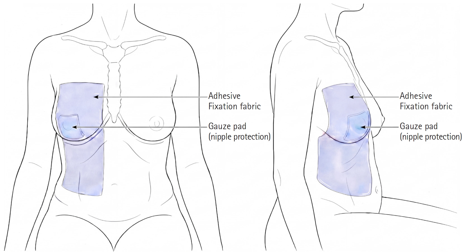
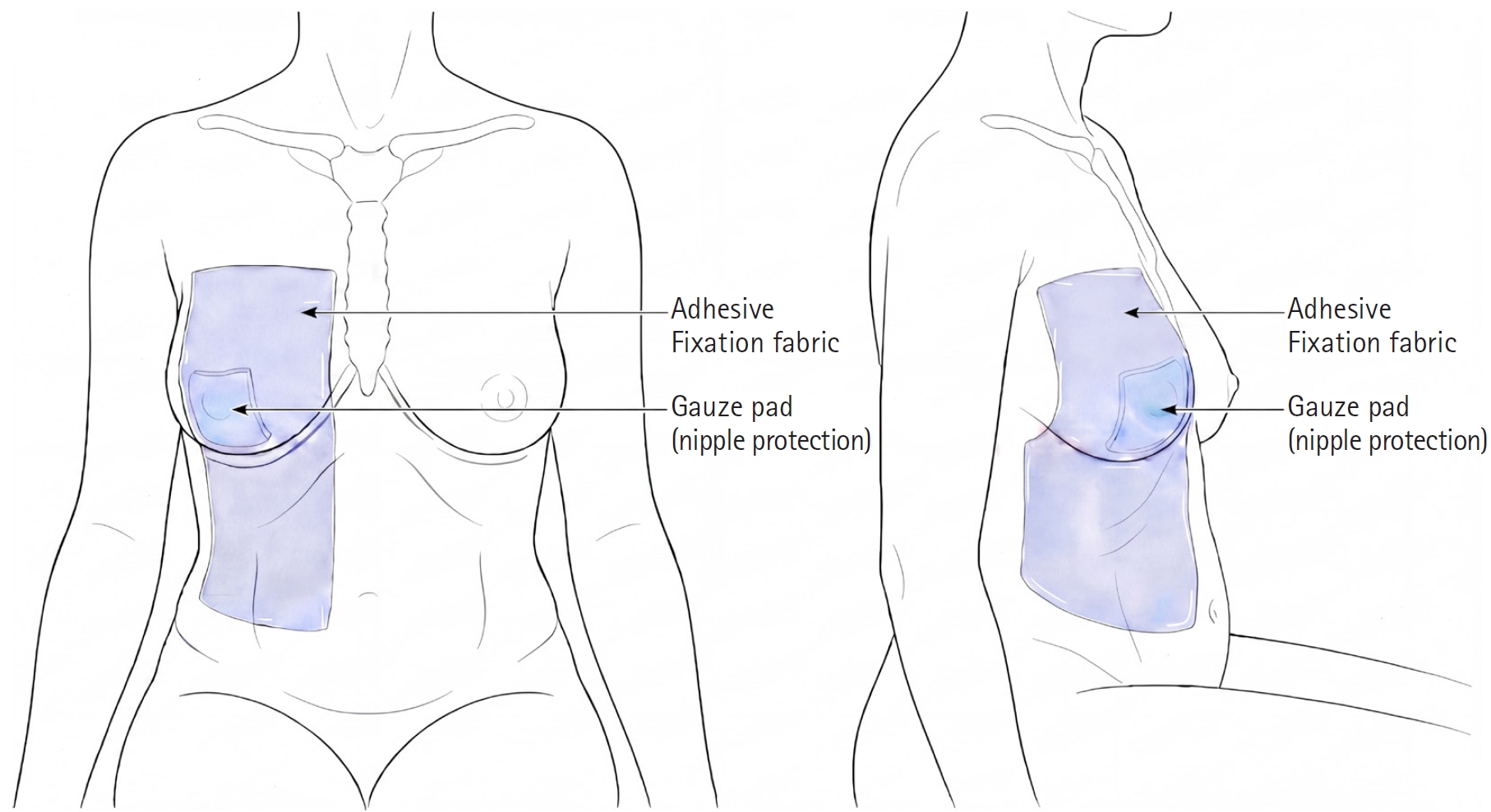
3], although the effect of breast fixation on these complications has not been evaluated. To reduce this positional migration, the ipsilateral breast is fixed to the abdominal wall with an adhesive nonwoven fabric while the patient is in the sitting position before draping (
Fig. 1). In an analysis of 118 women undergoing right axillary TIVAP placement, breast fixation was associated with significantly smaller changes in the distance ratio between the port chamber and the catheter tip on supine and erect chest radiographs [
3].
Technical Difficulties and Troubleshooting
Most technical difficulties are resolved with additional manipulation rather than a change of access route. When the axillary vein is small or collapses with respiration, a gentle end-expiratory breath-hold facilitates ultrasound-guided venipuncture. A wire or catheter that enters an unintended vein is redirected under fluoroscopic guidance. Catheter kinking at the cuff-catheter junction is corrected by dissecting the tissue between the puncture tract and the port pocket. Bending of the peel-away sheath, recognized as resistance to catheter passage, is overcome by withdrawing the sheath while advancing the catheter. If the axillary artery is punctured, the needle is removed, manual compression is applied, and the vein is repunctured under ultrasound guidance, or under venographic guidance if a hematoma has collapsed the vein. If access still cannot be obtained, the procedure is converted to either the IJV or the contralateral axillary vein, depending on tumor location and previous surgery or radiation therapy.
Clinical Outcomes
In the authors’ initial series of 216 patients, technical success was achieved in all cases. Immediate complications were limited to puncture-site hematoma in two patients (0.9%) [
1]. In a long-term analysis of 932 placements over 311,069 catheter days, no catheter fracture from pinch-off syndrome, catheter disconnection, or device inversion was identified [
4].
In a cohort of 4,773 patients, symptomatic axillary vein thrombosis developed in 18 patients (0.38%) at a median of 35.5 days after implantation, with pancreatic and lung cancer as independent risk factors [
5].
Conclusion
In the authors’ practice, single-incision TIVAP placement via the axillary vein has become the default approach. It avoids the drawbacks of subcutaneous tunneling, eliminates the risk of pinch-off syndrome, and has shown high technical success with a low rate of long-term complications, supporting it as a feasible alternative to the conventional two-incision technique.
Conflict of interest
No potential conflict of interest relevant to this article was reported.
Funding
None.
Acknowledgments
None.
Author contributions
Conceptualization: TSS, MGS. Data curation: WJY, MGS. Formal analysis: MGS, TSS. Investigation: MGS, TSS, WJY. Methodology: TSS, MGS, WJY. Project administration: MGS. Resources: MGS, TSS. Supervision: TSS, MGS. Validation: MGS, TSS. Visualization: WJY. Writing - original draft: WJY. Writing - review & editing: WJY, MGS, TSS.
Data availability statement
Data sharing does not apply to this article as no datasets were generated or analyzed during the current study.
Supplementary material
Video 1.
Step-by-step demonstration of the single-incision technique for totally implantable venous access port placement via the axillary vein.
Fig. 1.Ipsilateral breast fixation to reduce catheter tip migration after totally implantable venous access port placement in women with large pendulous breasts. (A) Frontal view showing the adhesive nonwoven fabric sheet applied from the inferior breast onto the upper abdominal wall, spanning the inframammary fold. (B) Lateral view demonstrating the breast held inferiorly against the abdominal wall by the fixation fabric. Fixation is performed in the sitting position to maximize gravitational descent of the breast tissue before adhesion. Gauze padding is placed over the nipple to reduce skin discomfort under the adhesive.
References
- 1. Seo TS, Song MG, Kang EY, Lee CH, Yong HS, Doo K. A single-incision technique for placement of implantable venous access ports via the axillary vein. J Vasc Interv Radiol. 2014;25:1439-1446. https://doi.org/10.1016/j.jvir.2013.12.571
- 2. Song MG, Seo TS, Kim YH, Cho SB, Chung HH, Lee SH, et al. Effect of catheter diameter on left innominate vein in breast cancer patients after totally implantable venous access port placement. J Vasc Access. 2018;19:615-619. https://doi.org/10.1177/1129729818765062
- 3. Song MG, Seo TS, Kim YH, Cho SB, Jung E, Chung HH, et al. Effectiveness of breast fixation to reduce migration of the tip of a totally implantable venous access port in women. J Vasc Access. 2016;17:348-352. https://doi.org/10.5301/jva.5000565
- 4. Seo TS, Song MG, Kim JS, Choi CW, Seo JH, Oh SC, et al. Long-term clinical outcomes of the single-incision technique for implantation of implantable venous access ports via the axillary vein. J Vasc Access. 2017;18:345-351. https://doi.org/10.5301/jva.5000751
- 5. Song MG, Seo TS, Yang WJ. Characteristics of symptomatic axillary vein thrombosis after placement of totally implanted venous access port in cancer patients. Iran J Radiol. 2020;17:e102754. https://doi.org/10.5812/iranjradiol.102754
Citations
Citations to this article as recorded by