Abstract
Complex hepatic cystic lesions with inconclusive imaging pose a recurring diagnostic problem: cyst-fluid cytology and conventional core-needle biopsy seldom capture the epithelial lining needed for a histological diagnosis, and current guidelines advise against simple cyst aspiration. We describe a sheath-assisted percutaneous forceps biopsy technique for direct cyst-wall sampling under ultrasound guidance, illustrated in two male patients (76 and 48 years). After Seldinger placement of an 8-Fr sheath into the cyst, rat-tooth forceps obtained direct biopsies of the wall epithelium, and the tract was embolized with Gelfoam. Technical success was achieved in both cases; histology showed columnar biliary epithelium consistent with mucinous cystic neoplasm, whereas fluid cytology was non-diagnostic. No complications occurred. This technique offers a feasible means of obtaining a histological diagnosis in complex hepatic cystic lesions when conventional methods are inconclusive.
-
Keywords: Mucinous cystic neoplasm; Percutaneous biopsy; Hepatic cystic lesion; Rat-tooth forceps; Ultrasound guidance; Cyst wall sampling
Introduction
Mucinous cystic neoplasm of the liver (MCN), historically termed biliary cystadenoma (BCA), is a rare cyst-forming epithelial tumor accounting for fewer than 5% of hepatic cystic lesions [
1-
3]. Under the current World Health Organization classification, the entity is defined by subepithelial ovarian-like stroma and distinguished from intraductal papillary neoplasm of the bile duct by the absence of biliary communication; lesions are graded from low-grade dysplasia to associated invasive carcinoma [
3]. Although MCN occurs predominantly in women, well-documented occurrence in men is recognized [
3]. The lesions are slow-growing, follow a stepwise tumorigenic progression, and carry a risk of malignant transformation, so complete surgical excision is the accepted standard and a tissue diagnosis materially influences management [
2,
4].
Preoperative distinction of MCN from simple, complicated, hydatid, and other cystic lesions is unreliable, as cross-sectional imaging features overlap substantially [
3,
5]. This uncertainty carries a clinical cost in both directions: a meaningful proportion of patients undergo major hepatectomy on presumptive grounds, while a cautious radiological differential of “rule out biliary cystadenoma” for what is in fact a simple cyst drives unnecessary surgery. In one tertiary series, 19 of 20 lesions resected for presumed BCA proved to be simple cysts [
6]. Tissue diagnosis would resolve this, yet conventional approaches fail: cyst-fluid cytology is limited by low cellularity and patchy epithelial desquamation, standard trucut biopsy is designed to core solid tissue, and contemporary multi-society guidance accordingly advises against cyto-aspiration or needle biopsy of complex cysts [
2,
7]. The diagnostic substrate—the columnar epithelial lining, and where present the ovarian-like stroma—is rarely captured by these methods.
Sampling tissue through an introducer sheath or working channel is a well-established principle, used routinely in endoscopy, cholangioscopy, and percutaneous transhepatic biliary procedures [
8-
11]. More recent endoscopic advances—such as endoscopic ultrasonography (EUS)–guided sampling of pancreatic cysts and cholangioscopy-guided forceps biopsy of biliary lesions—share the same underlying rationale: in cystic disease, the epithelial wall must be sampled directly to obtain a diagnosis [
11,
12]. Its application to percutaneous hepatic cyst-wall biopsy, however, has not been systematically described. We report a sheath-assisted percutaneous technique for direct cyst-wall sampling using rat-tooth forceps under ultrasound guidance, with technical and histopathological outcomes in two cases of complex hepatic cystic lesions confirmed as MCN in which conventional investigations were inconclusive. The contribution is not a new device but a reproducible adaptation of trans-sheath sampling to the percutaneous hepatic route, targeting the precise substrate that fluid cytology and core-needle biopsy miss.
Case Report
Case 1
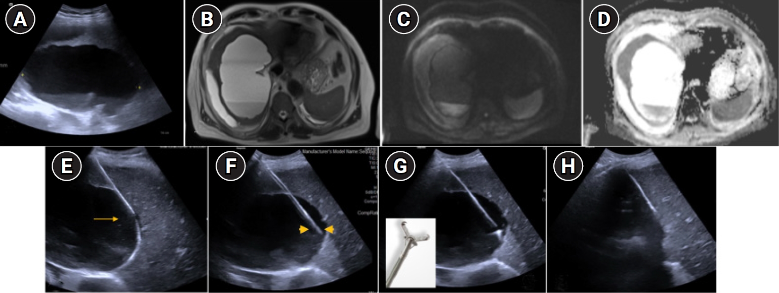
A 76-year-old man presented with a six-month history of right upper quadrant pain, intermittent fever, and rigors. Examination revealed mild right hypochondrial tenderness. Investigations showed leukocytosis (14.47 × 10³/µL), markedly elevated C-reactive protein (145.7 mg/L), and mildly raised gamma-glutamyl transferase (96 U/L); hydatid enzyme-linked immunosorbent assay serology was negative. Ultrasound demonstrated a multiloculated hypoechoic cyst (10 × 8 cm) in the right hepatic lobe with internal echogenic debris. Contrast-enhanced MRI confirmed a well-defined multiloculated cystic lesion (10.2 × 8.4 × 7.6 cm) involving segments VII, VIII, and IVa, with thin enhancing septations and a subcapsular component, and no solid component, diffusion restriction, or biliary communication (
Fig. 1A–
1D). The principal differentials were MCN, complicated simple cyst, and hydatid disease, the last considered unlikely given negative serology and absent biliary communication. Following multidisciplinary team (MDT) review, percutaneous biopsy was agreed as the safest route to tissue diagnosis.
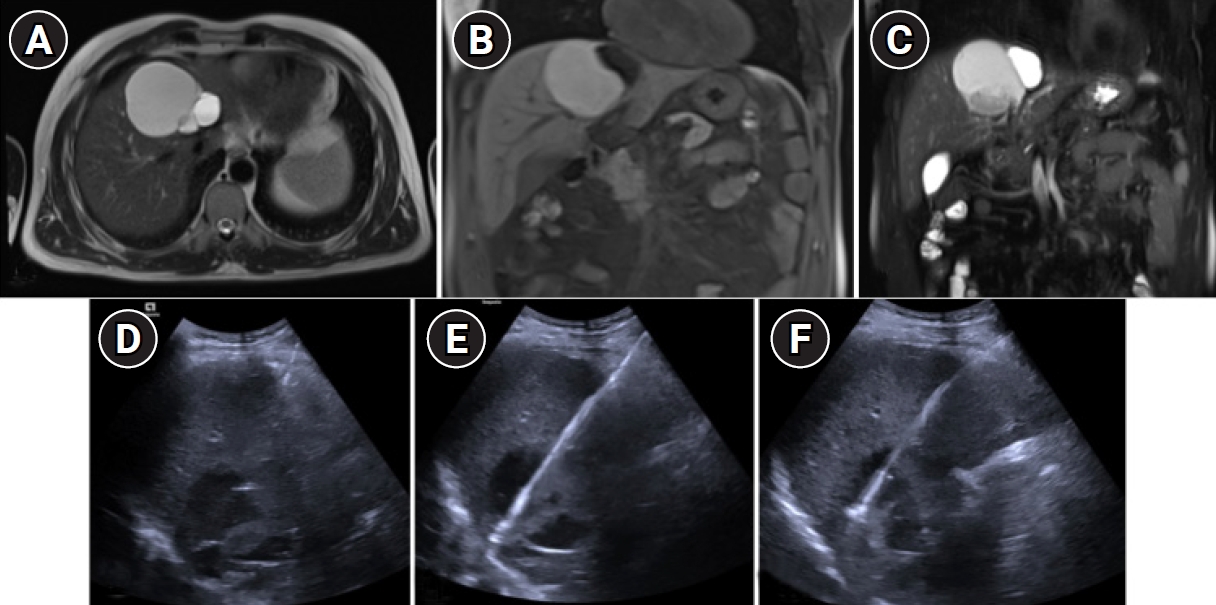
A 48-year-old man presented with right upper quadrant pain and progressive abdominal fullness. Investigations showed borderline IgG anti-Echinococcus antibody (12.83 IU/mL), mildly raised total bilirubin (2.2 mg/dL), and normal serum CA 19-9 (31.8 U/mL). Ultrasound showed a large multiloculated hypoechoic lesion in segment IVa with heterogeneous contents and dependent debris. MRI demonstrated a T2-hyperintense multiloculated cystic lesion (6.3 × 7.9 × 6.6 cm) in segments VIII/IVa with a T2-hypointense rim, delayed peripheral enhancement, and partial T1 hyperintensity indicating proteinaceous contents, without solid component, diffusion restriction, or biliary communication (
Fig. 2A–
2C). Given the borderline serology, an initial diagnosis of hydatid disease was made and oral albendazole (400 mg twice daily) commenced; after 12 weeks without clinical or radiological response, MDT review recommended percutaneous biopsy before surgical planning.
Both procedures were performed under local anesthesia with pre-procedural intravenous antibiotic prophylaxis, using continuous real-time ultrasound guidance and full aseptic precautions; local anesthetic was infiltrated to the level of the hepatic capsule. An access route traversing a cuff of intervening hepatic parenchyma was selected in both patients. An 18-gauge needle was used to puncture the anterior cyst wall under direct visualization; a 0.035-inch guidewire was advanced into the cavity and the tract serially dilated to accept an 8-Fr introducer sheath positioned with its tip just within the lumen (
Figs. 1E,
2). Rat-tooth biopsy forceps (Olympus FB-230U, Olympus Medical Systems, Tokyo, Japan) were advanced through the sheath and deployed against the inner wall under real-time ultrasound guidance (
Fig. 1F), obtaining multiple full-thickness punch biopsies of the epithelial lining (
Figs. 1G,
2E)—three samples in case 1 and four in case 2. Cyst fluid was then aspirated for cytology (
Figs. 1H,
2F), and the access tract was embolized with Gelfoam slurry to mitigate hemorrhage.
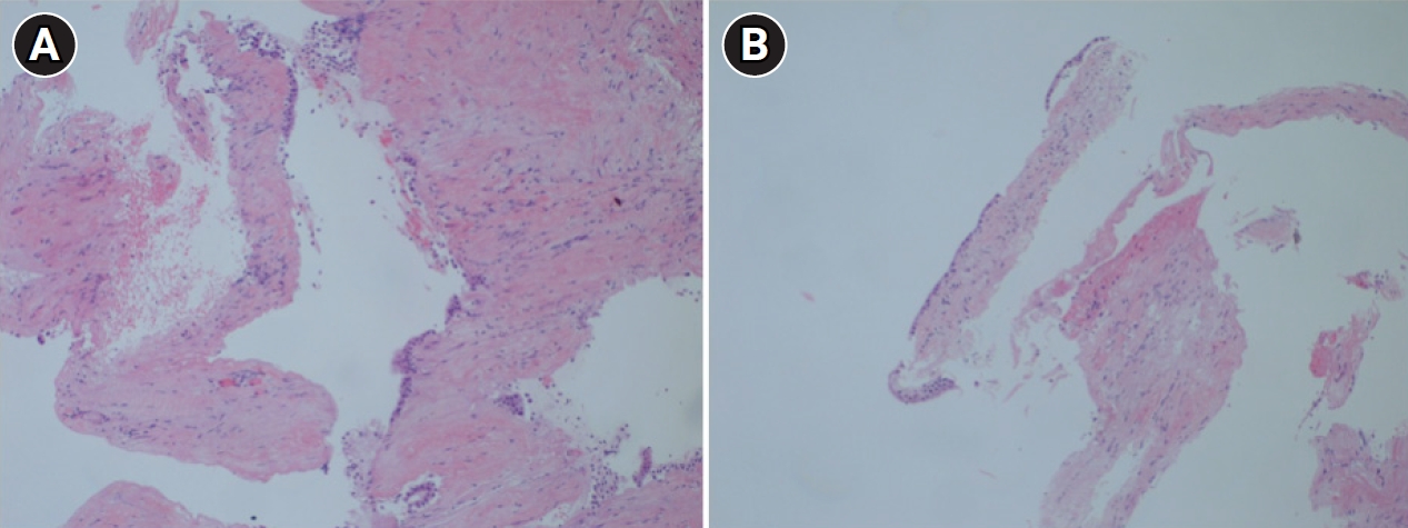
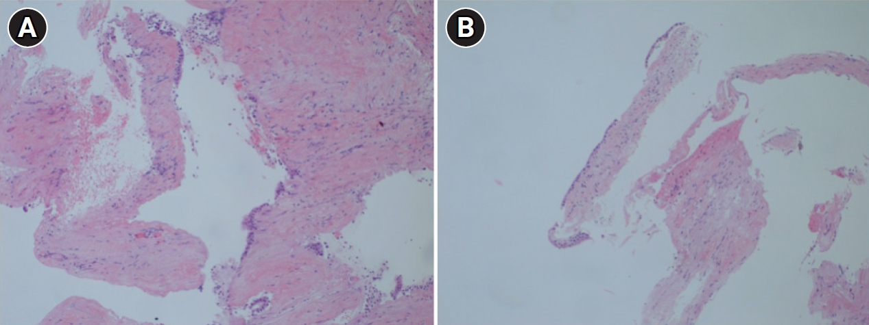
Technical success was achieved in both cases. Histopathology of the forceps specimens showed the cyst wall lined by a single layer of tall columnar biliary epithelium in both patients (
Fig. 3). In contrast, cyst-fluid cytology demonstrated only inflammatory cells with no diagnostic epithelial elements. The diagnosis was subsequently confirmed on the resection specimens, which demonstrated ovarian-like stroma diagnostic of mucinous cystic neoplasm of the liver. Both patients were monitored until surgery and developed no persistent abdominal pain, clinically significant bleeding, bile leak, sepsis, or other procedure-related complication. Each underwent elective hepatic resection within one week, with no intraoperative evidence of tract-related sequelae.
This report was conducted in accordance with the Declaration of Helsinki. The Institutional Review Board granted a waiver of formal approval for this retrospective report of two de-identified cases. Written informed consent for the procedure and for publication of clinical details and accompanying images was obtained from both patients.
Discussion
The histological diagnosis of MCN is a recognized challenge. On imaging, it presents as a multiloculated cystic lesion with enhancing septations, features shared by hydatid cysts, complicated, hemorrhagic simple cysts, cystic metastases, and hepatic abscess, so confident imaging-based diagnosis is unreliable [
3,
5]. Cyst-fluid cytology yields a diagnosis only when intact columnar epithelium is sampled—uncommon, given desquamation and patchy neoplastic distribution and serum and intracystic tumor markers lack specificity [
2,
7]. The described technique addresses this gap directly: whereas tru-cut biopsy traverses a cystic wall tangentially or exits into fluid, rat-tooth forceps advanced through an indwelling coaxial sheath can be deployed under real-time ultrasound control against the inner wall and withdrawn with a full-thickness epithelial bite, capturing the diagnostic epithelial lining—mechanistically analogous to forceps biopsy through a bronchoscope or cholangioscope working channel, transposed to a percutaneous hepatic route. In both cases, direct wall sampling yielded diagnostic columnar epithelium whereas cyst-fluid cytology showed only inflammatory cells, illustrating the potential diagnostic advantage of sampling the epithelial lining directly; this observation, based on two cases, requires confirmation in larger series.
Trans-sheath forceps biopsy is itself established in several clinical settings—and recent endoscopic experience with EUS through-the-needle microforceps for pancreatic cysts and cholangioscopy-guided forceps biopsy underscores the shared move toward direct epithelial sampling in cystic disease [
11,
12]. The novel element here is the percutaneous hepatic adaptation: Seldinger sheath placement into the cyst, intraluminal forceps deployment using ultrasound guidance for full-thickness epithelial bites, and Gelfoam tract embolization.
Regarding the site of sampling, no high-level evidence directly compares biopsy of the free surface of the cyst with a wall buttressed by parenchyma. By analogy with percutaneous liver-mass biopsy, a trajectory through a cuff of intervening normal parenchyma is conventionally preferred, the parenchyma providing a tamponade effect and limiting intraperitoneal leakage of cyst contents—of particular relevance to a potentially neoplastic mucinous lesion, in which peritoneal spillage is undesirable [
13]. Since cystic lesions have a thin wall, diagnostic yield would be higher when the sample is acquired from the free wall than from an area surrounded by liver parenchyma. In both cases, the route was planned through intervening parenchyma where feasible, with Gelfoam tract embolization, an established hemostatic adjunct [
14,
15]. A sheathed working channel may also reduce seeding relative to repeated needle passes, although this risk cannot be eliminated completely for a potentially malignant cystic neoplasm.
Nomenclature warrants comment. Current World Health Organization terminology designates this entity a mucinous cystic neoplasm, defined by ovarian-like stroma, reserving intraductal papillary neoplasm of the bile duct for lesions with biliary communication; the older term biliary cystadenoma persists in clinical and surgical usage, and the two are frequently used interchangeably [
1-
3]. Definitive subclassification rests on the resection specimen, as ovarian-like stroma and focal dysplasia may not be represented in a wall biopsy. Both patients here were male—an uncommon but documented presentation [
3]—underscoring that, where ovarian-like stroma is not demonstrated, classification as MCN should be made with caution and the differential of intraductal papillary neoplasm considered.
Complete surgical excision remains the cornerstone of management for mucinous cystic neoplasm, serving both as definitive treatment and as the only reliable means of excluding associated invasive carcinoma, since a non-targeted percutaneous biopsy may not sample the most atypical focus [
3]. Non-resective approaches such as aspiration, sclerotherapy, and fenestration carry high recurrence and are inappropriate for a neoplastic lesion [
3].
Establishing the necessity of tissue confirmation is central to the rationale for this technique. Because MCN cannot be reliably distinguished from simple, complicated, hydatid, or other cystic lesions on imaging, and because its malignant potential mandates resection whereas benign mimics do not, a preoperative histological diagnosis directly determines the management pathway. Confirmation of MCN averts two opposing errors: unnecessary major hepatectomy for a lesion that proves to be a simple cyst—reported in up to 19 of 20 resections for presumed BCA [
6]—and inappropriate non-resective treatment (aspiration or sclerotherapy) of a neoplastic lesion, which risks recurrence and peritoneal dissemination of mucinous contents. It also redirects management away from unnecessary medical therapy, as in case 2, where 12 weeks of empirical albendazole for presumed hydatid disease preceded a tissue-proven diagnosis of MCN. Accordingly, this technique is not a first-line investigation for all hepatic cysts but should be reserved—by MDT consensus involving interventional radiology, hepatobiliary surgery, and hepatology—for lesions that remain indeterminate after full imaging and cytological workup, where non-operative management has failed, or where histology will alter the management pathway.
This report is limited to two cases, insufficient to establish safety, efficacy, or generalizability, and includes no formal comparison with alternative techniques. The approach adds modest procedural complexity relative to simple aspiration. Larger prospective or multicenter series are needed to define diagnostic yield, the number of passes required for adequacy, complication and tract-seeding rates at follow-up, and the technique’s place relative to EUS-guided wall biopsy within the diagnostic algorithm.
Sheath-assisted percutaneous forceps biopsy of the cyst wall under ultrasound guidance is a technically feasible means of obtaining a histological diagnosis in complex hepatic cystic lesions when imaging and cytology are inconclusive. In two cases of MCN, direct epithelial sampling yielded diagnostic epithelium where fluid cytology did not, contributing to a histological diagnosis that informed surgical management. Prospective evaluation in larger cohorts is warranted to define its safety profile, optimal procedural parameters, and role within the diagnostic algorithm for complex hepatic cystic lesions.
Conflict of interest
No potential conflict of interest relevant to this article was reported.
Funding
None.
Acknowledgments
None.
Author contributions
Conceptualization: SSB. Data curation: VP. Formal analysis: VP, SY. Methodology: VP, SY, SSB. Writing of the original draft: SS. Writing of the review & editing: SS, AK, SSB.
Data availability statement
Data sharing does not apply to this article as no datasets were generated or analyzed during the current study.
Fig. 1.Case 1 (76-year-old man). (A) Grayscale ultrasound: multiloculated hypoechoic cyst (10 × 8 cm) in the right hepatic lobe with internal echogenic debris. (B–D) Axial MRI (T2-weighted, diffusion-weighted, and apparent diffusion coefficient mapping): well-defined multiloculated cystic lesion (10.2 × 8.4 × 7.6 cm) in segments VII/VIII/IVa with subcapsular extension; diffusion restriction, no solid component or biliary communication. (E) Seldinger access: guidewire (arrow) within the cyst lumen prior to 8-Fr sheath insertion. (F) Rat-tooth forceps advanced through the sheath (arrowheads) and deployed against the cyst wall. (G) Ultrasonography: Forceps jaw opening against the wall and withdrawal with tissue sample. (H) Post-biopsy cyst-fluid aspiration and the tract was embolized with Gelfoam.
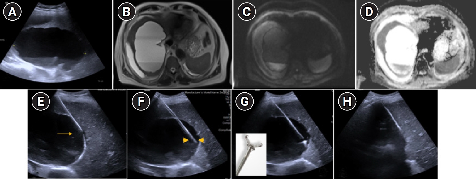
Fig. 2.Case 2 (48-year-old man). (A–C) Axial and coronal MRI: T2-hyperintense multiloculated cystic lesion (6.3 × 7.9 × 6.6 cm) in segments VIII/IVa with a T2-hypointense rim, delayed peripheral enhancement, and partial T1 hyperintensity consistent with proteinaceous contents; no solid component or biliary communication. (D) Grayscale ultrasound: multiloculated cyst with heterogeneous dependent debris. (E) Ultrasonography: Rat-tooth forceps deployed through the sheath for cyst-wall sampling. (F) Post-biopsy cyst-fluid aspiration (cytology: inflammatory cells only); the tract was embolized with Gelfoam.
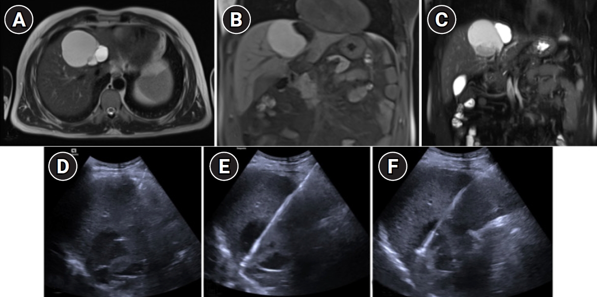
Fig. 3.Histopathology (H&E, ×100). Cyst-wall specimens from case 1 (A) and case 2 (B), each showing a single layer of tall columnar biliary epithelium lining the cyst wall, consistent with mucinous cystic neoplasm/biliary cystadenoma. Cyst-fluid cytology was non-diagnostic in both cases, confirming that direct wall sampling was essential for histological diagnosis.
References
- 1. Hutchens JA, Lopez KJ, Ceppa EP. Mucinous cystic neoplasms of the liver: epidemiology, diagnosis, and management. Hepat Med. 2023;15:33-41. https://doi.org/10.2147/HMER.S284842
- 2. Soares KC, Arnaoutakis DJ, Kamel I, Anders R, Adams RB, Bauer TW, et al. Cystic neoplasms of the liver: biliary cystadenoma and cystadenocarcinoma. J Am Coll Surg. 2014;218:119-128. https://doi.org/10.1016/j.jamcollsurg.2013.08.014
- 3. Lee MH, Katabathina VS, Lubner MG, Shah HU, Prasad SR, Matkowskyj KA, et al. Mucin-producing cystic hepatobiliary neoplasms: updated nomenclature and clinical, pathologic, and imaging features. Radiographics. 2021;41:1592-1610. https://doi.org/10.1148/rg.2021210011
- 4. Aziz H, Hamad A, Afyouni S, Kamel IR, Pawlik TM. Management of mucinous cystic neoplasms of the liver. J Gastrointest Surg. 2023;27:1963-1970. https://doi.org/10.1007/s11605-023-05709-6
- 5. Kim JY, Kim SH, Eun HW, Lee MW, Lee JY, Han JK, et al. Differentiation between biliary cystic neoplasms and simple cysts of the liver: accuracy of CT. AJR Am J Roentgenol. 2010;195:1142-1148. https://doi.org/10.2214/AJR.09.4026
- 6. Kumar A, Zendel A, Batres M, Gerber DA, Desai CS. Radiologic reporting of simple hepatic cyst versus biliary cystadenoma may lead to unnecessary surgery. Am Surg. 2023;89:1392-1395. https://doi.org/10.1177/00031348211054077
- 7. Pompili M, Ardito F, Brunetti E, Cabibbo G, Calliada F, Cillo U, et al. Benign liver lesions 2022: guideline for clinical practice of Associazione Italiana Studio del Fegato (AISF), Societa Italiana di Radiologia Medica e Interventistica (SIRM), Societa Italiana di Chirurgia (SIC), Societa Italiana di Ultrasonologia in Medicina e Biologia (SIUMB), Associazione Italiana di Chirurgia Epatobilio-Pancreatica (AICEP), Societa Italiana Trapianti d'Organo (SITO), Societa Italiana di Anatomia Patologica e Citologia Diagnostica (SIAPEC-IAP) - Part I - Cystic lesions. Dig Liver Dis. 2022;54:1469-1478. https://doi.org/10.1016/j.dld.2022.08.030
- 8. Liu Y, Zhou X, Kong L, Han X, Jiao D. Percutaneous transhepatic intraluminal forceps biopsy for patients with biliary stricture after endoscopic retrograde approach failure: a retrospective study. Quant Imaging Med Surg. 2023;13:2605-2619. https://doi.org/10.21037/qims-22-915
- 9. Pawlak KM, Juzwiak J, Brzosko M, Piatkowski J, Jackowski M, Jagielski M. Fluoroscopy-guided, sheath-assisted biopsy for indeterminate intraductal biliary masses. Endoscopy. 2026;58:E156-E157. https://doi.org/10.1055/a-2777-6802
- 10. Sakamoto H, Ishiwatari H, Yamamura M, Doi T, Sato J, Kakuda Y, et al. Utility of a novel sheath designed for mapping biopsy for preoperative malignant hilar biliary obstruction. Endosc Int Open. 2026;14:a27871325. https://doi.org/10.1055/a-2787-1325
- 11. Perez-Cuadrado-Robles E, Phillpotts S, Bronswijk M, Conrad CC, Binda C, Monino L, et al. PERcutaneous transhepatic CHOLangioscopy using a new single-operator short cholangioscope (PERCHOL): European feasibility study. Dig Endosc. 2024;36:719-725. https://doi.org/10.1111/den.14697
- 12. Guilabert L, Nikolic S, de-Madaria E, Vanella G, Capurso G, Tacelli M, et al. Endoscopic ultrasound for pancreatic cystic lesions: a narrative review. BMJ Open Gastroenterol. 2025;12:e001893. https://doi.org/10.1136/bmjgast-2025-001893
- 13. Thanos L, Zormpala A, Papaioannou G, Malagari K, Brountzos E, Kelekis D. Safety and efficacy of percutaneous CT-guided liver biopsy using an 18-gauge automated needle. Eur J Intern Med. 2005;16:571-574. https://doi.org/10.1016/j.ejim.2005.06.010
- 14. Singhal S, M DP, Inuganti S, Botcha S, Deepashree DT, Uthappa MC. Percutaneous ultrasound-guided plugged liver biopsy: a single-centre experience. Pol J Radiol. 2021;86:e239-e245. https://doi.org/10.5114/pjr.2021.105852
- 15. Nota T, Kageyama K, Yamamoto A, Jogo A, Sohgawa E, Yonezawa H, et al. Feasibility of gel-like radiopaque embolic material using gelatin sponge and contrast agent for tract embolization after percutaneous treatment. PLoS One. 2023;18:e0281384. https://doi.org/10.1371/journal.pone.0281384