Abstract
-
Purpose
This study aimed to evaluate the clinical efficacy and patency of stents placed for symptomatic iliac vein or IVC obstruction caused by malignant tumors.
-
Material and Methods
A total of 34 consecutive patients with malignant iliac vein or IVC obstruction underwent stent placement. We reviewed their electronic medical records, pre- and post-procedural CT scans, and angiograms retrospectively, and evaluated technical success, clinical success, and patient-based and vessel-based reocclusion rates confirmed by follow-up cross-sectional images. The reocclusion rate difference between IVC and iliac stents was also assessed.
-
Results
All patients complained of leg and/or trunk edema. The stents were placed in the following locations: IVC (n = 9); iliac veins (n = 19); and both IVC and iliac veins (n = 6). Technical success rate was 100% and the clinical success rate was 87.5% during hospitalization. Cross-sectional area of the affected thigh was significantly decreased after stent placement (mean 11.7%, p = 0.003). The median follow-up period was 65 days (Q1–Q3, 28.5–227.3; IQR, 198.8). The patient-based reocclusion rate was 42.8%. The vessel-based reocclusion rates for IVC (12.5%) tended to be lower than those of iliac veins (45.0%), though the difference was not statistically significant (p = 0.194).
-
Conclusion
Stent insertion for iliac vein or IVC obstruction secondary to malignancy is technically feasible and can help to improve patients’ symptoms. IVC stents tend to achieve a higher rate of patency than iliac vein stents.
-
Keywords: Iliac vein; Vena cava, inferior; Stents; Neoplasm; Venous thrombosis
Introduction
Iliocaval venous obstructions have been successfully treated over the past few decades with minimally invasive interventions such as stent deployment [
1,
2]. These obstructions are often secondary to cancers. Especially, malignant tumors and their metastases originating from the hepatobiliary system, gastrointestinal tract and genitourinary organs can cause iliocaval venous obstruction. Metastatic lymph nodes or the tumors themselves can cause venous outflow obstruction with or without subsequent venous thrombosis by invasion or extrinsic compression of the iliocaval venous system. Venous thrombosis occurs in more than 1% of cancer patients every year and patients diagnosed with a malignancy have a four-fold higher risk of developing a venous thrombosis [
3].
Venous outflow obstruction causes ambulatory venous hypertension, ascites, renal insufficiency, and chronic venous insufficiency [
4-
7]. Furthermore, venous outflow obstruction caused by venous thromboembolism not only erodes quality of life through symptoms that include swelling, pain, and venous stasis ulcer, but also can increase mortality in cancer patients [
8].
Traditionally, emergent radiation therapy had been considered the standard treatment for malignant SVC syndrome (SVCS) [
9]. In the past decades, endovascular treatment including stent placement, angioplasty, thrombolysis, and thrombectomy has emerged as a superior and faster alternative therapy [
10], and is widely accepted as first-line therapy for emergent cases.
Apart from SVCS, malignant iliocaval obstruction, which can be regarded as an analogue of SVCS for the lower extremities, has been investigated far less. In the literature, malignancy-related iliocaval obstruction is treated as a subset of venous obstructions that are due to various causes or that affect various anatomical locations [
2,
11,
12]. There are several clinical studies on the usefulness of stent placement for malignant IVC syndrome [
13-
18]. And only few studies dealt with iliac vein obstruction [
19,
20]. Therefore, the purpose of our study was to evaluate the clinical efficacy and patency of the stents placed for symptomatic iliac vein or IVC obstruction caused by tumor invasion or extrinsic compression.
Materials and Methods
This is a single-center, retrospective cohort study. The cases were identified using a searching system of our institution (Severance Clinical Research Analysis Portal). Our institutional review board (Severance Hospital Human Research Protection Center) approved this retrospective study (protocol number: 4-2018-0619), and written informed consent for the procedure was obtained from all patients. Written consent for inclusion in the retrospective study was waived as per our institution’s policies.
Patient Population
Thirty-four consecutive patients who underwent iliocaval venous stent placement from May 2003 to October 2014 due to symptomatic malignancy-related iliac vein or IVC occlusion were included in this study. Electronic medical records (EMR) were reviewed to identify basic characteristics, including sex, age, etiology, and the clinical symptoms and signs associated with venous obstruction before and after treatment.
Endovascular Treatment
The malignancy-related iliocaval venous obstructions were diagnosed by CT venography and were confirmed by catheter-based venography in all patients. For the patients with evident venous thrombosis on the pre-procedural CT venography, intravenous heparin infusion (dose, 18 IU/kg/h; target activated partial thromboplastin time range, 40 to 69 seconds) was initiated at least 6 hours before the endovascular procedure. One of two types of retrievable IVC filter (Günther Tulip until August 2014 and Celect from September 2014, Cook Medical, Bloomington, IN, USA) was inserted before the procedure to prevent pulmonary thromboembolism, at the operator’s discretion.
For the patient with patent external iliac and femoro-popliteal veins, the femoral fossa was prepared in the usual sterile manner with the patient in the supine position (n = 20). In cases with external iliac vein occlusion and/or thrombosis in the femoro-popliteal vein, we used popliteal access with the patient in the prone position (n = 14). After local anesthesia administration of 2% lidocaine, the common femoral or popliteal vein was punctured under ultrasonographic guidance with a 21-gauge micropuncture introducer set (Cook Medical). A 0.035-inch hydrophilic guide wire (Terumo, Tokyo, Japan) was advanced to the iliac vein, and the introducer sheath with variable size (8–14 Fr) was inserted. After advancing the 0.035-inch guide wire to the iliac vein, a 5-Fr angiography catheter (Omni flush, Angio-Dynamics, Latham, MD, USA, or Contralateral, Boston Scientific, Watertown, MA, USA) was inserted, and pull-back venography was performed.
After confirming the diagnosis and extent of the thrombus, a rotating angiographic catheter was used to inject a small amount of diluted contrast medium to macerate the thrombus, and repetitive aspiration thrombectomy using the 11-Fr percutaneous introducer catheter (Arrow-Flex percutaneous introducer set; Arrow International, Reading, MA, USA) was performed. The manner of aspiration thrombectomy is described elsewhere [
21].
For the patients with thrombosis, we injected 3,000 IU of heparin intravenously before aspiration thrombectomy. Heparin was not used during the procedure for the patients without thrombosis.
An endovascular stent was inserted to recanalize the obstructed segment. Four types of self-expandable stents were placed. These included Hercules (S&G Biotech, Seongnam, Korea), S.M.A.R.T. Control (Cordis, Miami Lakes, FL, USA), E-Luminexx (Bard, Murray Hill, NJ, USA), and Wallstent (Boston Scientific, Galway, Ireland). Stents were chosen individually based on site and size of venous obstruction. Generally, Hercules stents (20–28 mm in diameter) were used for IVC obstructions and the other types of stents (10–14 mm in diameter) were used for iliac venous obstruction. Usually only one stent was deployed for each site of venous obstruction. However, if the length of a stent was shorter than the entire length of malignant venous obstruction, an additional stent was deployed to cover the entire lesion. The additional stent was required in nine cases (IVC, 1; right iliac vein, 3; left iliac vein, 5). Post-stent balloon dilatation was performed in 32 cases of persistent residual stenosis after stent insertion (except for two patients who showed immediate full expansion of the stents). Follow-up CT scans were taken as needed, and at the physician’s discretion.
Unless contraindicated (conditions including recent surgery or major hemorrhage, and central nervous system metastases), postprocedural anticoagulation with warfarin and/or enoxaparin was generally administered for 1–3 months.
Follow-up
All patients were followed up by the referring physician till July 2014 or the patient’s death. Clinical outcomes were assessed by reviewing the EMR. Radiological follow-up was made at the physician’s decision to reevaluate underlying cancer.
Outcomes
The primary outcome of this study was clinical success, which was defined as the reported resolution or improvement of symptoms related to the iliocaval venous obstruction after the interventional procedure. We also reviewed technical success, change of cross-sectional area of mid-thigh, and reocclusion rate. Technical success was defined as successful recanalization of the target lesion and restoration of venous return at the end of the interventional procedure. For more objective response evaluation, pre- and post-procedural cross-sectional areas of ipsilateral mid-thigh (for unilateral iliac vein occlusion) or sum of bilateral mid-thigh (for IVC or both iliac vein occlusion) were measured in cases where both pre- and post-procedural CT scans within 1 month after the procedure covering mid-thigh are available. Occurrence of reocclusion was evaluated by reviewing available follow-up CT scans.
In accordance with the Society of Interventional Radiology Standards of Practice Committee Classification of Complications by Outcome [
22], minor and major complications related to the interventional procedure were reviewed retrospectively.
We evaluated patient-based reocclusion rate (number of patients who experienced reocclusion of any stented vessels/number of total patients enrolled) and vessel-based reocclusion rates (number of stented vessels that were reoccluded during follow-up/number of all the stented vessels in the cohort). The difference between IVC and iliac stent reocclusion rates was analyzed by using Fisher’s exact test. Pre- and post-procedural mid-thigh areas were compared using a Wilcoxon signed-rank test. Data are presented as the mean ± standard deviation or as the median (range) for continuous variables and proportions (%) for categorical variables. A two-sided significance level of 0.05 was used to evaluate statistical significance. A statistical software package (SPSS Statistics version 20.0.0.2 for Windows, IBM Corp., Armonk, NY, USA) was used for data analysis.
Results
The patients consisted of 18 men and 16 women. Median age was 62.5 years (range, 36 to 89 years). The iliocaval venous obstructions were sequelae of the following cancers: colorectal (n = 9); ureter and bladder transitional cell carcinoma (n = 6); cervical (n = 5); prostate (n = 3); pancreatic cancer (n = 3); and one case each of vaginal, testicular, ovarian, breast, mixed Mullerian tumor, renal cell carcinoma, liposarcoma, and metastasis of unknown origin. Before the interventional treatments, the patients complained of edema occurring in both legs (n = 16), the right leg (n = 8), the left leg (n = 9), or truncal edema (n = 1). One patient with bilateral leg edema also complained of scrotal swelling. Twelve patients (35.3%) had deep vein thrombosis (DVT) associated with the venous obstruction, while 22 patients (64.7%) had symptomatic venous obstruction without thrombosis.
The median time interval between preprocedural CT scan and intervention was 3.5 days (range, 1 to 30 days). Aspiration thrombectomy was performed for all the patients who had DVT (n = 12) and more than 90% of thrombi were removed angiographically in all those patients. The degree of thrombus removal was determined by visual estimation and consensus of two interventional radiologists. Sites of stent deployment were: IVC alone (n = 9); unilateral (n = 14) or bilateral iliac veins (n = 5); and both IVC and iliac vein (n = 6). (
Table 1).
During the follow-up period (median, 65 days; range, 1 to 3,650 days), 12 patients (35.3%) died, 21 patients (61.8%) were lost to follow-up, and one patient (2.9%) was alive. The technical success rate was 100% (34/34). Clinical information regarding symptom improvement after intervention was available in only 24 patients (70.6%), and the clinical success rate was 87.5% (21/24). There were no procedure-related complications.
Follow-up CT scans were available in 21 patients (61.8%), and the median time period of imaging follow-up was 65 days (range, 7 to 3,167 days). Both pre- and post-procedural CT scans (within 1 month after the procedure) covering the mid-thigh were available in only 16 patients. Fourteen patients had decreased cross-sectional area of the mid-thigh level and the other two patients did not show a decrease. Mean percent change of the area ([pre-procedural area – post-procedural area]/pre-procedural area) was 11.7% (median, 10.4; Q1–Q3, 5.6 to 17.3; IQR, 11.7; p = 0.003).
Patient-based reocclusion rate was 42.8% (9/21) during the follow-up period. The overall vessel-based reocclusion rate was 35.7% (10/28), where the reocclusion rate of IVC stents was 12.5% (1/8) and that of iliac vein stents was 45% (9/20) (
Table 2). The reocclusion rate of IVC stents tended to be lower than that of iliac vein stents, though the result was not statistically significant (p = 0.194) (
Figs. 1,
2). Six patients required reintervention (additional stent placement in five patients, sole balloon angioplasty in one patient) and secondary patency was achieved in all cases.
Discussion
Malignancy-related iliocaval venous obstructions have been frequently treated with stenting, yet there is a lack of research regarding the clinical efficacy of stent placement when compared with SVCS. Our study demonstrated a 100% technical success rate and an 87.5% clinical success rate. These results are comparable with previous case studies of stenting for malignant iliocaval obstruction, which reported 94%–100% and 77%–95% technical and clinical success rates, respectively [
19,
20,
23-
25].
Clinical success, which was defined as the improvement of symptoms, was not graded nor scored, and was only determined by the physicians’ report recorded in EMR. To present more objective results, cross-sectional areas of the mid-thigh were measured on pre- and post-procedural CT scans. Although the cross-sectional areas significantly decreased on post-procedural CT scans compared to those of pre-procedural CT scans, this data was derived from only 47.1% (16/34) of patients. Hence, this result should be considered only as supplementary data.
Reocclusion of the venous stents was not uncommon, and the reocclusion rate was over 40% during the 2-month median follow-up period. At first glance, this reocclusion rate seems to be higher than those of SVC stents, which have reported primary patency from 64% to 92% [
23-
25]. However, reocclusion of IVC stents occurred in only one patient out of eight in our study, and this rate is comparable with the reported primary patency rates of SVC stents. The previous studies dealing with iliocaval stenting also reported 36.8% of symptom recurrence [
19] and 68.8% of primary patency [
20], which are similar to the results of our study.
The reocclusion rate of iliac vein stents, though statistically not significant, tended to be higher than that of IVC stents (45% vs. 12.5%, p = 0.194). The low patency rate of iliac vein stents may be explained by the differences in stent diameters. The diameters of IVC stents (range, 20 to 28 mm) were much larger than those of iliac vein stents (range, 10 to 14 mm). Reocclusions are generally amenable to secondary intervention, and the literature reports secondary patency rates ranging from 93%–100% [
20,
23-
25]. Out of nine patients who experienced reocclusion, only 6 patients received reintervention. The remaining three were in poor general condition and did not want to receive further aggressive treatment.
The present study examined both the efficacy and patency of iliocaval stents, but there were many limitations. This was a retrospective study and included a relatively small number of patients. Because the types of malignancies causing the venous obstructions were so variable, and due to the small number of patients in the study, we could not evaluate the clinical treatment efficacy and stent patency based on cancer type. We experienced a shortage of follow-up data. The majority of enrolled patients were in advanced or terminal stages of cancer and many patients died within a few days of treatment or were transferred to hospice centers or nursing homes and subsequently lost to follow-up. Therefore, we could evaluate clinical improvement of symptoms in only 70.6% (24/34) of the patients, and follow-up CT scans were only available for 61.8% (21/34). Therefore, we cannot insist on the clinical success of our study as a concrete conclusion, because selection bias may have been introduced. Patients who were unavailable for follow-up, including those who died shortly after the procedure or were transferred to hospice care, may have had poorer clinical outcomes. Therefore, the reported clinical success rate may have been overestimated. For the purpose of showing the outcome as objectively as possible, we calculated the change in cross-sectional area of the mid-thigh. But this information was also available in only 16 patients, and the interval between procedure and area measurement varied extremely (29–395 days). The follow-up period, defined as the hospitalization period in this facility, was relatively short and variable because most patients had terminal illnesses and were lost to follow-up after being sent to nursing care facilities. The follow-up CT scans tended to be performed more frequently in patients with persistent or recurrent symptoms, and this may have contributed to an increased reocclusion rate. Another limitation is that the small sample size precluded an adequate statistical analysis to identify factors affecting stent patency.
In conclusion, endovascular stent deployment can be an effective treatment option for malignant iliocaval venous obstruction. This is particularly true in cases of IVC obstruction, where IVC stent placement demonstrates higher patency rates. Although there is a fairly high possibility of reocclusion, especially with iliac vein stent placement, the use of aggressive interventions, including stent deployment, can lead to relief of symptoms for patients suffering from malignant iliocaval venous obstruction. More studies are needed to confirm our results and assess long-term patency.
Conflict of interest
No potential conflict of interest relevant to this article was reported.
Funding
None.
Acknowledgments
None.
Author contributions
Conceptualization: GMK, WS. Data curation: GMK, WS. Formal analysis: WS. Investigation: GMK, KH, MDK, JHK, JYW, JP. Methodology: GMK. Validation: KH, MDK, JHK. Writing - original draft: WS, GMK. Writing - review & editing: KH, JP.
Data availability statement
Data are available upon reasonable request from the corresponding author, subject to ethical and privacy limits.
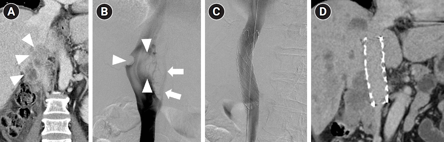
Fig. 1.A 68-year-old man with a history of right nephrectomy due to renal cell carcinoma presented with bilateral leg swelling. (A) CT scan shows a recurrent mass compressing the IVC (arrowheads). (B) Vena cavogram shows filling defects (arrowheads) at the same level. Note the collateral veins (arrows), suggesting IVC stenosis. (C) Vena cavogram shows disappearance of collateral veins, implying decreased pressure gradient, after the deployment of a 24 mm × 10 cm self-expandable stent (Hercules, S&G Biotech). (D) Follow-up CT scan (3 months after intervention) shows a widely patent IVC stent.
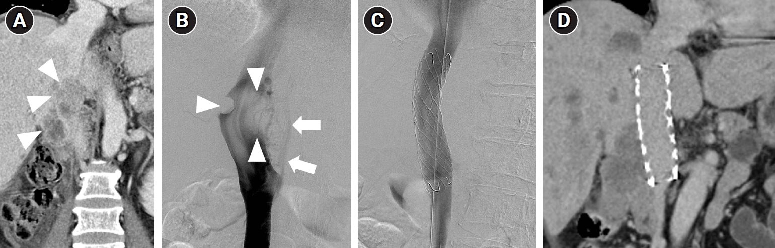
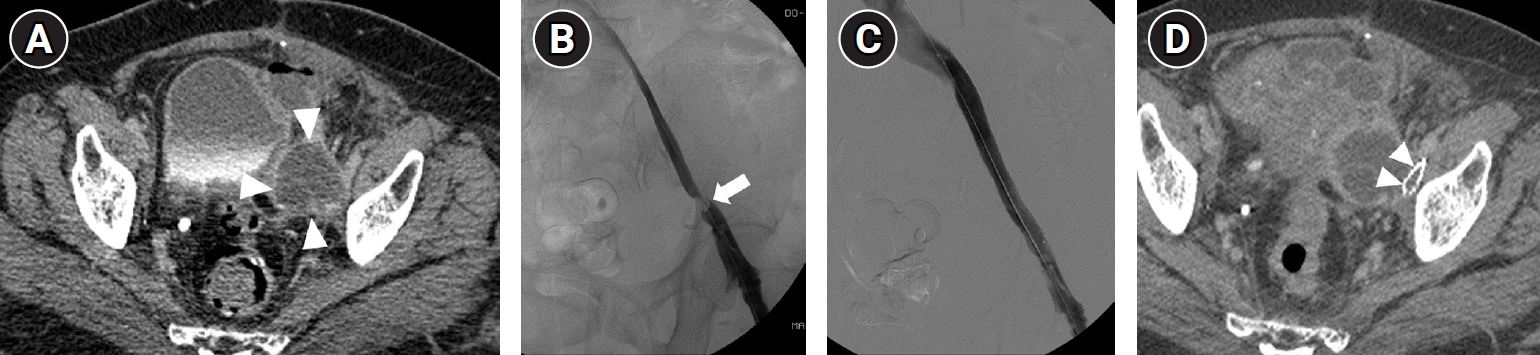
Fig. 2.A 72-year-old woman with a mixed Mullerian tumor complained of left leg swelling. (A) CT scan shows a metastatic lymph node at the left iliac chain (arrowheads). (B) Venography after aspiration thrombectomy shows extrinsic compression (arrow) of the left external iliac vein by the lymph node. (C) After a 14 mm × 10 cm self-expandable stent (E-Luminexx, Bard) was deployed, the venous flow was restored immediately and the edema improved gradually. (D) One month later, the patient complained of recurrent leg swelling. Follow-up CT scan shows a collapsed stent (arrowheads), and recurrent deep vein thrombosis.
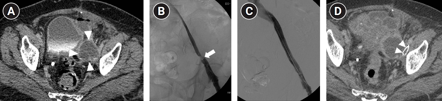
Table 1.
Table 1.
|
Characteristic |
Value |
|
Age (years) |
62.5 (36–89) |
|
Sex |
|
|
Male |
18 (52.9) |
|
Female |
16 (47.1) |
|
Etiology |
|
|
Colorectal cancer |
9 (26.5) |
|
Transitional cell carcinoma |
6 (17.6) |
|
Cervical cancer |
5 (14.7) |
|
Prostate cancer |
3 (8.8) |
|
Pancreatic cancer |
3 (8.8) |
|
Others |
8 (23.5) |
|
Cause of obstruction |
|
|
Extrinsic compression |
12 (35.3) |
|
Invasion |
22 (64.7) |
|
Sites of stent |
|
|
IVC |
9 (26.5) |
|
Iliac veins |
19 (55.9) |
|
IVC + iliac veins |
6 (17.6) |
Table 2.
Table 2.
|
Site of stents |
Reocclusion rate |
|
Patient-based |
|
|
Total |
42.8 (9/21) |
|
IVC |
20.0 (1/5) |
|
Bilateral iliac veins |
66.7 (2/3) |
|
Unilateral iliac veins |
60.0 (6/10) |
|
IVC + iliac veins |
0 (0/3) |
|
Vessel-based |
|
|
IVC |
12.5 (1/8) |
|
Iliac veins |
45.0 (9/20) |
References
- 1. AbuRahma AF, Perkins SE, Wulu JT, Ng HK. Iliofemoral deep vein thrombosis: conventional therapy versus lysis and percutaneous transluminal angioplasty and stenting. Ann Surg. 2001;233:752-760. https://doi.org/10.1097/00000658-200106000-00004
- 2. Titus JM, Moise MA, Bena J, Lyden SP, Clair DG. Iliofemoral stenting for venous occlusive disease. J Vasc Surg. 2011;53:706-712. https://doi.org/10.1016/j.jvs.2010.09.011
- 3. Horsted F, West J, Grainge MJ. Risk of venous thromboembolism in patients with cancer: a systematic review and meta-analysis. PLoS Med. 2012;9:e1001275. https://doi.org/10.1371/journal.pmed.1001275
- 4. Barnes RW, Collicott PE, Sumner DS, Strandness DE Jr. Noninvasive quantitation of venous hemodynamics in the postphlebitic syndrome. Arch Surg. 1973;107:807-814. https://doi.org/10.1001/archsurg.1973.01350230159029
- 5. Johnson BF, Manzo RA, Bergelin RO, Strandness DE Jr. Relationship between changes in the deep venous system and the development of the postthrombotic syndrome after an acute episode of lower limb deep vein thrombosis: a one- to six-year follow-up. J Vasc Surg. 1995;21:307-312. https://doi.org/10.1016/s0741-5214(95)70271-7
- 6. Markel A, Manzo RA, Bergelin RO, Strandness DE Jr. Valvular reflux after deep vein thrombosis: incidence and time of occurrence. J Vasc Surg. 1992;15:377-384. https://doi.org/10.1016/0741-5214(92)90259-B
- 7. Meissner MH, Moneta G, Burnand K, Gloviczki P, Lohr JM, Lurie F, et al. The hemodynamics and diagnosis of venous disease. J Vasc Surg. 2007;46 Suppl S:4S-24S. https://doi.org/10.1016/j.jvs.2007.09.043
- 8. Sorensen HT, Mellemkjaer L, Olsen JH, Baron JA. Prognosis of cancers associated with venous thromboembolism. N Engl J Med. 2000;343:1846-1850. https://doi.org/10.1056/NEJM200012213432504
- 9. Straka C, Ying J, Kong FM, Willey CD, Kaminski J, Kim DW. Review of evolving etiologies, implications and treatment strategies for the superior vena cava syndrome. Springerplus. 2016;5:229. https://doi.org/10.1186/s40064-016-1900-7
- 10. Rachapalli V, Boucher LM. Superior vena cava syndrome: role of the interventionalist. Can Assoc Radiol J. 2014;65:168-176. https://doi.org/10.1016/j.carj.2012.09.003
- 11. Nazarian GK, Austin WR, Wegryn SA, Bjarnason H, Stackhouse DJ, Castaneda-Zuniga WR, et al. Venous recanalization by metallic stents after failure of balloon angioplasty or surgery: four-year experience. Cardiovasc Intervent Radiol. 1996;19:227-233. https://doi.org/10.1007/BF02577640
- 12. Xiao L, Tong JJ, Shen J. Endoluminal treatment for venous vascular complications of malignant tumors. Exp Ther Med. 2012;4:323-328. https://doi.org/10.3892/etm.2012.589
- 13. Kuetting D, Thomas D, Wilhelm K, Pieper CC, Schild HH, Meyer C. Endovascular management of malignant inferior vena cava syndromes. Cardiovasc Intervent Radiol. 2017;40:1873-1881. https://doi.org/10.1007/s00270-017-1740-z
- 14. Brountzos EN, Binkert CA, Panagiotou IE, Petersen BD, Timmermans H, Lakin PC. Clinical outcome after intrahepatic venous stent placement for malignant inferior vena cava syndrome. Cardiovasc Intervent Radiol. 2004;27:129-136. https://doi.org/10.1007/s00270-003-0009-x
- 15. Devcic Z, Techasith T, Banerjee A, Rosenberg JK, Sze DY. Technical and anatomic factors influencing the success of inferior vena caval stent placement for malignant obstruction. J Vasc Interv Radiol. 2016;27:1350-1360. https://doi.org/10.1016/j.jvir.2016.02.030
- 16. Fletcher WS, Lakin PC, Pommier RF, Wilmarth T. Results of treatment of inferior vena cava syndrome with expandable metallic stents. Arch Surg. 1998;133:935-938. https://doi.org/10.1001/archsurg.133.9.935
- 17. Kishi K, Sonomura T, Fujimoto H, Kimura M, Yamada K, Sato M, et al. Physiologic effect of stent therapy for inferior vena cava obstruction due to malignant liver tumor. Cardiovasc Intervent Radiol. 2006;29:75-83. https://doi.org/10.1007/s00270-004-0324-x
- 18. Laing AD, Thomson KR, Vrazas JI. Stenting in malignant and benign vena caval obstruction. Australas Radiol. 1998;42:313-317. https://doi.org/10.1111/j.1440-1673.1998.tb00529.x
- 19. Maleux G, Vertenten B, Laenen A, De Wever L, Heye S, Clement P, et al. Palliative endovascular treatment of cancer-related iliocaval obstructive disease: technical and clinical outcomes. Acta Radiol. 2016;57:451-456. https://doi.org/10.1177/0284185115582059
- 20. Rabellino M, Moltini P, Di Caro V, Garcia-Monaco R. Symptomatic iliofemoral and iliocaval venous thrombosis in patients with cancer: endovascular treatment. Vasc Endovascular Surg. 2018;52:602-606. https://doi.org/10.1177/1538574418789018
- 21. Park SI, Lee M, Lee MS, Kim MD, Won JY, Lee DY. Single-session aspiration thrombectomy of lower extremity deep vein thrombosis using large-size catheter without pharmacologic thrombolysis. Cardiovasc Intervent Radiol. 2014;37:412-419. https://doi.org/10.1007/s00270-013-0676-1
- 22. Omary RA, Bettmann MA, Cardella JF, Bakal CW, Schwartzberg MS, Sacks D, et al. Quality improvement guidelines for the reporting and archiving of interventional radiology procedures. J Vasc Interv Radiol. 2003;14:S293-S295. https://doi.org/10.1097/01.rvi.0000094601.83406.e1
- 23. Fagedet D, Thony F, Timsit JF, Rodiere M, Monnin-Bares V, Ferretti GR, et al. Endovascular treatment of malignant superior vena cava syndrome: results and predictive factors of clinical efficacy. Cardiovasc Intervent Radiol. 2013;36:140-149. https://doi.org/10.1007/s00270-011-0310-z
- 24. Nagata T, Makutani S, Uchida H, Kichikawa K, Maeda M, Yoshioka T, et al. Follow-up results of 71 patients undergoing metallic stent placement for the treatment of a malignant obstruction of the superior vena cava. Cardiovasc Intervent Radiol. 2007;30:959-967. https://doi.org/10.1007/s00270-007-9088-4
- 25. Lanciego C, Pangua C, Chacon JI, Velasco J, Boy RC, Viana A, et al. Endovascular stenting as the first step in the overall management of malignant superior vena cava syndrome. AJR Am J Roentgenol. 2009;193:549-558. https://doi.org/10.2214/AJR.08.1904


 , Gyoung Min Kim2,*