Abstract
In a 58-year-old man with HCC undergoing TACE, a segment III tumor was supplied by an accessory left hepatic artery arising from the left gastric artery (LGA). The dominant technical challenge shifted from acute angulation at the LGA origin to inadequate back support in distal branches. To overcome repeated dislodgement, the catheter was exchanged for a 5-Fr catheter with a hand-cut side hole, positioned at the LGA orifice while the tip was anchored in the celiac trunk. This technique allowed a microcatheter to traverse in a single well-supported pass, enabling successful superselective TACE without immediate complications. This case emphasizes the importance of distinguishing ostial angulation from back-support failure and highlights the utility of proximal side-hole anchoring in complex multivessel selection.
-
Keywords: Chemoembolization, therapeutic; Carcinoma, hepatocellular; Hepatic artery; Anatomic variation; Catheterization
Introduction
Knowledge of variant hepatic arterial anatomy is essential for safe and effective TACE of HCC. An accessory or replaced left hepatic artery (LHA) arising from the left gastric artery (LGA) is among the most common hepatic arterial variants, reported in up to 16% of patients undergoing hepatic angiography [
1].
Selective catheterization of acutely angulated branches remains a technical challenge during TACE. When the primary obstacle is the acute takeoff angle itself, a shepherd’s hook-shaped microguidewire is often effective for engaging the ostium by allowing rotation and withdrawal maneuvers [
2,
3]. In contrast, when the main limitation is inadequate back support—particularly in small-caliber or tortuous distal vessels—the microcatheter-guidewire system tends to dislodge repeatedly despite successful initial engagement. In such situations, a catheter with a surgically created side hole can provide stable support by anchoring the main catheter shaft in a proximal, larger-caliber vessel while permitting the microcatheter to exit laterally through the side hole toward the target branch [
4-
7].
We report a case in which both problems occurred sequentially during a single TACE procedure: acute angulation at the LGA origin and subsequent back-support failure in the accessory LHA and its tumor feeder.
Case Report
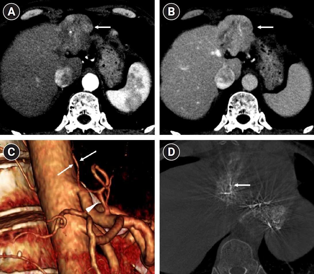
Institutional Review Board approval was waived for this case report in accordance with the policy of Seoul National University Hospital. A 58-year-old man with hepatitis B virus–related liver cirrhosis on entecavir and HCC (Child-Pugh class A5) presented for further locoregional treatment. He had previously undergone transarterial radioembolization, radiation therapy to segment II, and four prior sessions of conventional TACE. Contrast-enhanced CT demonstrated a hypervascular nodule in segment III, and three-dimensional CT angiography suggested an accessory LHA from the mid-portion of LGA (
Fig. 1).
Celiac arteriography was performed with a 5-Fr Rösch hepatic (RH) catheter (Grafia, Sungjin-Hitech, Suwon, Korea), which showed the LGA arising from the celiac trunk at an acute angle. A shepherd’s hook technique using a microcatheter (Progreat Lambda, 1.7-Fr, Terumo, Tokyo, Japan) and microguidewire (Transend, Boston Scientific, Marlborough, MA, USA) was used to select the LGA successfully. An accessory LHA arose from an intermediate portion of the LGA’s course; selecting this vessel caused the microcatheter-guidewire system to dislodge from the ostium on two occasions before successful selection of the accessory LHA was achieved on a third attempt (
Video 1).
From the accessory LHA, a segment III tumor-feeding branch arose, as confirmed by cone-beam CT obtained via the catheter positioned in the accessory LHA (
Fig. 1D). While advancing toward this feeder, the catheter could not provide adequate back support, and the microcatheter system repeatedly dislodged before reaching the feeder (
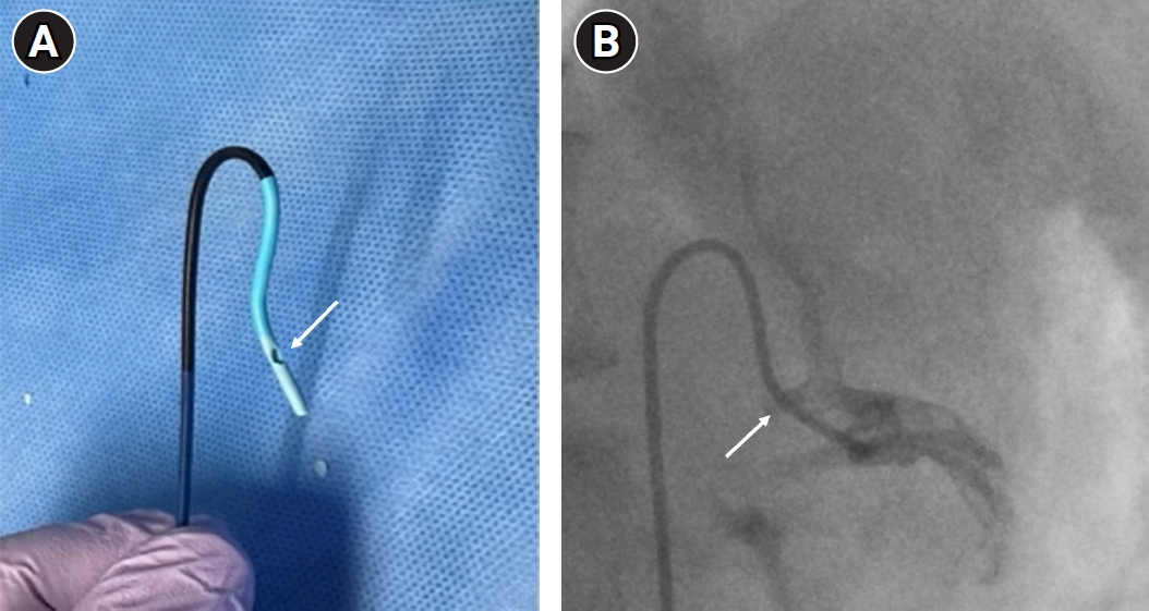
Video 1). The catheter was therefore exchanged for a 5-Fr Simmons type 1 catheter (J.S Angio Catheter, Simmons 1, A&A M.D., Seongnam, Korea), and a side hole approximately 3 mm in length was carefully and gradually incised into the catheter wall using a No. 11 scalpel blade (
Fig. 2A). In this case, the side hole was created approximately 7 mm proximal to the catheter tip; the exact location relative to the tip was individualized according to the distance and angular relationship between the celiac trunk and the LGA origin. After creation, the catheter was flushed once with heparinized saline to confirm patency. The microcatheter was subsequently advanced through the side hole to verify that the opening was adequately sized for smooth passage. No sharp edges were produced at the cut margin. The Simmons catheter was seated at the proximal portion of the celiac trunk, and the side hole was aligned with the orifice of the LGA (
Fig. 2B). The microcatheter was then advanced through the side hole over a 0.016-inch microguidewire (ASAHI Meister, Asahi Intecc, Seto, Japan). No resistance or shearing was encountered during microcatheter manipulation, and the system traversed the LGA and accessory LHA in a single, well-supported pass, achieving successful superselective catheterization of the segment III feeder (
Video 2). Angiography via the microcatheter confirmed a tumor blush corresponding to the segment III nodule.
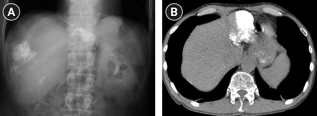
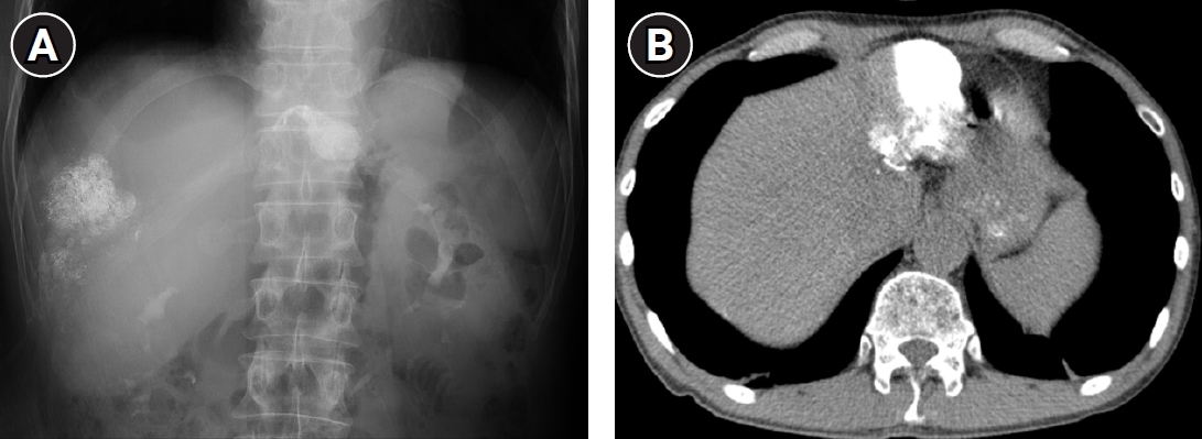
TACE was performed by infusing an emulsion of 5 mL of iodized oil (Lipiodol Ultra-Fluid, Guerbet, Paris, France) and 5 mg of idarubicin (Zavedos, Pfizer, New York, NY, USA) into the segment III feeder, followed by embolization with gelatin sponge particles (100–150 μm [PrimeGel, PLmicromed, Yansan, Korea] and 350–560 μm [EGgel, Engain, Hwaseong, Korea]) until flow stasis was achieved. The procedure was completed without complications. Follow-up CT demonstrated compact, dense lipiodol uptake confined to the treated tumor, consistent with a favorable treatment response (
Fig. 3).
Discussion
This case illustrates that the dominant technical challenge during selective catheterization can shift within a single TACE procedure, necessitating real-time recognition and adaptation of strategies. Initially, the acute angulation of the LGA origin from the celiac trunk was effectively overcome using the shepherd’s hook microguidewire technique [
2,
3]. However, once the accessory LHA was engaged, repeated dislodgement occurred during advancement toward the segment III feeder despite no sharp angulation at that level. This pattern clearly indicates inadequate back support rather than ostial angulation as the limiting factor. Such failure typically reflects an inability of the catheter shaft to transmit forward pushing force to the microcatheter: rather than advancing distally, the shaft buckles or loops within the larger, more compliant vessel just proximal to the target ostium. Differentiating these two mechanisms is clinically important because the optimal solution differs: shepherd’s hook reshaping primarily addresses engagement failure, whereas back-support failure requires enhanced stability during distal advancement [
4-
7].
The side-hole catheter technique was first introduced by Miyayama et al. [
4] for selective catheterization of the inferior phrenic artery and has since been applied to various difficult branches, including the internal mammary artery [
5], anomalous bronchial and subclavian arteries [
6], and hepatic arterial variants [
7-
9]. In most reported cases, the side hole was created near the catheter tip and positioned directly at or immediately adjacent to the target vessel ostium, with the catheter tip acting as a distal anchor [
4,
5,
8-
10]. In the present case, we applied a proximally anchored configuration, in which the side hole was aligned with the LGA orifice while the main catheter tip remained stable in the large-caliber celiac trunk. This allowed the microcatheter to traverse the LGA, accessory LHA, and segment III feeder in one continuous, well-supported pass without repeated re-engagement of the parent vessels. A Simmons type 1 catheter was chosen for the side-hole technique because its reformed, reverse-curve tip forms a self-retaining, hook-shaped anchor once seated in the celiac trunk, providing reliable back support for the side-hole maneuver by bracing the catheter shaft at this fixed distal anchor point. A similar proximal anchoring approach was described by Meng et al. [
7] using transradial access for a replaced LHA. Our case further demonstrates the utility of this proximal configuration in complex multivessel selection during conventional transfemoral TACE, especially when cumulative loss of back support develops across multiple distal branches.
The success of this technique also relies on precise orientation of the side hole toward the target ostium under fluoroscopic guidance. Creation of a side hole using a scalpel blade is technically simple and requires no specialized equipment [
9,
10]. Nevertheless, operators should be cautious of potential drawbacks, including weakening of the catheter shaft, risk of microcatheter or guidewire shearing at the sharp edges, and possible interaction with embolic materials. In this case, the microcatheter advanced smoothly without any resistance or shearing, and no procedure-related complications occurred.
In conclusion, systematic evaluation of the failure pattern after each catheterization step—whether the system fails to engage the ostium or dislodges during advancement—can guide timely selection between shepherd’s hook and side-hole techniques. When cumulative loss of back support across multiple distal branches is the main issue, a proximally anchored side-hole catheter offers a practical solution. This adaptive approach may improve procedural efficiency and safety in patients with challenging hepatic arterial anatomy during TACE.
Conflict of interest
No potential conflict of interest relevant to this article was reported.
Funding
None.
Acknowledgments
None.
Author contributions
Conceptualization: HCK. Data curation: MGK. Project administration: HCK. Resources: HCK. Supervision: HCK. Visualization: MGK, HCK. Writing - original draft: MGK. Writing - review & editing: HCK.
Data availability statement
The datasets generated or analyzed during the study are available from the corresponding author on reasonable request.
Supplementary material
Video 1.
The left gastric artery (LGA) was selected using the shepherd’s hook technique, and the microcatheter then advanced directly into the accessory left hepatic artery (LHA). However, while advancing the microcatheter toward the segment III feeder, inadequate catheter back support caused the microcatheter-guidewire system to repeatedly dislodge from the feeder ostium.
Video 2.
After a side hole was created in the Simmons catheter, the catheter was seated in the celiac trunk with the side hole aligned with the orifice of the left gastric artery (LGA). The microcatheter then received sufficient back support to traverse the LGA and accessory left hepatic artery (LHA) and reach the segment III tumor feeder.
Fig. 1.A 58-year-old man was referred for TACE of HCC in liver segment III. Preprocedural contrast-enhanced CT shows a typical HCC (arrow) with early arterial enhancement (A) and washout on the portal venous phase (B). (C) Volume-rendered CT angiography shows the left gastric artery (LGA) (arrowhead) arising from the celiac trunk at an acute angle, with the accessory left hepatic artery (LHA) (arrows) originating from the mid-portion of the LGA. (D) Cone-beam CT obtained via the catheter positioned in the accessory LHA after shepherd’s hook selection shows the feeding vessel (arrow) of the segment III tumor, corresponding to the lipiodol deposition in the same tumor shown in
Fig. 3.
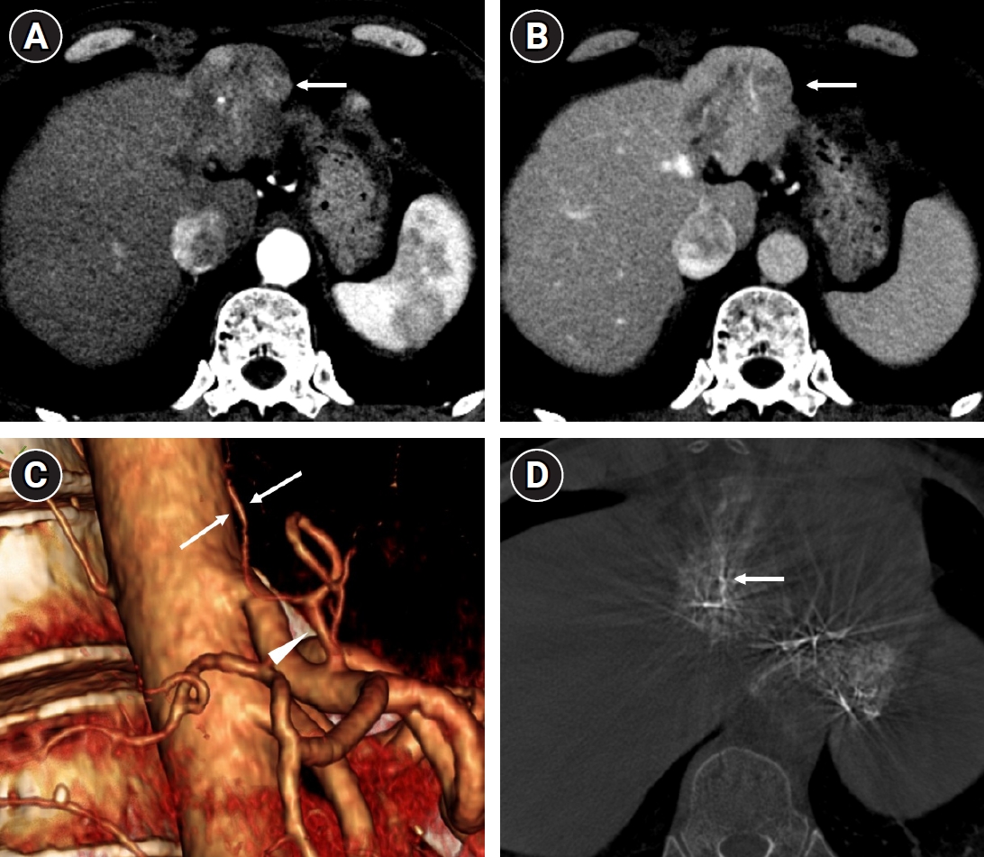
Fig. 2.(A) A side hole (arrow) was created near the tip portion of a 5-Fr Simmons catheter using a No. 11 scalpel blade. (B) The side hole was aligned precisely with the orifice (arrow) of the left gastric artery.
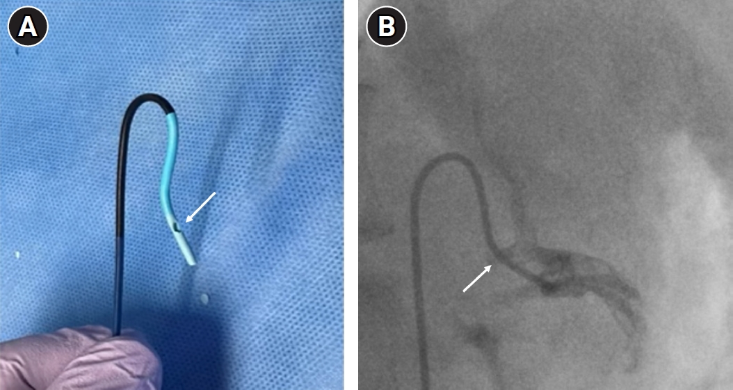
Fig. 3.(A) Spot fluoroscopic image obtained at the end of the procedure shows dense, compact lipiodol uptake confined to the segment III tumor. (B) Follow-up CT shows compact, dense lipiodol deposition confined to the treated tumor, consistent with a favorable treatment response.
References
- 1. Choi TW, Chung JW, Kim HC, Lee M, Choi JW, Jae HJ, et al. Anatomic variations of the hepatic artery in 5625 patients. Radiol Cardiothorac Imaging. 2021;3:e210007. https://doi.org/10.1148/ryct.2021210007
- 2. Baek JH, Chung JW, Jae HJ, Lee W, Park JH. A new technique for superselective catheterization of arteries: preshaping of a micro-guide wire into a shepherd's hook form. Korean J Radiol. 2007;8:225-230. https://doi.org/10.3348/kjr.2007.8.3.225
- 3. Kim HC, Chung JW. Shepherd's hook technique for superselective catheterization of hepatic arteries. J Vasc Interv Radiol. 2014;25:1993-1996. https://doi.org/10.1016/j.jvir.2014.07.029
- 4. Miyayama S, Matsui O, Akakura Y, Yamamoto T, Fujinaga Y, Koda W, et al. Use of a catheter with a large side hole for selective catheterization of the inferior phrenic artery. J Vasc Interv Radiol. 2001;12:497-499. https://doi.org/10.1016/s1051-0443(07)61890-9
- 5. Oh JS, Choi BG, Chun HJ, Lee HG. A side-hole catheter for catheterization of a difficult internal mammary artery. Acta Radiol. 2017;58:307-310. https://doi.org/10.1177/0284185116642633
- 6. Won JH, Park SI, Park KJ, Oh YJ, Hwang SC. Microcatheter placement through a side hole created in a 5-F catheter into proximal subclavian arterial branches causing hemoptysis. J Vasc Interv Radiol. 2004;15:881-884. https://doi.org/10.1097/01.RVI.0000136996.50373.E4
- 7. Meng XX, Liao HQ, Liu HC, Jiang HL, Gu YF, Li X, et al. Application of side-hole catheter technique for transradial arterial chemoembolization in patients with hepatocellular carcinoma. Abdom Radiol (NY). 2019;44:3195-3199. https://doi.org/10.1007/s00261-019-02072-6
- 8. Kim HC, Miyayama S, Choi JW, Kim GM, Chung JW. Hepatocellular carcinoma supplied by the inferior phrenic artery or cystic artery: anatomic and technical considerations. Radiographics. 2023;43:e220076. https://doi.org/10.1148/rg.220076
- 9. Miyayama S, Yamashiro M, Nagai K, Yokka A. Use of a catheter with a large side hole or cleft in selective catheterization of small branch arteries. Interv Radiol (Higashimatsuyama). 2016;1:67-74. https://doi.org/10.22575/interventionalradiology.1.2_67
- 10. Miyayama S, Yamashiro M, Okuda M, Aburano H, Shigenari N, Morinaga K, et al. Creation of a cleft in an angiography catheter to facilitate catheterization of branches of the aorta arising at an acute angle. J Vasc Interv Radiol. 2008;19:1769-1771. https://doi.org/10.1016/j.jvir.2008.08.024
Citations
Citations to this article as recorded by