Abstract
Variant right inferior adrenal arterial supply to caudate lobe HCC is rare and may be overlooked. We report a 53-year-old man with recurrent caudate lobe HCC after 12 TACE sessions. During the 13th TACE, cone-beam CT of the common hepatic artery did not reveal tumor vascularity. At the 14th TACE, performed 3 days later, the right inferior phrenic artery, the most common extrahepatic collateral feeder to caudate lobe HCC, showed no tumor supply. Angiography and cone-beam CT from a small aortic common trunk giving rise to a variant right inferior adrenal artery and the right renal capsular artery revealed predominant supply from the variant right inferior adrenal artery. Embolization through these branches achieved compact iodized oil uptake without immediate complications.
-
Keywords: Carcinoma, hepatocellular; Chemoembolization, therapeutic; Angiography; Collateral circulation
Introduction
HCC in the caudate lobe is technically challenging to treat because of its deep location and variable arterial anatomy, which can limit surgical or percutaneous approaches and make intra-arterial therapy an important treatment option [
1]. Caudate arterial branches are usually small, may be multiple, and often communicate with one another; therefore, the tumor-feeding artery can differ according to the subsegmental location of the caudate tumor [
2]. Recurrent caudate lobe HCC, particularly after repeated TACE, may also recruit extrahepatic collateral arteries [
2,
3]. The right inferior phrenic artery (RIPA) is the most frequent extrahepatic collateral artery supplying caudate lobe HCC [
3]. In contrast, inferior adrenal arterial supply to caudate lobe HCC has only rarely been described [
3,
4]. We report a case of recurrent caudate lobe HCC after multiple prior TACE sessions in which the dominant tumor feeder was an adrenal branch interpreted as a variant right inferior adrenal artery.
Case Report
Institutional Review Board approval was waived for this case report in accordance with the policy of Seoul National University Hospital. A 53-year-old man with HCC was referred for treatment of recurrent HCCs, including a recurrent caudate lobe lesion and a separate recurrent lesion in segment IV. He had hepatitis B virus-related liver cirrhosis. Between December 2016 and September 2025, he had undergone 12 sessions of TACE for multifocal recurrent HCC, as well as radiofrequency ablation and stereotactic ablative radiotherapy. The prior TACE sessions included chemoembolization via several segmental hepatic arteries and the RIPA.
The caudate lobe HCC relevant to this report was first treated in November 2024. At that time, the tumor was located in the inferior portion of the Spiegel lobe. Angiography demonstrated tumor supply from a caudate artery, and selective TACE resulted in dense accumulation of iodized oil. In September 2025, marginal recurrence developed in the adjacent caudate process. This recurrence was again supplied by a caudate artery and was treated with selective TACE, resulting in dense accumulation of iodized oil.
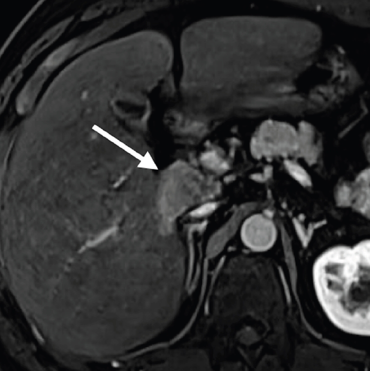
In April 2026, follow-up liver MRI demonstrated a 1-cm nodule in segment IV and a 3.6-cm recurrent lesion involving both the Spiegel lobe and caudate process. Both lesions represented recurrence within a previously TACE-treated site. Because the prior stereotactic ablative radiotherapy had been directed to a separate segment IV lesion and the caudate lesion itself had not been treated with radiation, the caudate lesion was assessed using the Liver Imaging Reporting and Data System (LI-RADS) CT/MRI Nonradiation Treatment Response Algorithm v2024. Mass-like arterial phase hyperenhancement within the treated caudate lesion (
Fig. 1) met the criteria for the LI-RADS Treatment Response Viable. The lesion also showed washout on delayed-phase imaging, hypointensity on hepatobiliary-phase imaging, and high signal intensity on diffusion-weighted imaging. Before the planned TACE, the patient had preserved liver function, with Child-Pugh class A5 and an Eastern Cooperative Oncology Group performance status of 0. Preprocedural laboratory tests showed total bilirubin, 0.7 mg/dL; albumin, 4.0 g/dL; prothrombin time/INR, 0.89; platelet count, 240×10³/µL; serum creatinine, 1.01 mg/dL; AFP, 2805.69 ng/mL; and PIVKA, 1026 mAU/mL.
The 13th TACE session was performed in June 2026. Through right common femoral arterial access, cone-beam CT of the common hepatic artery showed a small enhancing tumor in segment IV, but no enhancement of the recurrent caudate lobe tumor. Because the operator thought that the caudate lobe tumor had no tumor vascularity, he performed chemoembolization through the segment IV hepatic artery and recommended external beam radiation therapy for the caudate lobe tumor. He did not interrogate the RIPA.
Immediate postembolization unenhanced CT scan after the 13th TACE demonstrated iodized oil uptake in the segment IV lesion, indicating a technically satisfactory immediate embolization result for the segment IV nodule. At the multidisciplinary team meeting 2 days after the 13th TACE, further treatment for the caudate HCC was discussed. Occult extrahepatic collateral supply to the caudate lobe tumor was suspected based on the discordance between the preprocedural MRI findings and the angiographic findings during the 13th TACE session. The definite arterial phase hyperenhancement on preprocedural MRI indicated arterial vascularity that was not demonstrated on common hepatic arteriography or cone-beam CT. The history of repeated caudate-directed TACE and prior RIPA chemoembolization further supported the possibility of occult extrahepatic collateral supply. The 14th TACE session was performed 3 days later to evaluate and treat the caudate lobe tumor. The RIPA was first selectively catheterized; however, angiography and cone-beam CT showed no tumor staining. Selective angiography of the left inferior phrenic artery and right middle adrenal artery also showed no tumor supply. Careful catheter exploration along the abdominal aorta was then performed using a 5-Fr RH catheter (GRAFIA, Sungjin-Hitech Co., Ltd., Suwon, Korea) to identify small periaortic branches (
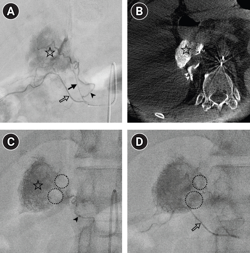
Video 1). This revealed a separate, more caudal aortic common trunk giving rise to an adrenal branch and the right renal capsular artery. The adrenal branch was interpreted as a variant right inferior adrenal artery based on its caudal origin, common trunk with the right renal capsular artery, and course toward the inferior aspect of the right adrenal gland. Selective angiography from this common trunk demonstrated intense tumor staining (
Fig. 2A), and cone-beam CT confirmed enhancement corresponding to the target recurrent caudate lobe HCC (
Fig. 2B).
A hypertrophied tumor-feeding branch from the variant right inferior adrenal artery was then superselectively catheterized with a 1.7-Fr Progreat Lambda microcatheter (Terumo, Tokyo, Japan) over a 0.016-inch ASAHI Meister 16 guidewire (Asahi Intecc, Seto, Japan). TACE was performed using a mixture of 5 mL of iodized oil and 5 mg of idarubicin, followed by smaller calibrated gelatin particles (Prime-Gel, 100–150 μm, PL Micromed Co., Ltd., Yangsan, Korea) for distal embolization. Because residual arterial flow persisted, larger particles (EGgel 350–560 μm, ENGAIN Co., Ltd., Hwaseong, Korea) were subsequently administered to provide additional, relatively proximal flow reduction until angiographic stasis was achieved. Most of the tumor showed dense accumulation of iodized oil, with a small residual unstained portion (
Fig. 2C). The residual portion was supplied by fine branches from the right renal capsular artery. These branches were subsequently superselectively embolized using the same sequence of the iodized oil–idarubicin mixture, smaller calibrated gelatin particles, and larger particles until angiographic stasis was achieved, resulting in iodized oil uptake in the residual tumor portion (
Fig. 2D).
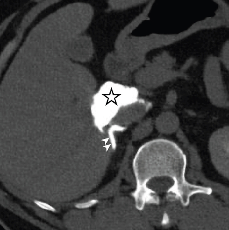
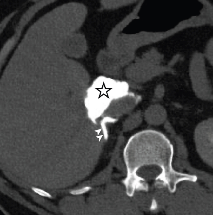
Postprocedural noncontrast CT demonstrated compact iodized oil uptake within the target recurrent caudate lobe HCC (
Fig. 3). Focal iodized oil deposition was also noted in the right adrenal gland and was interpreted as non-target adrenal parenchymal embolization through the variant right inferior adrenal arterial route. Direct adrenal involvement was considered unlikely because preprocedural MRI showed no adrenal mass, no definite continuity between the caudate lobe tumor and the right adrenal gland, and no imaging findings suggestive of adrenal parenchymal tumor involvement. Nevertheless, microscopic adrenal involvement could not be definitively excluded in the absence of histologic confirmation. Throughout and immediately after the procedure, the patient’s vital signs remained stable, and no immediate adrenal-related or other procedure-related complication was observed. The patient was discharged on postprocedural day 1. CT scan 1 month after 14th TACE showed no viable tumor, and longer-term imaging follow-up to formally assess the treatment response of both the caudate lobe and segment IV lesions was scheduled.
Discussion
This case illustrates a rare extrahepatic collateral pathway to recurrent caudate lobe HCC. The caudate lobe has variable arterial supply, and caudate arteries can arise from both right and left hepatic arterial systems. These arteries are frequently small and interconnected, and the dominant feeder can differ according to the subsegmental location of the tumor [
2]. Therefore, careful evaluation of caudate arterial supply is essential during TACE.
The RIPA is the most common extrahepatic collateral artery supplying caudate lobe HCC. Woo et al. [
3] reported that 31 of 35 caudate lobe HCCs treated through extrahepatic collateral arteries were supplied by the RIPA, whereas non-RIPA collaterals were uncommon and included the gastroduodenal, dorsal pancreatic, right gastric, and left gastric arteries. Our case differed from this usual pattern. During the 13th TACE session, the operator did not suspect extrahepatic collateral supply to the caudate lobe HCC, and regarded it as a hypovascular tumor that was not a good candidate for TACE, and recommended external beam radiation therapy. At the 14th TACE session, the RIPA did not supply the tumor. Instead, the tumor was supplied by a small common trunk arising from the abdominal aorta, which gave rise to a variant right inferior adrenal artery and right renal capsular artery. This variant right inferior adrenal artery served as the dominant tumor feeder, with fine ancillary supply from the right renal capsular branch.
Several factors may have contributed to this unusual collateral pathway. Repeated caudate-directed TACE may have attenuated the usual intrahepatic caudate arterial supply, a mechanism that has been suggested to promote extrahepatic collateralization, particularly through the RIPA, in caudate lobe HCC [
3]. In our patient, however, the potential RIPA route may have been limited by previous RIPA chemoembolization, which has been associated with a higher incidence of non-RIPA extrahepatic collateral supply [
3]. Although prior stereotactic ablative radiotherapy had been directed to a separate segment IV lesion rather than the caudate lesion, a possible contribution of regional radiation-related vascular injury could not be entirely excluded. However, its role in the development of this collateral pathway remains uncertain [
5]. In addition, the recurrent tumor involved the lower Spiegel lobe and caudate process, placing it close to the retroperitoneal space, right adrenal gland, and right kidney. This location may have favored recruitment of the variant right inferior adrenal and renal capsular arteries, consistent with adjacent-organ collateralization and known communications among the inferior phrenic, adrenal, and retroperitoneal arterial networks [
6,
7].
Non-target embolization of the adrenal gland is a potential concern when the adrenal artery is used as a collateral route. A small preliminary series of transarterial embolization/TACE performed through adrenal arteries for HCC reported no embolization-related complications [
8]; however, the available evidence remains limited. Therefore, careful superselective catheterization is important to minimize non-target adrenal embolization. In this case, focal iodized oil deposition was noted in the right adrenal gland, but no immediate adrenal-related or other procedure-related complication was observed. This case underscores the importance of superselective catheterization and careful monitoring when performing TACE through an adrenal arterial feeder.
This case provides a practical angiographic lesson. In recurrent caudate lobe HCC after repeated TACE, extrahepatic collateral supply should be kept in mind when cone-beam CT of the hepatic artery fails to demonstrate tumor blush. For tumors involving the lower Spiegel lobe or caudate process, especially in patients with prior caudate-directed and RIPA chemoembolization, small periaortic adrenal–renal capsular collateral pathways, including variant adrenal arterial branches, should be considered after exclusion of the usual caudate arterial and RIPA routes. Recognition of this rare pathway may help identify occult tumor supply and enable complete tumor embolization with superselective TACE.
Conflict of interest
No potential conflict of interest relevant to this article was reported.
Funding
None.
Acknowledgments
None.
Author contributions
Conceptualization: DK, HCK. Data curation: DK. Formal analysis: DK, HCK. Investigation: DK, HCK. Methodology: DK, HCK. Project administration: HCK. Resources: HCK. Supervision: HCK. Validation: HCK. Visualization: DK. Writing - original draft: DK. Writing - review & editing: DK, HCK.
Data availability statement
The datasets generated or analyzed during the study are not publicly available due to patient privacy but are available from the corresponding author on reasonable request.
Supplementary material
Video 1.
Catheter exploration along the abdominal aorta during the 14th TACE session. After angiography of the right inferior phrenic artery showed no tumor supply, small periaortic branches were carefully evaluated using a 5-Fr RH catheter, leading to identification of a common trunk giving rise to an adrenal branch and the right renal capsular artery. The adrenal branch was interpreted as a variant right inferior adrenal artery.
Fig. 1.Preprocedural liver MRI of hepatic arterial phase shows the recurrent tumor involving the Spiegel lobe and caudate process (arrow).
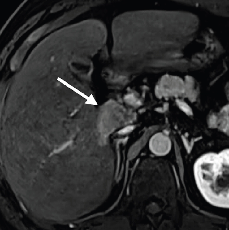
Fig. 2.Angiographic and cone-beam CT findings during the 14th TACE session. (A) Selective angiography from a small common trunk arising from the abdominal aorta demonstrates a variant right inferior adrenal artery (arrow), a hypertrophied tumor-feeding branch arising near its origin (arrowhead), and the right renal capsular artery (open arrow). Intense tumor staining corresponding to the recurrent caudate lobe HCC is also identified (asterisk). (B) Cone-beam CT from the same common trunk confirms enhancement of the bulk of the target caudate lobe tumor (asterisk). Concomitant enhancement of the right adrenal gland is also noted (double arrowhead). (C) After superselective catheterization of a hypertrophied tumor-feeding branch from the variant right inferior adrenal artery (arrowhead), TACE shows dense iodized oil accumulation in most of the tumor (asterisk), with a small residual unstained portion (dashed circles). (D) Subsequent superselective embolization through fine branches from the right renal capsular artery (open arrow) results in iodized oil accumulation within the residual tumor portion (dashed circles).
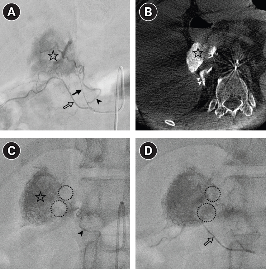
Fig. 3.Postprocedural noncontrast CT after the 14th TACE session. Noncontrast CT demonstrates compact iodized oil uptake throughout the target recurrent caudate lobe HCC (asterisk), indicating technically successful superselective TACE. Concomitant focal iodized oil deposition is also noted in the right adrenal gland (double arrowhead).
References
- 1. Kim HC, Chung JW, Jae HJ, Yoon JH, Lee JH, Kim YJ, et al. Caudate lobe hepatocellular carcinoma treated with selective chemoembolization. Radiology. 2010;257:278-287. https://doi.org/10.1148/radiol.10100105
- 2. Kim HC, Miyayama S, Chung JW. Selective chemoembolization of caudate lobe hepatocellular carcinoma: anatomy and procedural techniques. Radiographics. 2019;39:289-302. https://doi.org/10.1148/rg.2019180110
- 3. Woo S, Kim HC, Chung JW, Jung HS, Hur S, Lee M, et al. Chemoembolization of extrahepatic collateral arteries for treatment of hepatocellular carcinoma in the caudate lobe of the liver. Cardiovasc Intervent Radiol. 2015;38:389-396. https://doi.org/10.1007/s00270-014-0929-7
- 4. Rash C, Lee E, Klinsky S, Angel W. Case study of inferior adrenal artery as an extrahepatic collateral arterial supply to hepatocellular carcinoma. Radiol Case Rep. 2025;20:129-132. https://doi.org/10.1016/j.radcr.2024.10.026
- 5. Park HJ, Griffin RJ, Hui S, Levitt SH, Song CW. Radiation-induced vascular damage in tumors: implications of vascular damage in ablative hypofractionated radiotherapy (SBRT and SRS). Radiat Res. 2012;177:311-327. https://doi.org/10.1667/rr2773.1
- 6. Kim HC, Chung JW, Lee W, Jae HJ, Park JH. Recognizing extrahepatic collateral vessels that supply hepatocellular carcinoma to avoid complications of transcatheter arterial chemoembolization. Radiographics. 2005;25 Suppl 1:S25-S39. https://doi.org/10.1148/rg.25si055508
- 7. Kim HC, Miyayama S, Choi JW, Kim GM, Chung JW. Hepatocellular carcinoma supplied by the inferior phrenic artery or cystic artery: anatomic and technical considerations. Radiographics. 2023;43:e220076. https://doi.org/10.1148/rg.220076
- 8. Hu S, Tu J, Jia Z, Huang Y, Jiang G. Transarterial embolization/chemoembolization therapy for hepatocellular carcinoma fed by adrenal artery: preliminary results. Medicine (Baltimore). 2016;95:e5762. https://doi.org/10.1097/MD.0000000000005762