Abstract
Duodenal perforation is an uncommon but potentially fatal complication of pancreatic surgery. We report a 73-year-old man who presented with hematochezia after laparoscopic distal pancreatectomy with splenectomy for pancreatic body cancer, complicated by a postoperative pancreatic fistula that led to gastroduodenal artery bleeding treated with a stent-graft. Contrast-enhanced CT showed active duodenal bleeding with a surrounding hematoma. The bleeding duodenal branch was embolized, and the hematoma was drained percutaneously; a fistulous communication between the periduodenal collection and the duodenal lumen was subsequently demonstrated. Through the drainage tract, an 8-Fr Foley catheter was advanced into the duodenal lumen and its balloon inflated to secure a controlled fistula, while a percutaneous gastrojejunostomy provided distal enteral feeding. The catheter was maintained for approximately 2 months, and the tract was embolized at the time of removal to minimize residual leakage. The patient recovered fully. This case demonstrates the feasibility of staged interventional management for postoperative duodenal perforation when reoperation is not feasible.
-
Keywords: Intestinal perforation; Postoperative complications; Drainage; Case reports
Introduction
Duodenal perforation is a full-thickness disruption of the duodenal wall that constitutes a life-threatening emergency associated with substantial morbidity and mortality. It arises from diverse etiologies, including peptic ulcer disease, iatrogenic injury during ERCP or endoscopic ultrasound, blunt or penetrating trauma, malignancy, and abdominal surgery. Iatrogenic perforation is uncommon; post-ERCP perforation occurs in fewer than 1% of procedures but carries a reported mortality of 4.2%–37% depending on perforation type, timing of diagnosis, and management strategy [
1]. The predominantly retroperitoneal location of the duodenum and its proximity to the pancreas and biliary tree contribute to atypical presentation, delayed diagnosis, and persistent leakage, which together account for the disproportionate morbidity relative to perforations elsewhere in the gastrointestinal tract.
Classical management has centered on open surgical repair—primary closure with an omental patch, peritoneal lavage, and drainage. Although effective for source control, open repair imposes considerable physiological stress, particularly in elderly patients, those with multiple comorbidities, or those presenting late, and reported complication rates reach 42%–58% with hospital stays of 5–15 days [
2,
3]. In the postoperative setting, reoperation is further constrained by adhesions, tissue friability, and inflammation within a recently manipulated surgical field, and revision surgery has been required in up to 67% of retroperitoneal duodenal perforations managed surgically [
4]. These limitations underscore the need for less invasive alternatives in selected patients.
Advances in interventional radiology and endoscopy have established minimally invasive techniques as viable alternatives or adjuncts to surgery. Image-guided percutaneous drainage achieves source control with reported success of 77%–88% [
5,
6], covered self-expandable metal stents attain technical success approaching 100% and clinical healing of 87%–93% [
7,
8], and endoscopic vacuum therapy has reported 100% technical and 80% clinical closure [
9]. A randomized trial of combined endoscopic and radiologic management versus surgery for perforated peptic ulcer showed shorter hospital stay (1 vs. 7 days) and lower complication rates (24% vs. 58%) in the interventional arm [
3], while a meta-analysis of post-ERCP perforations found no significant difference in mortality between non-operative and surgical management [
10]. Beyond reduced physiological insult, these techniques permit staged, patient-tailored management—stabilizing the patient first and combining drainage with defect sealing according to perforation size, location, and degree of contamination—thereby enabling an individualized rather than uniformly operative strategy.
Duodenal perforation following pancreatic surgery represents a particularly challenging scenario, as early reoperation within a fresh operative field carries high risk and is often technically difficult or infeasible. Herein, we report a case of postoperative duodenal perforation managed by percutaneous drainage followed by catheter-based tract sealing, illustrating the feasibility of a staged, minimally invasive approach when reoperation is not a favorable option.
Case Report
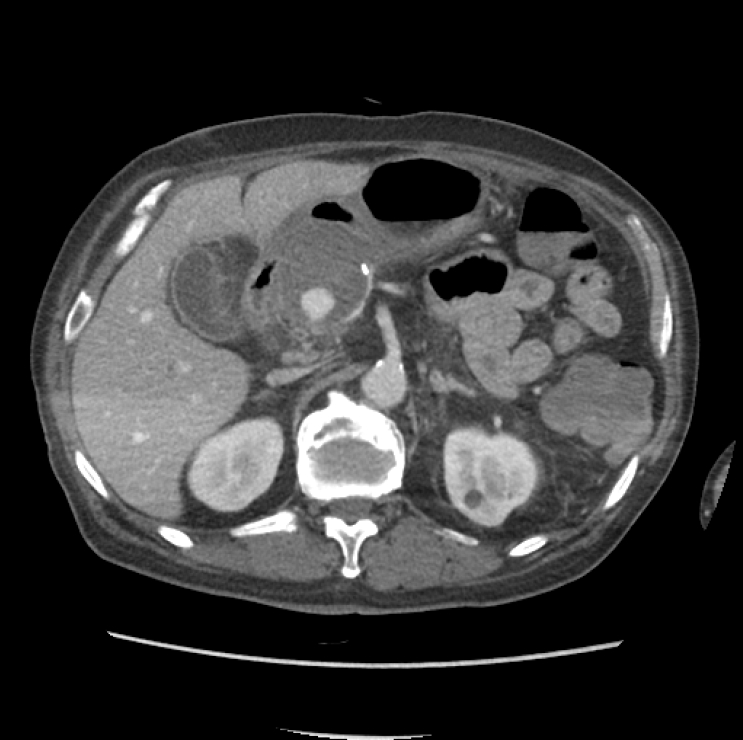
A 73-year-old man presented with hematochezia 5 weeks after laparoscopic distal pancreatectomy with splenectomy for pancreatic body cancer. His postoperative course had been complicated by a postoperative pancreatic fistula (POPF). Three weeks after surgery, enzymatic erosion associated with the POPF resulted in bleeding from the gastroduodenal artery (GDA), which was treated by stent-graft placement. On arrival, he was hemodynamically unstable, with a blood pressure of 99/46 mmHg and a heart rate of 105 beats/min. Initial laboratory studies showed a hemoglobin level of 9.5 g/dL, a hematocrit of 30.6%, and a platelet count of 369 × 10³/μL; coagulation studies were within normal limits. Contrast-enhanced multiphasic abdominal CT demonstrated active bleeding from the duodenum with a surrounding hematoma (
Fig. 1).
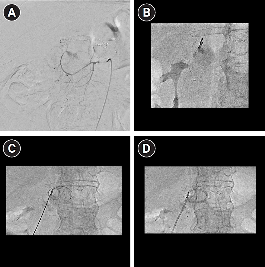
Emergency transarterial embolization of the bleeding duodenal branch was performed with a Concerto detachable coil (Medtronic, Minneapolis, MN, USA) and a 1:1 mixture of N-butyl cyanoacrylate (Histoacryl, B. Braun, Melsungen, Germany) and ethiodized oil (Lipiodol, Guerbet, Villepinte, France). Percutaneous catheter drainage (PCD) of the hematoma was then undertaken under combined ultrasonographic and cone-beam CT guidance. Because the left hepatic lobe and the gastric antrum lay anterior to the collection, a transhepatic route was selected, and cone-beam CT was used to plan and confirm a trajectory that traversed the left hepatic lobe while avoiding the stomach. The aspirated fluid was dark brown, bile-tinged, and purulent on gross inspection. Fluid amylase and lipase levels were 112 U/L and 1,007 U/L, respectively, and culture grew
Klebsiella pneumoniae. Fluid bilirubin was not measured, so a biliary contribution could not be quantified biochemically; however, the bile-tinged appearance was consistent with an infected collection receiving enteric content, in the setting of the previously documented POPF. The biochemical findings alone could not establish direct communication with the pancreatic fistula. Daily drainage was approximately 300 mL/day immediately after PCD placement and persisted at this level over the subsequent 5 days (
Fig. 2).
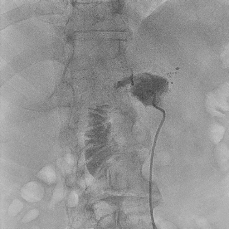
Three days later, a tubogram through the PCD demonstrated a fistulous communication between the drainage cavity and the duodenum, with injected contrast passing into the duodenal lumen (
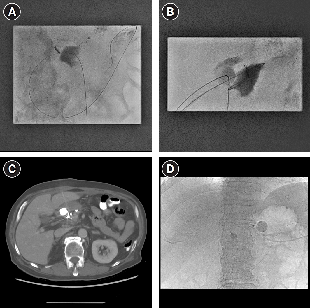
Fig. 3). Because the enteric output persisted despite adequate drainage, a catheter-directed approach to occlude the fistula was undertaken on day 5 after PCD placement. The fistula was cannulated through the PCD tract using a 5-Fr Kumpe catheter (Cook Medical, Bloomington, IN, USA) and a 0.035-inch guidewire. After access into the duodenal lumen was achieved, an 8-Fr silicone-coated latex Foley balloon catheter (SEWOON Medical, Cheonan, Korea) was positioned in exchange for the existing drainage catheter, the balloon was inflated within the lumen, and slight tension was applied to secure the tube. Output thereafter declined to approximately 20 mL/day and remained minimal until removal. Because a separate drainage catheter was not maintained, the catheter and the residual cavity were monitored by tubography every 2 days for the first week and weekly thereafter; no re-accumulation was observed. A percutaneous gastrojejunostomy was subsequently created to enable enteral feeding beyond the duodenal fistula (
Fig. 4).
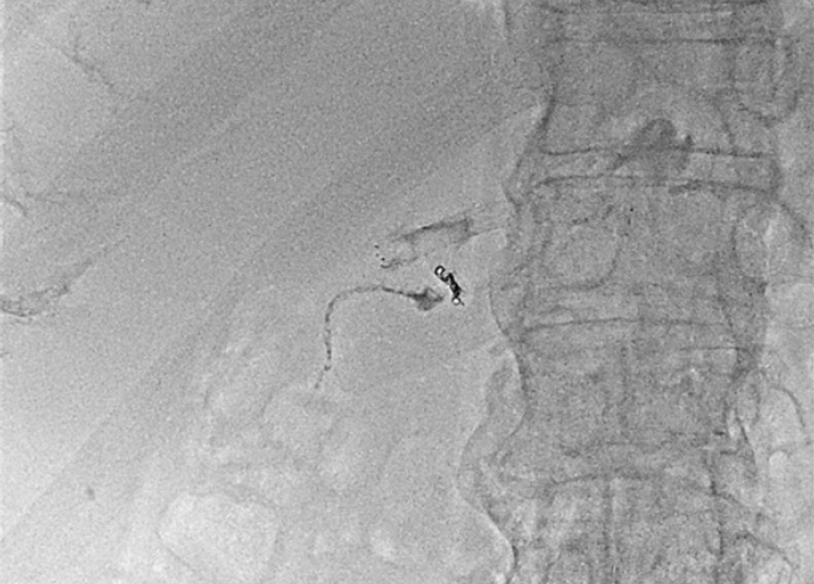
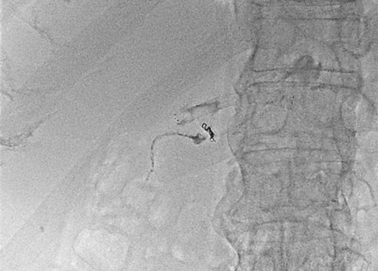
The catheter was maintained for approximately 2 months, during which the patient received enteral feeding through the gastrojejunostomy and underwent rehabilitation therapy. The patient recovered, and the gastrojejunostomy was removed once oral feeding was tolerated. At the time of catheter removal, tubography confirmed a mature fistulous tract without extraluminal leakage. To minimize residual leakage along the tract after catheter withdrawal, the tract was embolized with a 1:1 mixture of N-butyl cyanoacrylate and ethiodized oil. After balloon deflation, the catheter was withdrawn until its tip lay immediately outside the duodenal wall, and approximately 1 mL of the mixture was injected while the catheter was gradually withdrawn, so that embolization was confined to the extraluminal tract outside the fistula and no glue was delivered into the duodenal lumen. Dextrose flushing was not used (
Fig. 5). After the absence of leakage or abdominal pain was confirmed, the patient was discharged the following day. No procedure-related adverse events occurred during the course of interventional management. At the outpatient visit 2 weeks after discharge, the patient was asymptomatic and tolerated oral intake, with no clinical evidence of recurrent leakage; follow-up was clinical, and no follow-up imaging was obtained.
The requirement for institutional review board review was waived by the Institutional Review Board of Seoul National University Bundang Hospital because of the retrospective nature of this case report (IRB No. B-2608-1065-102). Written informed consent for publication of this case and the accompanying images was obtained from the patient.
Discussion
Duodenal fistula arising after pancreatic and periduodenal surgery is an uncommon but potentially lethal complication, and its management in the postoperative setting is particularly problematic. Early reoperation within a recently operated field is technically demanding because of altered anatomy, dense adhesions, and tissue friability, and primary repair or omental patch closure frequently fails under these conditions. Reported complication rates after surgical management of duodenal perforation range from 42% to 58% [
2,
3], and revision surgery has been required in up to 67% of retroperitoneal duodenal perforations managed operatively [
4]. A meta-analysis of post-ERCP perforations found no mortality advantage for initial surgery over non-operative management [
10], supporting consideration of less invasive alternatives in appropriately selected patients.
Advances in endoscopy and interventional radiology have expanded the non-operative options for duodenal perforation. Covered self-expandable metal stents can seal iatrogenic defects with high technical and clinical success [
7,
8], and endoscopic vacuum therapy has yielded promising closure rates in early series [
9]. These endoscopic approaches, however, depend on adequate luminal access, may require repeated endoscopic sessions for device exchange or removal, and are supported mainly by small, heterogeneous reports. In the postoperative abdomen with distorted anatomy, repeated transluminal intervention may be impractical, increasing the appeal of a purely percutaneous, image-guided strategy.
The mechanism of the duodenal fistula in this case appears multifactorial. The patient had an established POPF, and enzyme-rich pancreatic fluid is itself capable of eroding adjacent vascular and enteric structures; indeed, the GDA bleeding 3 weeks after surgery is attributable to this process. Superimposed on this, the duodenal wall had been subjected to sequential loss of arterial inflow—first stent-graft exclusion of the GDA and, approximately 2 weeks later, embolization of a duodenal branch with a liquid agent that penetrates to the distal arteriolar level. Although the pancreaticoduodenal arcades usually provide robust collateral supply, cumulative devascularization within a field already compromised by enzymatic inflammation and hematoma may reduce mural perfusion beyond the compensatory capacity of the arcade. The fistula was demonstrated only three days after embolization, a sequence compatible with post-embolization mural ischemia in addition to enzymatic and operative injury. The relative contributions of these mechanisms cannot be established from a single case; nevertheless, the sequence is clinically instructive, since embolization of duodenal branches in a patient whose proximal inflow has already been excluded may warrant anticipatory surveillance for mural injury, for which percutaneous techniques such as those described here offer a means of management without reoperation.
The core principle of interventional management is to eliminate the fluid collection and convert an uncontrolled leak into a controlled, externally drained fistula using image-guided PCD [
5,
6,
11]. Percutaneous Foley catheter placement has been reported as a safe and effective means of achieving this conversion in postoperative duodenal stump leakage, with successful luminal placement and no procedure-related complications in a consecutive series [
12]. The duodenum, however, is a high-pressure, high-motility segment continuously exposed to bile and pancreatic secretions, so drainage alone may be insufficient to divert luminal content and allow the tract to close. In the present case, a Foley catheter was advanced across the fistula and its balloon inflated within the duodenal lumen, reducing free efflux of enteric content into the tract. The objective effect of this maneuver was reflected in the drainage output, which fell from approximately 300 mL/day, sustained over the preceding 5 days despite adequate drainage, to approximately 20 mL/day after balloon occlusion. We additionally considered whether the tissue reactivity of latex-based catheters, relative to all-silicone drains, might favor fibrous tract maturation, an inference extrapolated from urological biocompatibility data in which latex catheters provoke greater inflammatory reaction and tissue trauma than silicone [
13,
14]. This rationale remains speculative. It has not been validated for enteric fistula tracts; the catheter used here was silicone-coated latex, so the tissue-contacting surface was not bare latex; and in a single case the contribution of catheter material cannot be separated from those of prolonged indwelling and mechanical balloon occlusion. We therefore regard balloon occlusion as the principal mechanism and any material-related effect as unproven and hypothesis-generating. A conceptually similar catheter-based approach has been reported for post-ERCP duodenal perforation [
1].
To further support healing, a percutaneous gastrojejunostomy was placed so that enteral nutrition could be delivered distal to the fistula, maintaining nutritional status while minimizing passage of ingested content across the defect—a consideration of particular relevance for proximal, higher-output fistulas [
11]. Tract embolization at the time of catheter removal was performed to minimize residual leakage along the mature tract rather than as salvage for failed tract formation, and we regard it as an integral final step of the technique. Under this combined strategy the patient recovered and was discharged approximately 2 months after the initial drainage. This report describes a single case and should be regarded as proof of concept; the technique requires validation in larger series, and criteria for patient selection and for conversion to surgery when interventional management fails remain to be defined. Nonetheless, the case illustrates that a staged, entirely percutaneous strategy—collection drainage, balloon-assisted controlled fistula formation, distal enteral feeding, and tract embolization at catheter removal—can be a feasible option for complex postoperative duodenal fistula or perforation, including cases in which ischemic injury after sequential arterial interventions may have contributed, when reoperation is difficult or infeasible.
Conflict of interest
No potential conflict of interest relevant to this article was reported.
Funding
None.
Acknowledgments
None.
Author contributions
Conceptualization: JHL, CJY. Investigation: JHL, KYK. Supervision: CJY. Writing - original draft: JHL. Writing - review & editing: JHL, KYK, CJY.
Data availability statement
Data sharing does not apply to this article as no datasets were generated or analyzed during the current study.
Fig. 1.A 73-year-old man presented with hematochezia 5 weeks after laparoscopic distal pancreatectomy with splenectomy for pancreatic body cancer. Contrast-enhanced CT shows active contrast extravasation from a duodenal branch, with an adjacent perilesional hematoma.
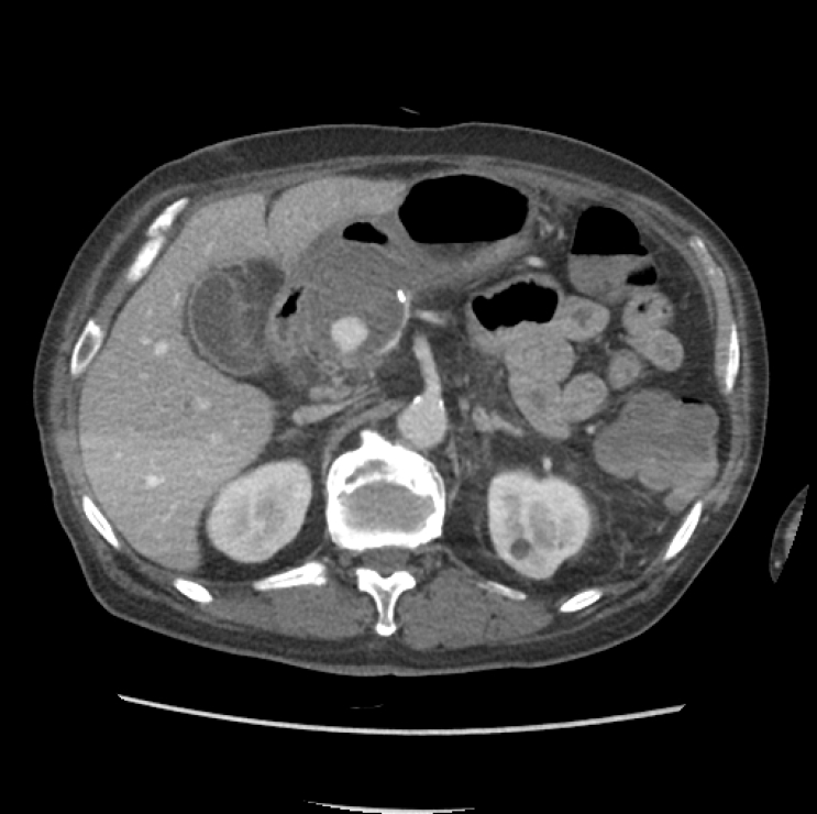
Fig. 2.Emergency transarterial embolization and percutaneous drainage. (A) Selective angiography demonstrates active bleeding from the duodenal branch. (B) Post-embolization spot image after microcoil placement and injection of a 1:1 glue-lipiodol mixture shows cessation of bleeding. (C) The hematoma is punctured under combined ultrasonographic and cone-beam CT guidance via a transhepatic route, avoiding the gastric antrum, and (D) a percutaneous drainage catheter is placed.
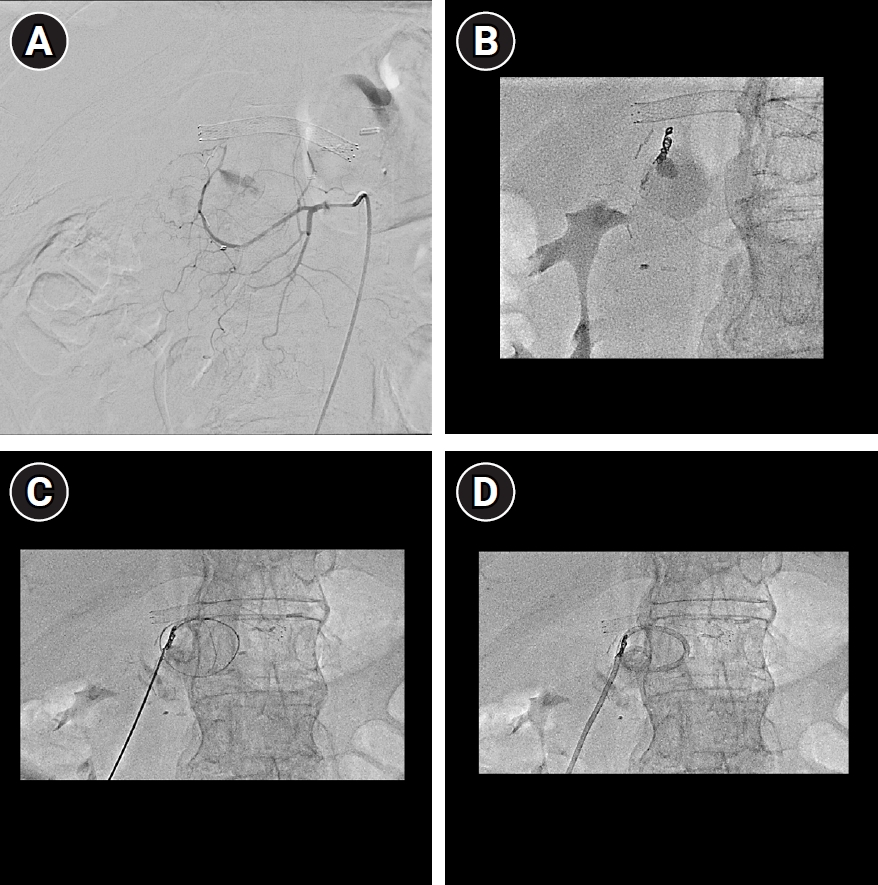
Fig. 3.Tubogram obtained 3 days after drainage. Contrast injected through the percutaneous drainage catheter opacifies the cavity and passes into the duodenal lumen through a fistulous communication, confirming a duodenal fistula.
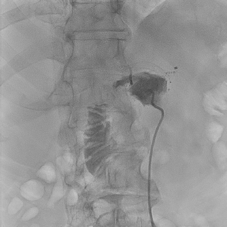
Fig. 4.Catheter-directed controlled fistula formation. (A) Through the drainage tract, the fistula was cannulated with a 5-Fr Kumpe catheter and a 0.035-inch guidewire, and access into the duodenal lumen was achieved. (B) An 8-Fr silicone-coated latex Foley balloon catheter (SEWOON Medical, Cheonan, Korea) was positioned across the fistula and its balloon inflated within the duodenal lumen to secure the tract. (C) Axial CT after the procedure demonstrates the balloon within the duodenal lumen. (D) A percutaneous gastrojejunostomy was created to deliver enteral nutrition distal to the fistula.
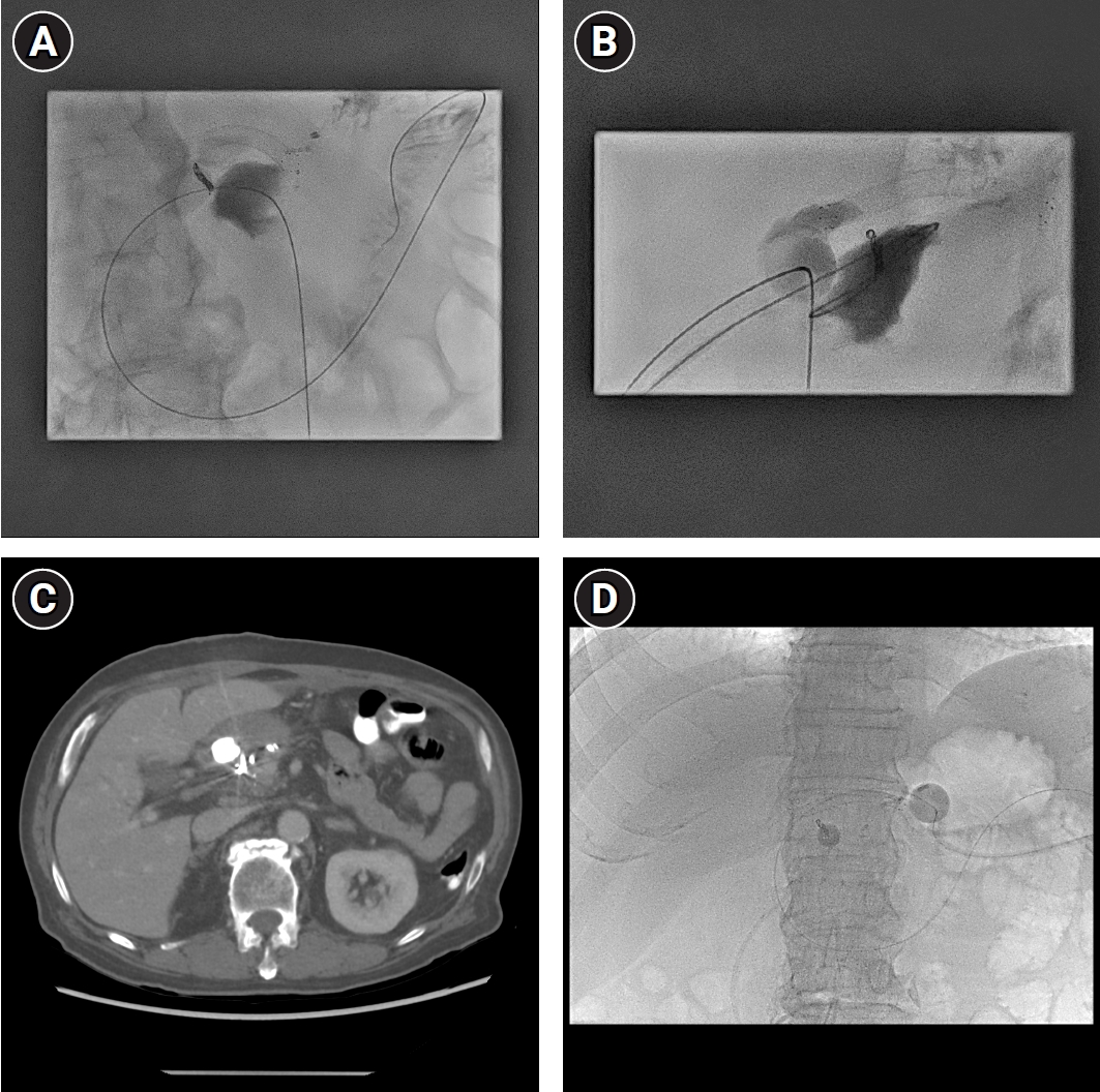
Fig. 5.Removal of the Foley catheter and embolization of the percutaneous tract. Tubography demonstrated a mature fistulous tract without extraluminal leakage. The tract was then embolized with a 1:1 mixture of N-butyl cyanoacrylate and ethiodized oil while the catheter was gradually withdrawn, to minimize residual leakage.
References
- 1. Ma Y, Bi Y, Wei C, Ding X, Zhou G, Yin M, et al. Interventional three-tube method for the management of duodenal perforation after endoscopic retrograde cholangiopancreatography for choledocholithiasis: a case series. Quant Imaging Med Surg. 2025;15:7563-7571. https://doi.org/10.21037/qims-2024-2650
- 2. Arroyo Vazquez JA, Khodakaram K, Bergstrom M, Park PO. Stent treatment or surgical closure for perforated duodenal ulcers: a prospective randomized study. Surg Endosc. 2021;35:7183-7190. https://doi.org/10.1007/s00464-020-08158-3
- 3. Negm S, Mohamed H, Shafiq A, AbdelKader T, Ismail A, Yassin M, et al. Combined endoscopic and radiologic intervention for management of acute perforated peptic ulcer: a randomized controlled trial. World J Emerg Surg. 2022;17:24. https://doi.org/10.1186/s13017-022-00429-9
- 4. Yurttas C, Thiel C, Wichmann D, Horvath P, Strohaker J, Bongers MN, et al. Retrospective analysis of different therapeutic approaches for retroperitoneal duodenal perforations. Sci Rep. 2022;12:10243. https://doi.org/10.1038/s41598-022-14278-8
- 5. McCarthy CJ, Butros SR, Dawson SL, Arellano RS. Image-guided percutaneous management of duodenal perforation following endoscopic retrograde cholangiopancreatography (ERCP): assessment of efficacy and safety. Clin Radiol. 2018;73:319.e9-319.e15. https://doi.org/10.1016/j.crad.2017.09.014
- 6. Rahman MM, Al Mamun A, Hossain MD, Das MK. Peptic ulcer perforation: management of high-risk cases by percutaneous abdominal drainage. Trop Doct. 2005;35:30-31. https://doi.org/10.1258/0049475053001822
- 7. Sbrozzi-Vanni A, Galloro G, Piciucchi M, Castellani D, Germani U, Dell'Amico I, et al. Over-the-scope technique for partially covered self-expandable metal stent placement to treat duodenal perforation during EUS. J Gastrointestin Liver Dis. 2023;32:65-69. https://doi.org/10.15403/jgld-4625
- 8. Bergstrom M, Arroyo Vazquez JA, Park PO. Self-expandable metal stents as a new treatment option for perforated duodenal ulcer. Endoscopy. 2013;45:222-225. https://doi.org/10.1055/s-0032-1325885
- 9. Chevallay M, Lorenz F, Bichard P, Frossard JL, Schmidt T, Goeser T, et al. Outcome of endoscopic vacuum therapy for duodenal perforation. Surg Endosc. 2023;37:1846-1853. https://doi.org/10.1007/s00464-022-09686-w
- 10. Samara A, Diamantis A, Perivoliotis K, Mavrovounis G, Simeonidis D, Baloyiannis I, et al. Initial management of duodenal perforations after ERCP: a systematic review and meta-analysis. HPB (Oxford). 2022;24(Suppl 1):S541. https://doi.org/10.1016/j.hpb.2022.05.1172
- 11. Ghimire P. Management of enterocutaneous fistula: a review. JNMA J Nepal Med Assoc. 2022;60:93-100. https://doi.org/10.31729/jnma.5780
- 12. Oh JS, Lee HG, Chun HJ, Choi BG, Lee SH, Hahn ST, et al. Percutaneous management of postoperative duodenal stump leakage with Foley catheter. Cardiovasc Intervent Radiol. 2013;36:1344-1349. https://doi.org/10.1007/s00270-012-0518-6
- 13. Liedberg H. Catheter induced urethral inflammatory reaction and urinary tract infection: an experimental and clinical study. Scand J Urol Nephrol Suppl. 1989;124:1-43.
- 14. Nickel JC, Olson ME, Costerton JW. In vivo coefficient of kinetic friction: study of urinary catheter biocompatibility. Urology. 1987;29:501-503. https://doi.org/10.1016/0090-4295(87)90037-9