Abstract
Acute synthetic portal graft thrombosis post–living donor liver transplantation (LDLT) is a rare, life-threatening complication. A 32-year-old woman with Budd-Chiari syndrome and chronic Yerdel grade IV portal thrombosis underwent LDLT using an expanded polytetrafluoroethylene jump graft from the superior mesenteric vein to the donor portal vein. Five months post-transplant, she developed acute graft failure and contrast-enhanced computed tomography confirmed complete occlusion with absent portal flow. Due to prohibitive surgical risk and lytic bleeding hazards, emergency transhepatic percutaneous mechanical thrombectomy with balloon angioplasty restored immediate portal flow without lytics. Liver function rapidly normalized, and the graft remained patent on Doppler ultrasound at 12-month follow-up. Emergency mechanical thrombectomy with angioplasty offers a feasible, effective endovascular salvage strategy for acute synthetic portal graft failure.
-
Keywords: Liver transplantation; Thrombectomy; Portal vein; Graft occlusion, vascular; Angioplasty
Introduction
Portal vein thrombosis (PVT) in living donor liver transplantation (LDLT) presents a substantial surgical and interventional challenge. When native venous tissue is unusable due to extensive diffuse thrombosis (Yerdel grade IV), extra-anatomic jump grafts using synthetic expanded polytetrafluoroethylene (ePTFE) may be constructed to establish adequate portal inflow [
1,
2]. These synthetic conduits can successfully salvage liver transplantation in complex portal pathology, but they carry a significant risk of late conduit thrombosis.
Due to the extreme rarity of synthetic portal graft usage—utilized in as few as 0.3% of liver transplant recipients in large series [
2]—standardized management algorithms for acute graft occlusion do not exist. Surgical revision carries prohibitive morbidity and mortality due to severe dense postoperative adhesions and portal hypertension. Catheter-directed thrombolysis (CDT) and systemic thrombolysis entail prolonged treatment durations and substantial bleeding risks in coagulopathic post-transplant patients. We report a case of acute ePTFE portal graft thrombosis successfully managed with urgent percutaneous mechanical thrombectomy (PMT), balloon angioplasty of an underlying anastomotic stenosis, and long-term anticoagulation, highlighting this combination as a viable emergency endovascular salvage strategy [
3,
4].
Case Report
A 32-year-old female patient underwent LDLT for decompensated cirrhosis secondary to Budd-Chiari syndrome. Preoperative imaging revealed chronic Yerdel grade IV PVT [
1]. A 12-mm ringed ePTFE jump graft was used to establish inflow from the recipient superior mesenteric vein (SMV) to the donor portal vein. Post-transplantation, the patient was maintained on oral anticoagulation with warfarin (target INR, 2.0 to 2.5). Early postoperative graft patency was confirmed on routine bedside color Doppler ultrasound surveillance prior to discharge; however, routine baseline CT prior to the acute thrombotic presentation was not performed, and static early sonograms were not archived in PACS. She maintained regular outpatient compliance, with serial INR measurements ranging between 1.8 and 2.2 prior to the acute presentation.
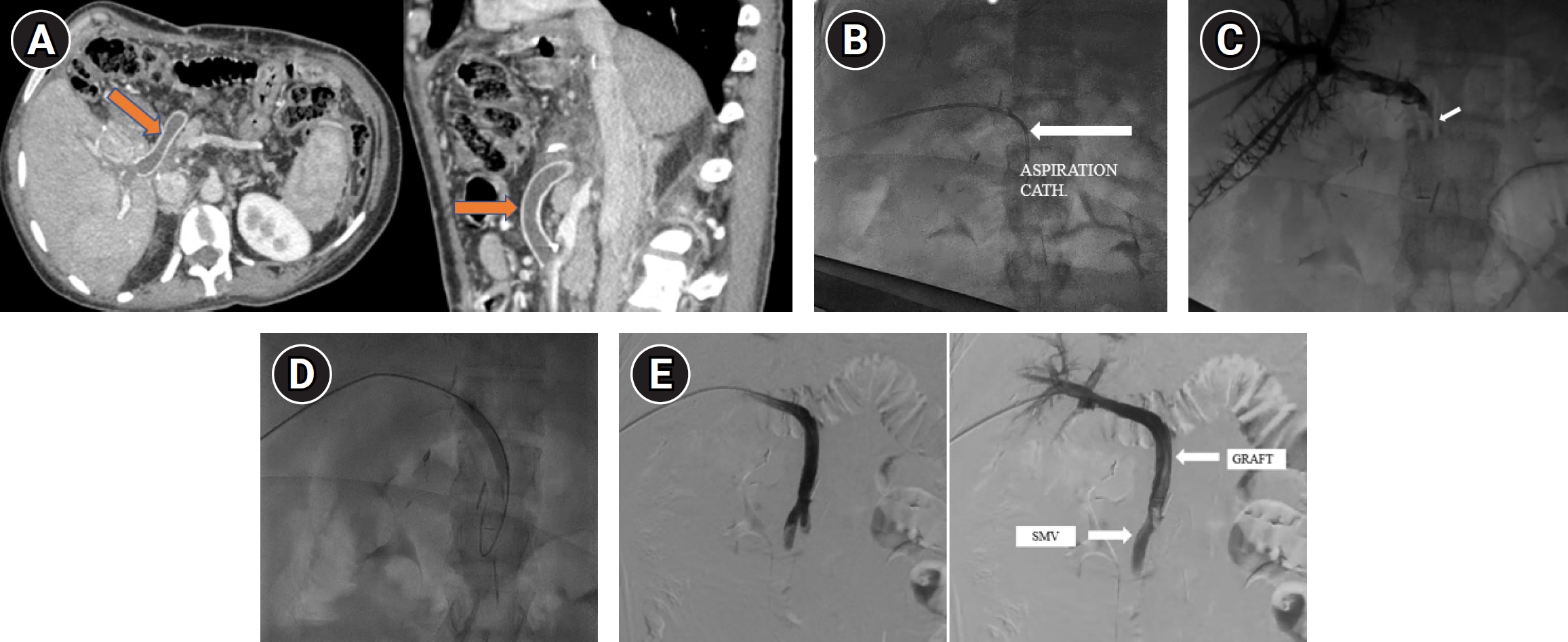
Five months after transplantation, the patient presented with acute abdominal discomfort, jaundice, and marked malaise. Laboratory testing demonstrated severe acute graft dysfunction: total bilirubin 4.5 mg/dL, AST 142 U/L, ALT 264 U/L, and INR 1.97. Urgent contrast-enhanced computed tomography confirmed a large filling defect completely occluding the ePTFE graft, with no portal flow (
Fig. 1A).
A multidisciplinary team meeting involving transplant surgeons and interventional radiologists evaluated treatment options. Surgical revision was rejected due to prohibitive mortality risks associated with adhesions, portal hypertension, and coagulopathy. Systemic thrombolysis and CDT were avoided due to the prolonged timeframe required for clot dissolution (24–48 hours) during ongoing acute hepatic ischemia, as well as the high bleeding risk associated with transhepatic track instrumentation in a coagulopathic state (INR, 1.97). Consequently, emergency percutaneous endovascular salvage was chosen.
Under ultrasound guidance, percutaneous transhepatic access into a right portal vein branch was performed using a 22-gauge Chiba needle (Cook Medical, Bloomington, IN, USA). A 6-Fr vascular sheath (Terumo, Tokyo, Japan) was placed, and a 0.035-inch stiff hydrophilic guidewire (Radifocus Guide Wire M, Terumo) navigated across the thrombosed graft into the patent main SMV. Initial venography confirmed total occlusion.
To accommodate a large-bore mechanical aspiration system, the initial 6-Fr sheath was exchanged over a stiff wire for an 8-Fr vascular sheath (Terumo). An 8-Fr aspiration catheter (Indigo Aspiration System, CAT8, Penumbra Inc., Alameda, CA, USA) was advanced into the graft. Three passes were made to extract large, organized thrombus fragments without using lytic agents (
Fig. 1B). Subsequent venography showed significant thrombus debulking and flow restoration, but revealed an underlying ~70%–80% focal mechanical stenosis at the proximal graft-to-SMV anastomosis (
Fig. 1C). This was successfully treated with an 8 mm × 40 mm high-pressure Mustang balloon catheter (Boston Scientific, Pulau Pinang, Malaysia) angioplasty (
Fig. 1D). Completion venography confirmed complete anatomical recanalization with brisk hepatopetal flow into intrahepatic branches (
Fig. 1E). Additional stent placement was intentionally withheld because balloon angioplasty achieved an optimal anatomical and hemodynamic result without elastic recoil or flow-limiting dissection, thereby avoiding permanent metallic coverage within a synthetic conduit in a young patient. The transhepatic parenchymal track was embolized with Gelfoam slurry (Pfizer, New York, NY, USA) upon sheath withdrawal to ensure hemostasis.
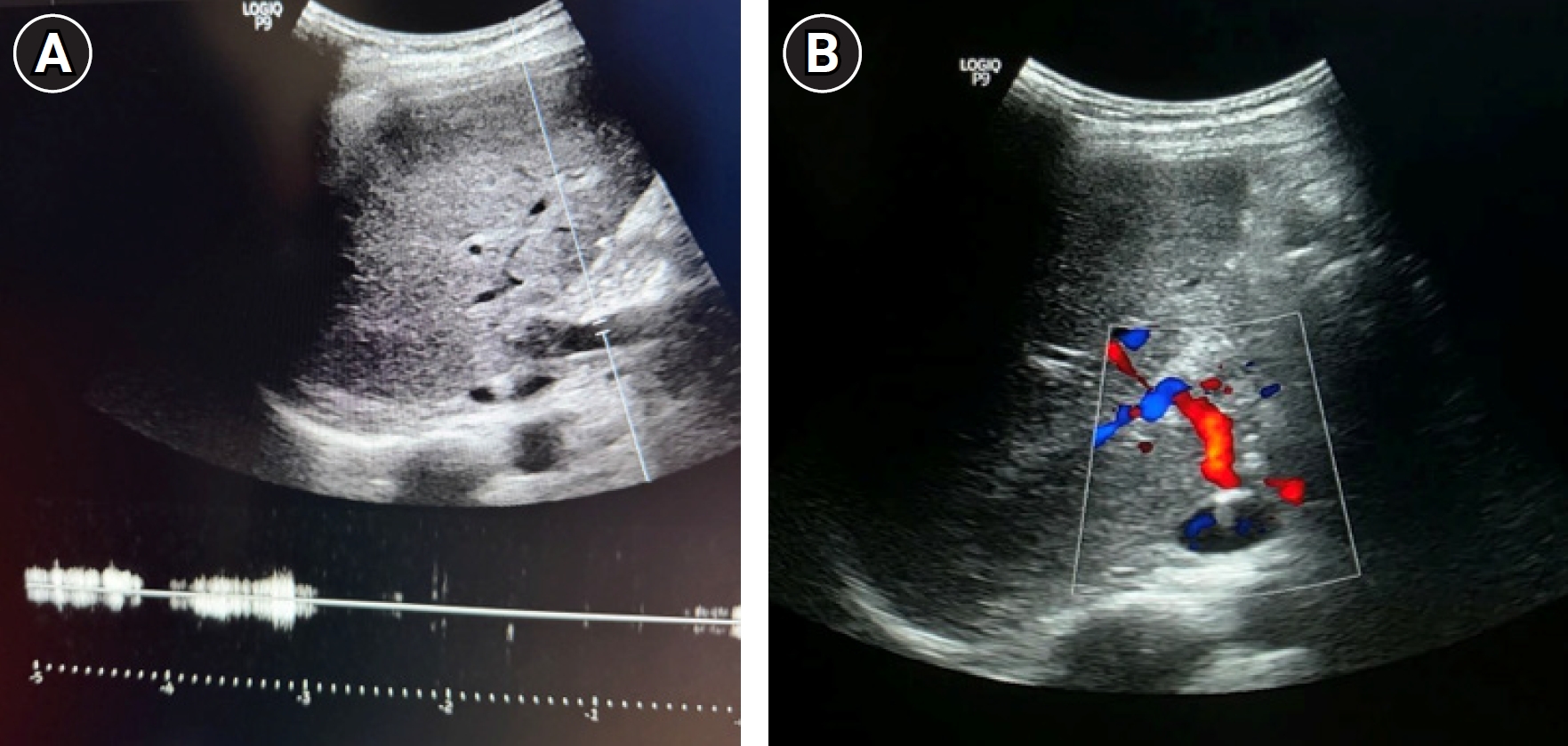
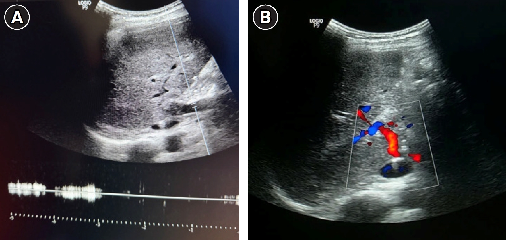
The patient's clinical recovery was rapid. By post-procedure day 3, serum total bilirubin decreased to 0.8 mg/dL and AST/ALT levels normalized. Therapeutic oral anticoagulation with warfarin was re-initiated on post-procedure day 1. The patient was discharged on day 5 with an INR of 2.1. Routine outpatient follow-up with color Doppler ultrasound at 12 months confirmed sustained patency of the ePTFE portal jump graft with normal hepatopetal flow velocity (
Fig. 2A,
2B).
This study was conducted in accordance with the CARE guidelines and the Declaration of Helsinki. The requirement for Institutional Review Board approval was waived by the Institutional Ethics Committee of Mahatma Gandhi Medical College and Hospital (MGUMST), Jaipur, India, for this anonymized case report. Written informed consent was obtained from the patient.
Discussion
Acute thrombosis of an ePTFE portal graft is a life-threatening emergency. The management of such cases is complicated by the "rebalanced hemostasis" of liver failure, where patients are simultaneously pro-thrombotic and prone to bleeding [
5].
The etiology of graft failure in synthetic conduits is often multifactorial. While ePTFE grafts provide life-saving venous reconstruction when native veins are unviable, their non-endothelialized synthetic surface is inherently thrombogenic compared to autologous veins. In our patient, despite maintained compliance with oral anticoagulation (INR, 1.8–2.2), the primary mechanical trigger for acute thrombosis was an underlying ~70%–80% proximal anastomotic stenosis. Flow disturbance, turbulence, and relative stasis across the narrowed anastomosis likely precipitated acute thrombus propagation along the synthetic conduit.
This case demonstrates the utility of distinguishing between available interventional options for portal graft revascularization: (1) Systemic thrombolysis: Highly discouraged due to diffuse systemic bleeding risks, severe intracranial hemorrhage risks, and low local efficacy in large-vessel portal occlusion. (2) CDT: Effective for dissolving microscopic thrombi in peripheral intrahepatic portal venous radicles, but requires 24–48 hours of continuous infusion. In acute organ-threatening ischemia, this delay may result in irreversible hepatic necrosis. Furthermore, prolonged vascular sheath retention and continuous fibrinolysis significantly elevate bleeding risks from the transhepatic access track in coagulopathic patients. (3) PMT: Provides immediate bulk thrombus removal without lytic agents, rapidly reversing hepatic ischemia without disrupting the hemostatic balance [
4]. When combined with balloon angioplasty to correct underlying mechanical stenoses, PMT offers an effective, single-session salvage technique.
Regarding technical considerations, transhepatic access remains safe in coagulopathic patients (INR >1.5) provided that the parenchymal track is embolized with Gelfoam or microcoils upon catheter removal. Furthermore, avoiding metallic stenting when balloon angioplasty yields acceptable hemodynamic results preserves long-term options and avoids stent-related intimal hyperplasia or metal fatigue within extra-anatomic jump conduits.
In conclusion, acute thrombosis of an ePTFE portal jump graft is a rare emergency. While long-term oral anticoagulation is essential for maintaining synthetic conduit patency, acute graft occlusion triggered by anatomical stenosis can be successfully treated with emergency PMT and balloon angioplasty, offering a feasible and effective endovascular salvage option.
Conflict of interest
No potential conflict of interest relevant to this article was reported.
Funding
None.
Acknowledgments
None.
Author contributions
Conceptualization: NG, HR. Data curation: NG, SD. Project administration: HR. Resources: NG, HR, SD. Supervision: HR. Visualization: NG, SD. Writing - original draft: NG. Writing - review & editing: NG, HR.
Data availability statement
All relevant data supporting the findings of this case report are included within the article. Further anonymized clinical details are available from the corresponding author upon reasonable request.
Fig. 1.Diagnostic and interventional revascularization sequence of the thrombosed expanded polytetrafluoroethylene (ePTFE) portal jump graft. (A) Pre-procedural contrast-enhanced computed tomography of the abdomen in axial (left) and coronal (right) views obtained 5 months post-transplantation showing a non-enhancing filling defect (arrows) completely occluding the synthetic ePTFE graft lumen. (B) Intraprocedural fluoroscopy image demonstrating the 8-Fr aspiration catheter (Indigo Aspiration System, Penumbra Inc.) advanced transhepatically across the thrombosed graft to perform mechanical aspiration thrombectomy (arrow). (C) Selective portogram obtained immediately following mechanical aspiration thrombectomy demonstrating substantial thrombus extraction but a persistent focal ~70%–80% stenosis at the proximal graft-to-superior mesenteric vein (SMV) anastomosis (arrow). (D) Intraprocedural fluoroscopy demonstrating balloon dilation across the proximal anastomotic stenosis using an 8 mm × 40 mm high-pressure balloon catheter. (E) Completion percutaneous transhepatic portogram following mechanical aspiration thrombectomy and balloon angioplasty. Sequential DSA frames illustrate the temporal progression of antegrade portal flow: the early phase image (left) shows contrast opacification of the inflow SMV with full luminal expansion across the successfully angioplastied proximal graft-to-SMV anastomosis; the late phase image (right) captures continuous, brisk hepatopetal flow through the widely patent tubular ePTFE jump graft with complete opacification of intrahepatic portal vein branches.
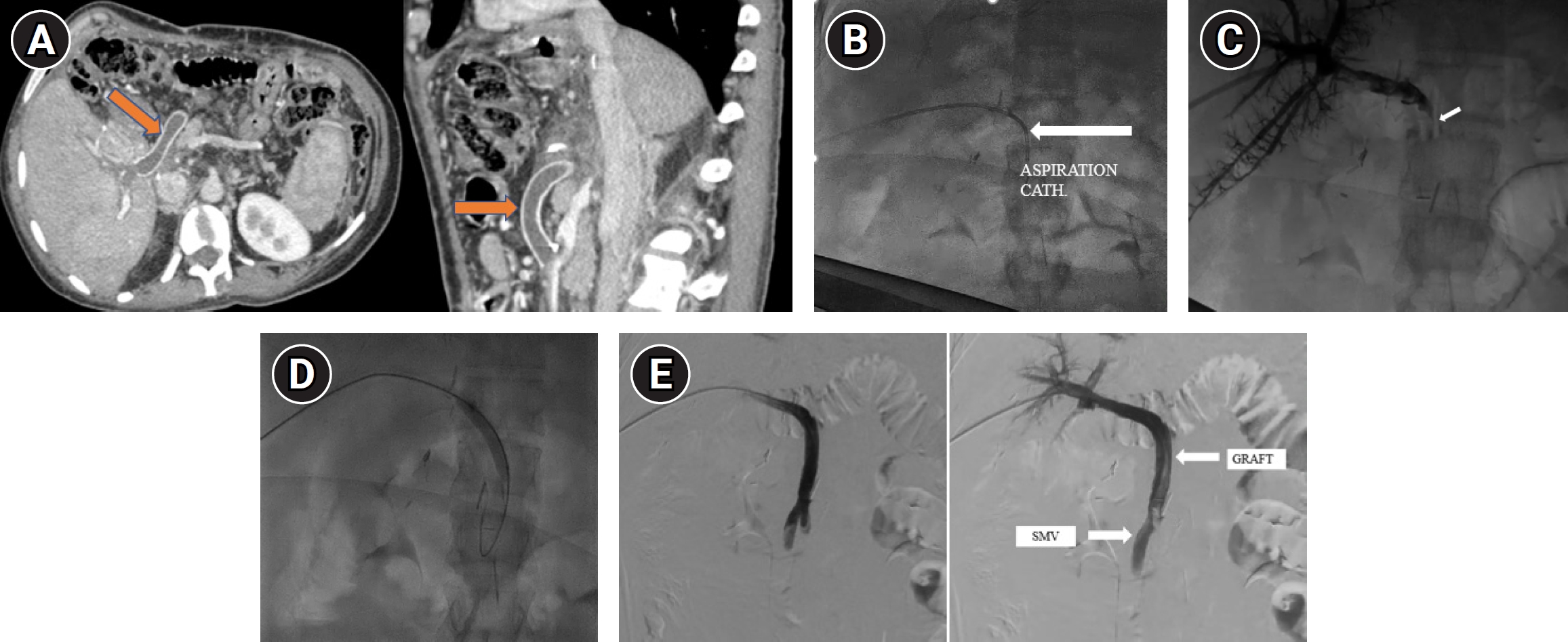
Fig. 2.(A) Follow-up spectral (pulsed-wave) Doppler ultrasound waveform at 12 months demonstrates continuous, monophasic, antegrade hepatopetal flow through the widely patent tubular expanded polytetrafluoroethylene (ePTFE) jump graft, traversing the perihepatic space without evidence of luminal narrowing, thrombosis, or perigraft fluid collection. (B) Follow-up color Doppler image at 12 months shows robust hepatopetal color filling within the synthetic ePTFE jump graft.
References
- 1. Yerdel MA, Gunson B, Mirza D, Karayalcin K, Olliff S, Buckels J, et al. Portal vein thrombosis in adults undergoing liver transplantation: risk factors, screening, management, and outcome. Transplantation. 2000;69:1873-1881. https://doi.org/10.1097/00007890-200005150-00023
- 2. Thorat A, Jeng LB, Hsu SC, Li PC, Yeh CC, Chen TH, et al. Reconstruction of the portal vein with expanded polytetrafluoroethylene jump graft in living donor liver transplantation recipients with complete portal vein thrombosis: a feasible and safe alternative. Surg Gastroenterol Oncol. 2018;23:16-20. https://doi.org/10.21614/sgo-23-1-16
- 3. Murali N, Nezami N, Latich I, Brown J, Mojibian H. Simultaneous proximal embolic protection and inferior vena cava mechanical thrombectomy using the FlowTriever system. Diagn Interv Radiol. 2020;26:345-348. https://doi.org/10.5152/dir.2019.19458
- 4. Frisoli JK, Sze D. Mechanical thrombectomy for the treatment of lower extremity deep vein thrombosis. Tech Vasc Interv Radiol. 2003;6:49-52. https://doi.org/10.1053/tvir.2003.36439
- 5. Parikh S, Shah R, Kapoor P. Portal vein thrombosis. Am J Med. 2010;123:111-119. https://doi.org/10.1016/j.amjmed.2009.05.023
Citations
Citations to this article as recorded by