Abstract
A 66-year-old man with pancreatic acinar cell carcinoma developed a hepatic metastasis that enlarged to 6.6 cm despite neoadjuvant FOLFIRINOX, pancreaticoduodenectomy, adjuvant chemoradiotherapy, and gemcitabine plus cisplatin. CT showed a heterogeneously hypoenhancing mass in the periphery of segment VI, an enhancement pattern predicting insufficient coverage by conventional chemoembolization alone. In a single session, 15 mL of an ethanol–doxorubicin–ethiodized oil mixture (1:2) was infused into the segment VI hepatic artery, and the resulting ethiodized oil deposition served as a fluoroscopic target for MWA guided by ultrasonography and fluoroscopy. A purulent perihepatic collection with pneumoperitoneum developed and resolved after percutaneous drainage. Complete response was sustained on follow-up CT, indicating that combined transarterial and thermal ablation can eradicate a large chemotherapy-refractory metastasis.
-
Keywords: Pancreatic neoplasms; Neoplasm metastasis; Chemoembolization, therapeutic; Microwave ablation; Case reports
Introduction
Pancreatic cancer remains among the most lethal solid tumors, and the liver is the most common site of distant recurrence after curative-intent resection. Multi-agent systemic chemotherapy, principally FOLFIRINOX (folinic acid, fluorouracil, irinotecan, and oxaliplatin) or gemcitabine-based regimens, is the established standard for metastatic disease and has improved survival relative to single-agent therapy [
1,
2]. Nevertheless, most patients eventually progress, and a clinically important subgroup does so in an oligoprogressive fashion, with a single dominant hepatic lesion enlarging while the remainder of the disease is controlled. Continued systemic therapy alone does not address that lesion, and the therapeutic question becomes whether local control can be achieved without interrupting systemic treatment.
Conventional options for a chemotherapy-refractory hepatic metastasis from pancreatic cancer are limited. Hepatic metastasectomy is rarely indicated and is further constrained after pancreaticoduodenectomy by adhesions and by the reduced reserve of a heavily pretreated patient. Percutaneous thermal ablation, either radiofrequency or microwave, is repeatable and spares parenchyma, but its efficacy falls sharply as tumor diameter increases; beyond 3 cm a circumferential ablative margin is difficult to obtain, and perfusion-mediated cooling from adjacent vessels further truncates the ablation zone. Transarterial therapies face the mirror-image problem: pancreatic cancer liver metastases are typically hypovascular, so conventional chemoembolization achieves only sparse retention of the ethiodized oil–anthracycline emulsion and rarely produces complete necrosis. Radioembolization and stereotactic body radiotherapy are alternatives, but availability, dosimetric constraints, and prior radiation fields restrict their use. Each modality, in other words, fails at a different part of the tumor.
This complementarity is the rationale for combining a transarterial and a percutaneous ablative approach in the same session. The combination has been most extensively validated in HCC, where chemoembolization plus thermal ablation improves tumor response, progression-free survival, and in several series overall survival compared with either modality alone [
3-
5]. Three mechanisms underlie the synergy. First, arterial embolization reduces tumor blood flow and therefore the heat-sink effect, so the same energy deposition yields a larger and more spherical coagulation zone. Second, the transarterial agent reaches the peritumoral rim, microscopic infiltration, and satellite nodules that lie outside a thermal ablation margin, whereas the thermal ablation destroys the central bulk that an embolic agent alone would not devitalize. Third, retained ethiodized oil renders the tumor radiopaque and converts a sonographically inconspicuous lesion into a fluoroscopically visible target, which has been shown to make ablation feasible in tumors that are invisible or poorly visible on ultrasonography [
6-
8]. In practice the two guidance modalities are used together: ultrasonography defines a safe puncture path and provides real-time monitoring of antenna advancement, while fluoroscopy confirms that the antenna is positioned within the ethiodized-oil–marked tumor and that overlapping ablation zones cover it in full. Because coverage is thus optimized from two independent routes, the combined approach can be expected to reduce local tumor progression relative to either approach alone.
A further refinement concerns the transarterial component itself. Adding absolute ethanol to the ethiodized oil–anthracycline emulsion converts chemoembolization into transarterial chemoablation, in which cytotoxicity no longer depends on tumor hypervascularity or on drug retention alone but on the direct ablative effect of ethanol delivered to the arteriolar and peritumoral portal venular level [
9-
14]. This property is particularly relevant to a hypovascular metastasis. Herein we report a large, chemotherapy-refractory hepatic metastasis from pancreatic acinar cell carcinoma treated in a single session by ethanol-based transarterial chemoablation followed by fluoroscopy- and ultrasonography-guided MWA.
Case Report
A 66-year-old man was referred for locoregional treatment of an enlarging solitary hepatic metastasis. He had been receiving medical treatment for type 2 diabetes mellitus and hypertension, and had no history of chronic liver disease. Pancreatic acinar cell carcinoma had been diagnosed in early 2023. He received neoadjuvant FOLFIRINOX for 12 cycles between April and September 2023, followed by open pylorus-preserving pancreaticoduodenectomy. Adjuvant treatment consisted of concurrent chemoradiotherapy and subsequent capecitabine from December 2023 to August 2024. During further follow-up a hepatic metastasis developed and enlarged progressively despite three cycles of gemcitabine plus cisplatin, reaching a maximum diameter of 6.6 cm, at which point locoregional therapy was requested. At referral his Eastern Cooperative Oncology Group performance status was 1, with preserved liver function (total bilirubin 0.4 mg/dL, albumin 4.4 g/dL, INR 1.0, platelet count 441 × 10³/μL).
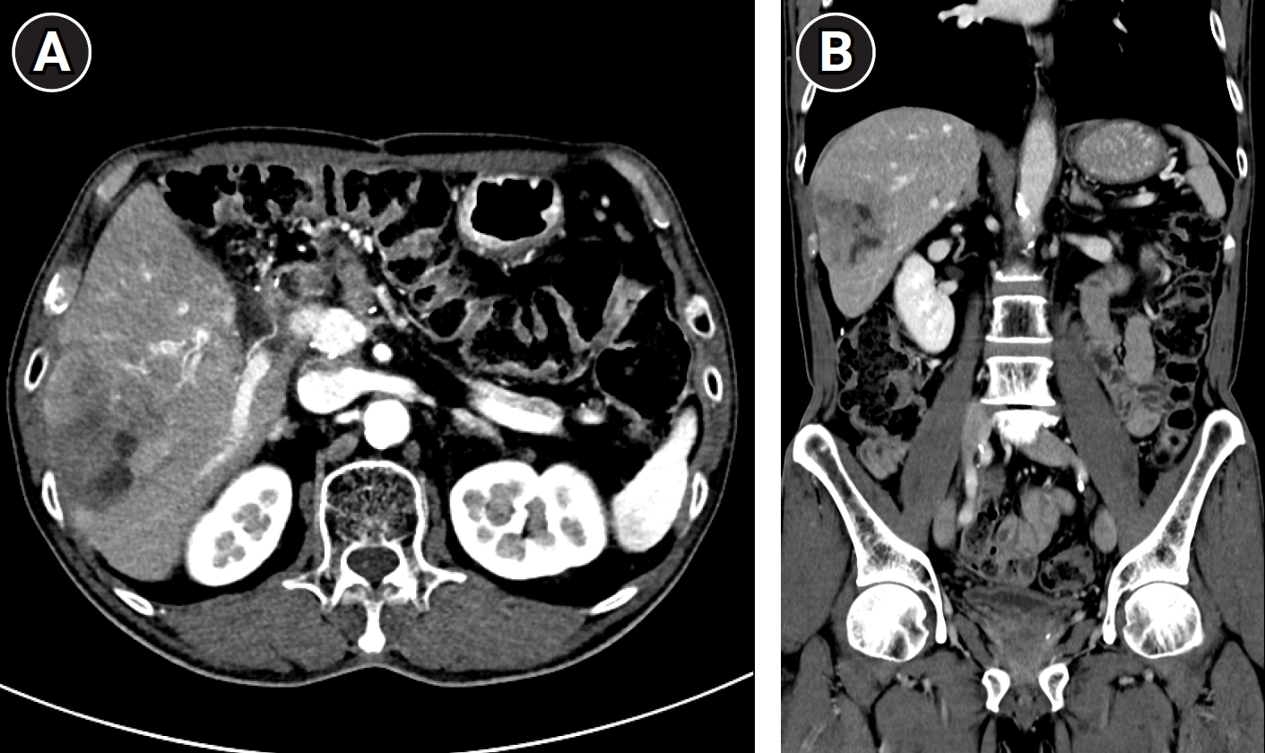
Pre-procedural contrast-enhanced liver CT demonstrated a 6.6-cm heterogeneous, predominantly hypoenhancing mass occupying the peripheral portion of segment VI, abutting the inferior hepatic surface. There was no invasion of the major hepatic vessels or of the portal vein, and no other intrahepatic or extrahepatic metastasis was identified (
Fig. 1A,
1B). Two features determined the treatment plan. The tumor size made complete coverage by percutaneous thermal ablation alone with an adequate circumferential margin unlikely, while the heterogeneous, weakly enhancing pattern predicted patchy retention of an ethiodized oil emulsion and therefore an insufficient effect from conventional chemoembolization alone. Ethanol-based transarterial chemoablation followed by percutaneous MWA of the same target in a single session was planned, and written informed consent was obtained.
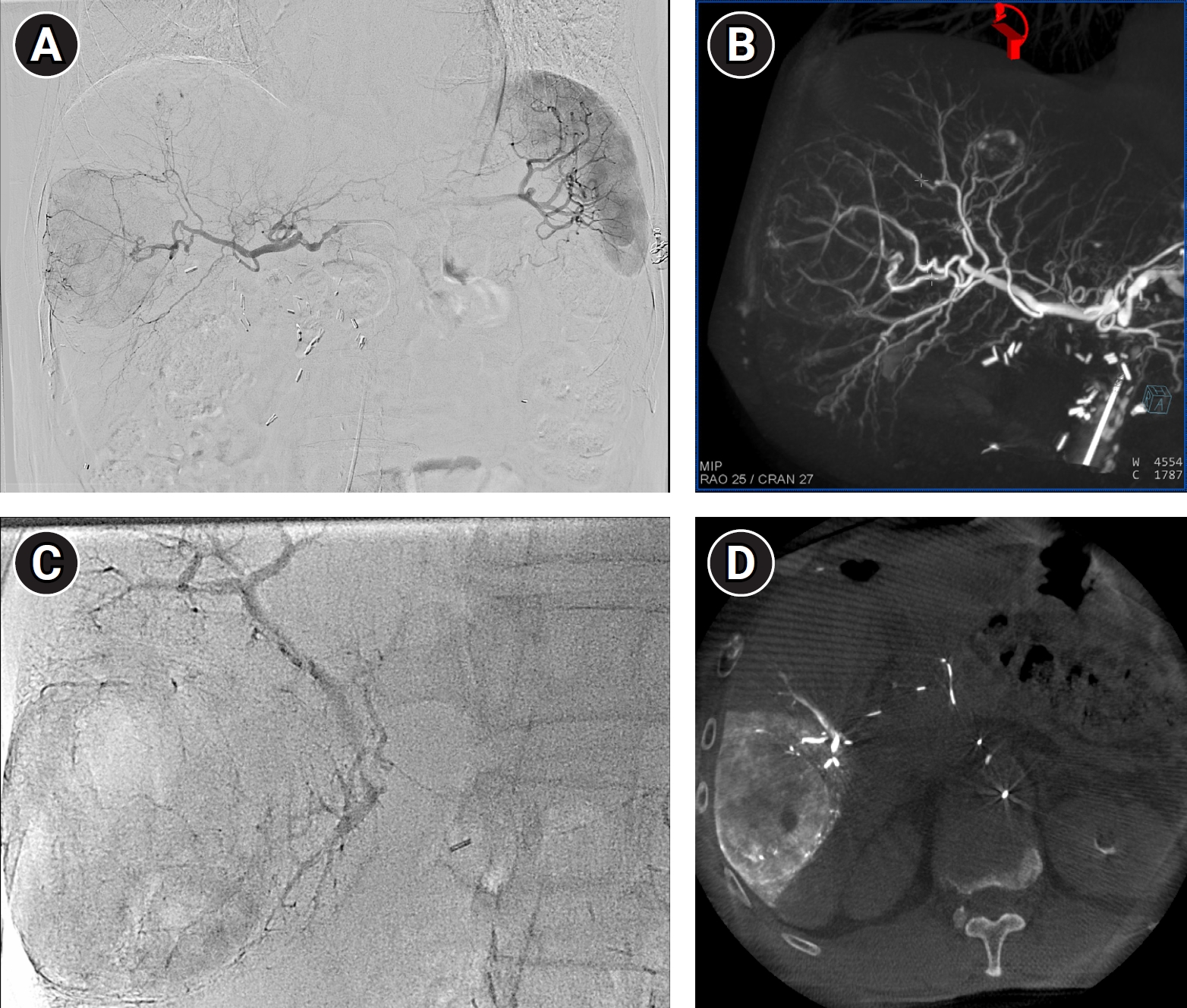
The procedure was performed on 19 March 2026. Through a right common femoral arterial approach, celiac and superior mesenteric arteriography confirmed the arterial anatomy (
Fig. 2A), and rotational cone-beam CT hepatic arteriography was obtained for tumor-feeder detection and treatment planning (
Fig. 2B). A microcatheter was advanced as close to the tumor as possible. Doxorubicin (Adriamycin, Ildong, Seoul, Korea) dissolved in iodinated contrast material was emulsified with ethiodized oil (Lipiodol, Guerbet, Villepinte, France), and the emulsion was then mixed with 99.5% dehydrated ethanol at an ethanol-to-emulsion volume ratio of 1:2, yielding a final ethanol concentration of approximately 33%. Approximately 15 mL of this mixture was infused slowly into the segment VI hepatic artery until the tumor vasculature was completely opacified and arterial flow within the tumor feeders had stagnated (
Fig. 2C). Immediate post-infusion cone-beam CT confirmed dense ethiodized oil deposition throughout the tumor, most compact at its periphery (
Fig. 2D).
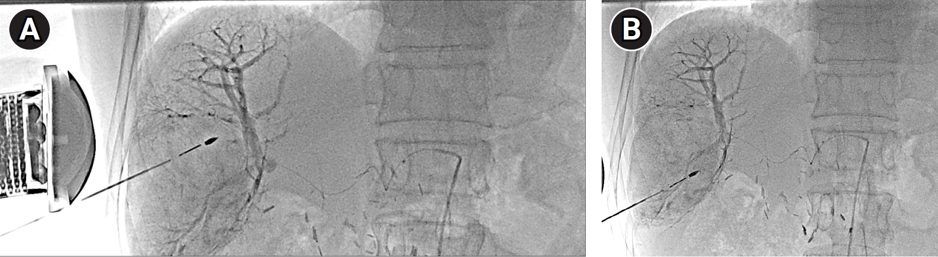
Percutaneous MWA was then performed in the same session, using the ethiodized-oil–laden tumor as the target. Under local anesthesia and conscious sedation, ultrasonography was used to select an intercostal approach avoiding the colon and the pleural reflection and to monitor advancement of the antenna in real time, and fluoroscopy confirmed that the antenna tip was positioned within the radiopaque tumor. A 13-gauge internally cooled microwave antenna connected to a STARwave microwave generator (STARmed, Goyang, Korea) was used, and four overlapping ablations were performed at 65 W for 8 minutes at each antenna position, with repositioning between ablations under combined ultrasonographic and fluoroscopic guidance until the ethiodized-oil–marked tumor and a surrounding margin were covered (
Fig. 3A,
3B). The needle tract was ablated during withdrawal. No immediate procedure-related adverse event occurred.
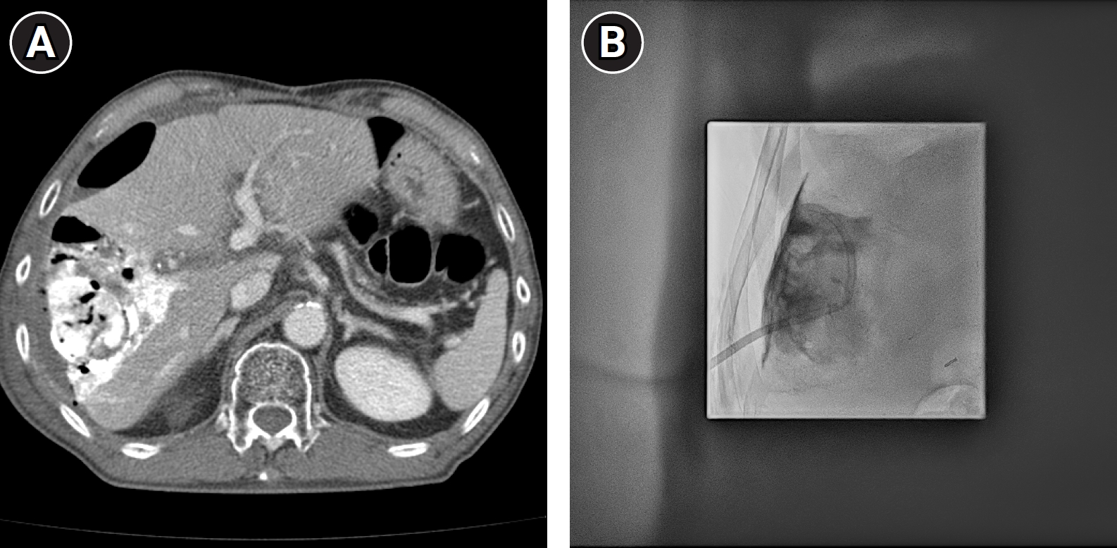
Contrast-enhanced CT obtained 2 days after the procedure showed the expected devascularized necrotic tumor, but also demonstrated air within the necrotic tissue, a new perihepatic fluid collection, and perihepatic pneumoperitoneum (
Fig. 4A). The patient complained of right upper quadrant pain and had a fever, without peritoneal irritation signs or hemodynamic instability. The findings were interpreted as post-ablation necrosis decompressing into the perihepatic space rather than hollow viscus perforation, and conservative management with broad-spectrum antibiotics and bowel rest was begun. Because the collection persisted, percutaneous catheter drainage was performed 11 days after the procedure under ultrasonographic guidance, and an 8.5-Fr pigtail catheter was placed through a route that avoided the ablation cavity and the colon (
Fig. 4B). The aspirated fluid was frankly purulent. Daily drainage was approximately 40–50 mL/day immediately after catheter placement and decreased gradually thereafter. The catheter and the residual cavity were monitored by tubography and follow-up CT, and no re-accumulation was observed.
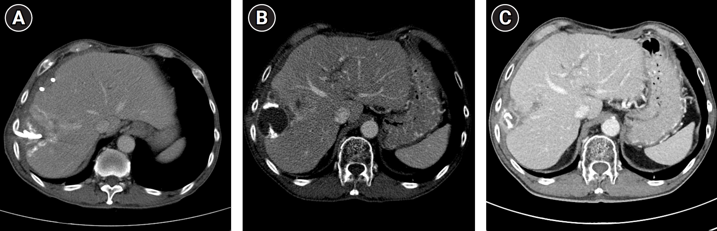
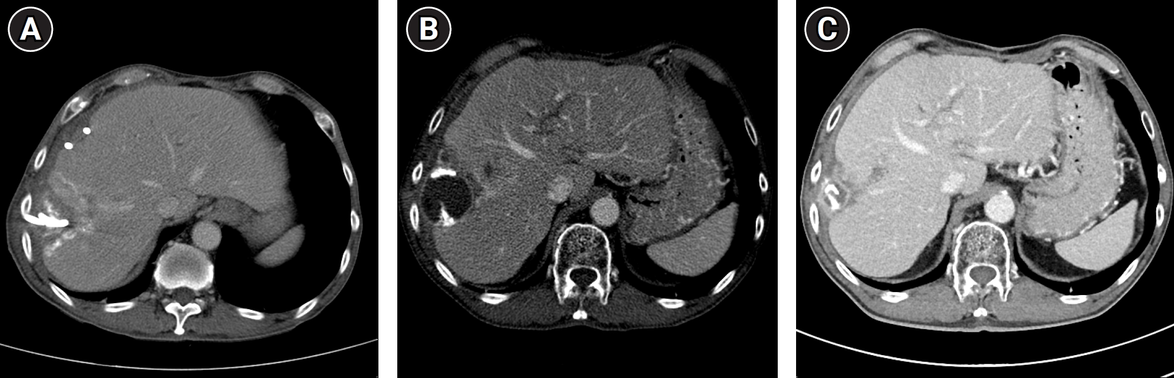
The patient's symptoms and inflammatory markers normalized over the following weeks. Follow-up CT approximately 1 month after the procedure showed disappearance of the treated tumor with no residual enhancing component, together with resolution of the perihepatic fluid collection and pneumoperitoneum; the drainage catheter was removed and the patient was discharged (
Fig. 5A). Systemic chemotherapy was resumed thereafter. CT at 1.5 months demonstrated a residual gas-containing post-ablation cavity at the treated site without any enhancing tumor component (
Fig. 5B), and by 3.5 months the cavity had involuted completely, leaving no residual or recurrent tumor and no fluid collection (
Fig. 5C). At the time of writing the patient remains on systemic chemotherapy without evidence of local progression.
The requirement for institutional review board review was waived by the Institutional Review Board of Seoul National University Bundang Hospital because of the retrospective nature of this case report (IRB No. B-2108-705-305). Written informed consent for publication of this case and the accompanying images was obtained from the patient.
Discussion
This case illustrates that a large hepatic metastasis from pancreatic cancer, progressing through multiple lines of systemic chemotherapy, can be brought to a sustained radiologic complete response by combining a transarterial and a percutaneous ablative approach in a single session. The two routes fail in different places and therefore compensate for one another. Arterial embolization reduces tumor perfusion and thus the convective heat loss that truncates a thermal ablation zone, so that a larger coagulation volume is achieved for the same energy delivery. Conversely, thermal ablation destroys the central tumor bulk that an embolic agent alone frequently fails to devitalize, while the transarterial agent reaches the peritumoral rim and any microscopic satellite disease lying beyond the thermal margin. The combination therefore optimizes coverage from two independent directions, the mechanism by which combined therapy is thought to lower local tumor progression [
3,
5]. The ethiodized oil distribution on the post-infusion cone-beam CT in this case, dense peripherally and less compact centrally, illustrates that complementarity directly.
A third and more practical advantage concerns targeting. Retained ethiodized oil converts the tumor into a radiopaque structure that can be targeted fluoroscopically, which is decisive when a lesion is poorly conspicuous on ultrasonography. Fluoroscopy-guided ablation of ethiodized-oil–marked tumors yields local control comparable to that of ultrasonography-guided ablation, and combined chemoembolization and ablation has been used specifically for ultrasonography-invisible early HCC [
6,
7]. Combining the two modalities exploits the strengths of each: ultrasonography gives a real-time, radiation-free view of the needle path and of adjacent bowel and pleura, whereas fluoroscopy verifies the relationship of the antenna to the tumor and to previously created ablation zones. A similar rationale underlies combined ultrasonographic and CT guidance for tumors in the hepatic dome [
8].
The clinical evidence supporting the combined approach comes almost entirely from HCC. A randomized trial showed that RFA combined with chemoembolization improved overall and recurrence-free survival compared with RFA alone [
3], and subsequent series have consistently found higher response rates and longer progression-free survival for the combination across early, intermediate, and recurrent disease [
4,
5]. The same pattern holds for microwave energy: in recurrent small HCC, chemoembolization followed by MWA produced a markedly higher objective response rate and 5-year progression-free survival than chemoembolization alone, and halved the number of repeat chemoembolization sessions [
5]. Data for hepatic metastases are far sparser and are dominated by colorectal primaries; for pancreatic cancer liver metastasis, experience with combined transarterial and percutaneous ablation is essentially anecdotal, which is why the present case is reported.
The transarterial component used here was not conventional chemoembolization but ethanol-based transarterial chemoablation, and this distinction is central to the rationale. Conventional chemoembolization depends on tumor hypervascularity and on retention of an ethiodized oil–anthracycline emulsion within the tumor bed; a hypovascular metastasis such as this one retains the emulsion poorly, and the expected cytotoxic effect is correspondingly weak. Adding absolute ethanol changes the mechanism of action. Ethanol produces immediate protein denaturation and coagulative necrosis independent of drug delivery kinetics; because the low-viscosity mixture penetrates to the arteriolar level and, through the peribiliary plexus, into the peritumoral portal venules, it embolizes both the afferent arterial and the peritumoral portal supply, a dual embolization that particle-based chemoembolization does not achieve [
9,
11]. Resected specimens after transarterial ethanol ablation have confirmed complete necrosis in lesions judged to be in complete response on CT [
11]. Clinically, a randomized trial found significantly higher per-tumor complete response rates and longer intralesional progression-free survival for transarterial ethanol ablation than for conventional chemoembolization [
10]; a phase I study of ablative chemoembolization reported a 100% complete response rate [
12]; and in tumors of 10 cm or more the ethanol-based technique achieved a complete response rate of 52.6% versus 16.7% for conventional chemoembolization, with a median overall survival of 21.6 versus 9.9 months [
13]. The specific formulation used in this case, a doxorubicin–ethiodized oil emulsion mixed with 99.5% ethanol at a 1:2 ratio, corresponds to that previously applied at our institution as superselective ablative chemo-ethanol embolization, which achieved a 100% complete response at 1 month in recurrent HCC without biliary complications or hepatic abscess [
15].
That same ablative potency, however, is the source of the complications observed here. When a large peripheral tumor undergoes near-complete necrosis, the necrotic cavity may lose its capsular integrity and decompress into the peritoneal cavity, producing exactly the constellation seen on the day-2 CT: intralesional gas, a perihepatic fluid collection, and pneumoperitoneum. The critical clinical judgment is to distinguish this sequela from hollow viscus injury, and in the present patient the absence of peritoneal signs and of hemodynamic compromise, together with a subcapsular necrotic cavity contiguous with the collection, made bowel perforation unlikely. The collection was nonetheless frankly purulent, so what occurred was an infected post-ablation necrosis rather than a sterile one. The prior pylorus-preserving pancreaticoduodenectomy is directly relevant to this: a biliary–enteric anastomosis renders the biliary tree permanently colonized and pneumobilia common, which both reduces the specificity of intralesional gas as a sign of infection and is a well-recognized independent risk factor for hepatic abscess after arterial embolization or thermal ablation. In such patients the volume of devitalized tissue created by a deliberately aggressive combined ablation should be assumed to be at risk of secondary infection, which argues for prophylactic antibiotic coverage, early cross-sectional imaging, and a low threshold for percutaneous drainage rather than for avoiding the combined approach. Reported adverse events attributable to the ethanol component itself include post-embolization syndrome and prolonged fever, most often with large or multifocal tumors, transient respiratory decompensation after high-volume infusion into bulky tumors, and, rarely, alcohol intoxication; biliary stricture and hepatic abscess have not been observed at an ethanol concentration of approximately 33% [
9,
14,
15]. Percutaneous catheter drainage converted an uncontrolled process into a controlled one and allowed the patient to be managed without reoperation.
Several limitations must be acknowledged. This is a single case with a follow-up of only 3.5 months, so durability of the local response is unproven, and because systemic chemotherapy was continued throughout, the local effect of the combined ablation cannot be isolated from that of systemic treatment. The complete response was defined radiologically and was not confirmed histologically. The optimal sequence and interval between the two components, the ethanol concentration and volume appropriate to a hypovascular metastasis, and the criteria identifying patients likely to benefit all remain undefined, and the complication observed here indicates that a larger necrotic burden carries a real, if manageable, cost. Within these limits, the case demonstrates that combined ethanol-based transarterial chemoablation and fluoroscopy- and ultrasonography-guided MWA can achieve complete necrosis of a large, chemotherapy-refractory pancreatic cancer liver metastasis, and that the resulting post-ablation cavity and perihepatic collection can be managed conservatively by percutaneous drainage.
Conflict of interest
Jae Hwan Lee and Kun Yung Kim, contributing editors of the Korean Journal of Interventional Radiology, were not involved in the editorial evaluation or decision to publish this article. The remaining author has declared no conflicts of interest.
Funding
None.
Acknowledgments
None.
Author contributions
Conceptualization: JHL, CJY. Investigation: JHL, KYK. Supervision: CJY. Writing - original draft: JHL. Writing - review & editing: JHL, KYK, CJY.
Data availability statement
Data sharing does not apply to this article as no datasets were generated or analyzed during the current study.
Fig. 1.Pretreatment contrast-enhanced CT in a 66-year-old man with a progressive hepatic metastasis from pancreatic acinar cell carcinoma. (A) Axial arterial phase image shows a 6.6-cm heterogeneous, weakly and irregularly enhancing mass in the peripheral portion of segment VI. (B) Coronal portal venous phase image shows the mass abutting the inferior surface of the right hepatic lobe, without invasion of the major hepatic veins or portal vein. Surgical clips from the previous pylorus-preserving pancreaticoduodenectomy are seen in the pancreatic bed.
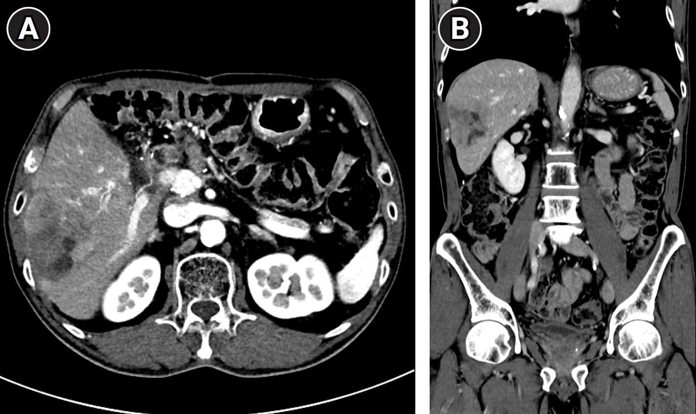
Fig. 2.Transarterial chemoablation of the segment VI metastasis. (A) Celiac arteriography demonstrates the hepatic arterial anatomy without a discrete hypervascular tumor stain. (B) Maximum-intensity-projection image reconstructed from rotational cone-beam CT hepatic arteriography, obtained for tumor-feeder detection and treatment planning, shows the candidate feeding branches arising from the segment VI hepatic artery (crosshairs). (C) Spot image after infusion of approximately 15 mL of a doxorubicin–ethiodized oil emulsion mixed with 99.5% ethanol at a 1:2 ethanol-to-emulsion ratio demonstrates a faint, well-demarcated tumor stain corresponding to the hypovascular metastasis. (D) Cone-beam CT obtained immediately after infusion of the ethanol-emulsion mixture shows dense ethiodized oil deposition throughout the tumor, most compact at its periphery.
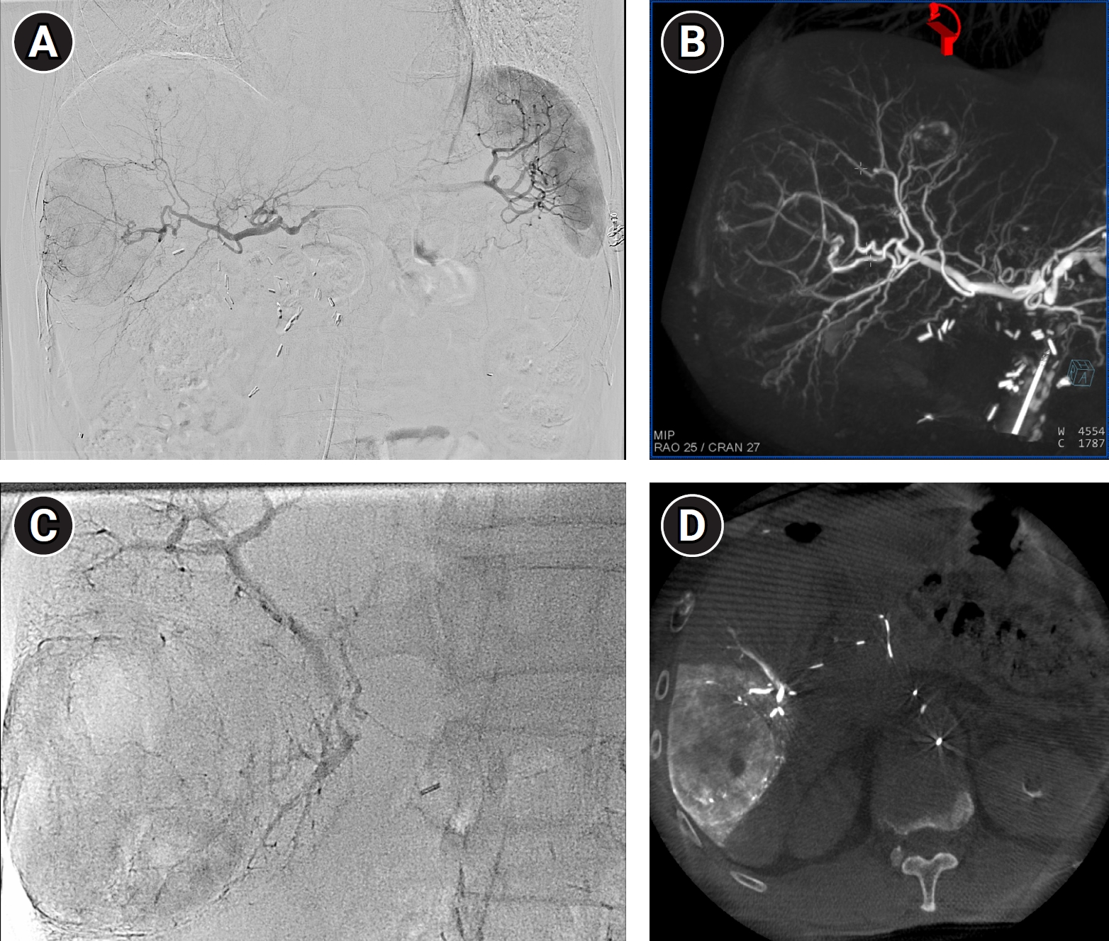
Fig. 3.Percutaneous MWA performed in the same session under combined ultrasonographic and fluoroscopic guidance, using the ethiodized-oil–marked tumor as the target. (A, B) Fluoroscopic images obtained in two different projections show the 13-gauge microwave antenna advanced through an intercostal approach with its tip positioned within the target tumor. Four overlapping ablations were performed at 65 W for 8 minutes at each antenna position.
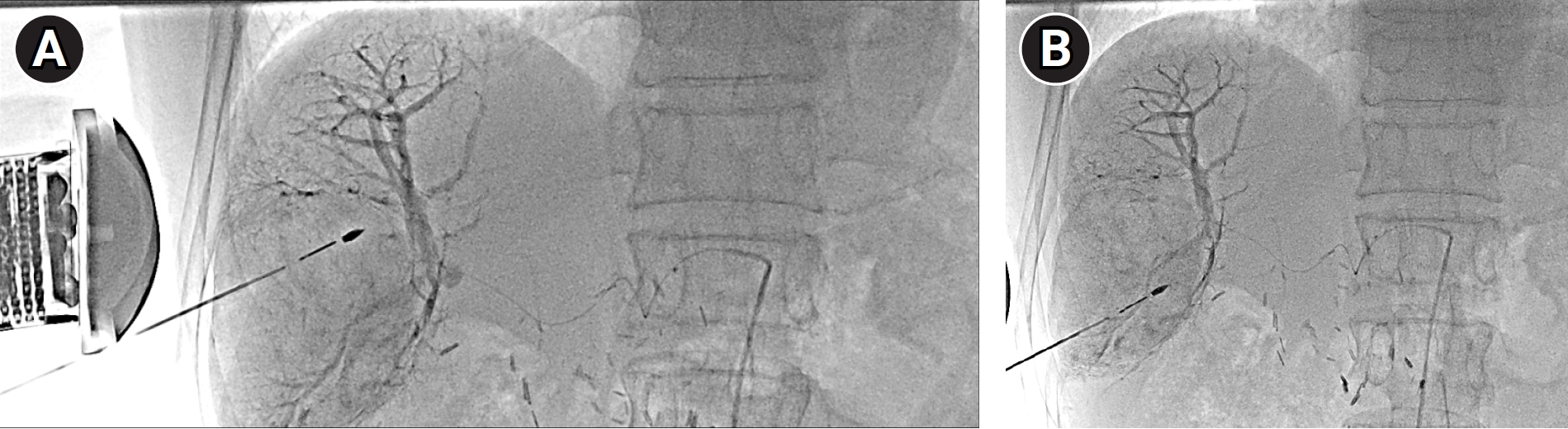
Fig. 4.Post-procedural complication and its management. (A) Contrast-enhanced CT obtained 2 days after the procedure shows the devascularized necrotic tumor containing gas, together with a perihepatic fluid collection and perihepatic pneumoperitoneum. (B) Fluoroscopic image obtained during percutaneous catheter drainage performed 11 days after the procedure shows an 8.5-Fr pigtail catheter positioned within the perihepatic collection; the aspirated fluid was frankly purulent.
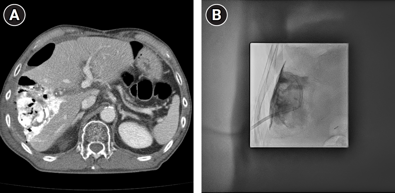
Fig. 5.Serial follow-up contrast-enhanced CT. (A) At 1 month, the treated tumor has disappeared with no residual enhancing component, and the perihepatic fluid collection and pneumoperitoneum have resolved; only a small focus of retained ethiodized oil remains. (B) At 1.5 months, a residual gas-containing post-ablation cavity is present at the treated site without an enhancing tumor component. (C) At 3.5 months, the cavity has involuted completely, with no residual or recurrent tumor and no fluid collection.
References
- 1. Conroy T, Desseigne F, Ychou M, Bouche O, Guimbaud R, Becouarn Y, et al. FOLFIRINOX versus gemcitabine for metastatic pancreatic cancer. N Engl J Med. 2011;364:1817-1825. https://doi.org/10.1056/NEJMoa1011923
- 2. Conroy T, Hammel P, Hebbar M, Ben Abdelghani M, Wei AC, Raoul JL, et al. FOLFIRINOX or gemcitabine as adjuvant therapy for pancreatic cancer. N Engl J Med. 2018;379:2395-2406. https://doi.org/10.1056/NEJMoa1809775
- 3. Peng ZW, Zhang YJ, Chen MS, Xu L, Liang HH, Lin XJ, et al. Radiofrequency ablation with or without transcatheter arterial chemoembolization in the treatment of hepatocellular carcinoma: a prospective randomized trial. J Clin Oncol. 2013;31:426-432. https://doi.org/10.1200/JCO.2012.42.9936
- 4. Wang C, Liao Y, Qiu J, Yuan Y, Zhang Y, Li K, et al. Transcatheter arterial chemoembolization alone or combined with ablation for recurrent intermediate-stage hepatocellular carcinoma: a propensity score matching study. J Cancer Res Clin Oncol. 2020;146:2669-2680. https://doi.org/10.1007/s00432-020-03254-2
- 5. Ji J, Yang W, Shi HB, Liu S, Zhou WZ. Transcatheter arterial chemoembolization alone versus combined with microwave ablation for recurrent small hepatocellular carcinoma after resection: a retrospective comparative study. BMC Gastroenterol. 2022;22:321. https://doi.org/10.1186/s12876-022-02387-7
- 6. Lee H, Yoon CJ, Seong NJ, Jeong SH, Kim JW. Comparison of combined therapy using conventional chemoembolization and radiofrequency ablation versus conventional chemoembolization for ultrasound-invisible early-stage hepatocellular carcinoma (Barcelona Clinic Liver Cancer stage 0 or A). Korean J Radiol. 2018;19:1130-1139. https://doi.org/10.3348/kjr.2018.19.6.1130
- 7. Kim J, Yoon CJ, Seong NJ, Jeong SH, Kim JW. Fluoroscopy-guided radiofrequency ablation for small hepatocellular carcinoma: a retrospective comparison with ultrasound-guided ablation. Clin Radiol. 2015;70:1009-1015. https://doi.org/10.1016/j.crad.2015.05.008
- 8. Kan X, Wang Y, Han P, Yao Q, Qian K, Xiong B, et al. Combined ultrasound/computed tomography guidance in percutaneous radiofrequency ablation after transarterial chemoembolization for hepatocellular carcinoma in the hepatic dome. Cancer Manag Res. 2019;11:7751-7757. https://doi.org/10.2147/CMAR.S212127
- 9. Yu SC, Hui EP, Wong J, Wong H, Mo F, Ho SS, et al. Transarterial ethanol ablation of hepatocellular carcinoma with lipiodol ethanol mixture: phase II study. J Vasc Interv Radiol. 2008;19:95-103. https://doi.org/10.1016/j.jvir.2007.08.038
- 10. Yu SC, Hui JW, Hui EP, Chan SL, Lee KF, Mo F, et al. Unresectable hepatocellular carcinoma: randomized controlled trial of transarterial ethanol ablation versus transcatheter arterial chemoembolization. Radiology. 2014;270:607-620. https://doi.org/10.1148/radiol.13130498
- 11. Yu SC, Lau TW, Tang P, Chan SK, Chu CC, Hui JW, et al. Mechanism and natural course of tumor involution in hepatocellular carcinoma following transarterial ethanol ablation. Cardiovasc Intervent Radiol. 2016;39:1136-1143. https://doi.org/10.1007/s00270-016-1360-z
- 12. Yu SC, Chan SL, Lee KF, Hui JW, Hui EP, Chu CM, et al. Ablative chemoembolization for hepatocellular carcinoma: a prospective phase I case-control comparison with conventional chemoembolization. Radiology. 2018;287:340-348. https://doi.org/10.1148/radiol.2017170154
- 13. Yu SC, Hui JW, Li L, Cho CC, Hui EP, Chan SL, et al. Comparison of chemoembolization, radioembolization, and transarterial ethanol ablation for huge hepatocellular carcinoma (≥ 10 cm) in tumour response and long-term survival outcome. Cardiovasc Intervent Radiol. 2022;45:172-181. https://doi.org/10.1007/s00270-021-02777-6
- 14. Yu SC, Hui JW, Chong CC, Chu CM, Cheung S, Wong J, et al. Transarterial ethanol ablation for small hepatocellular carcinoma (≤ 3 cm): a comparative study versus radiofrequency ablation. Cardiovasc Intervent Radiol. 2020;43:732-739. https://doi.org/10.1007/s00270-020-02426-4
- 15. Lee JH, Kim KY, Lee CH, Kim M, Yoon CJ. Superselective ablative chemo-ethanol embolization for recurrent single hepatocellular carcinoma: a 6-month outcome analysis. J Liver Cancer. 2024;24:217-223. https://doi.org/10.17998/jlc.2024.05.08