중심단어
Parapelvic cyst, Hydronephrosis, Sclerotherapy, Internal drainage
국문 초록
신우주위 낭종은 신우신배와의 연결이 없고, renal hilum 및 collecting system에 가까이 위치하고 있어 폐색, 감염, 고혈압, 통증, 결석 등의 합병증을 일으킬 수 있다. 무증상의 신우주위 낭종은 치료가 필요 없으나, 이와 연관된 증상이 발생하면 치료가 필요하다. 본 증례에서는 68세 남자 환자가 좌측 옆구리 통증으로 내원하여 촬영한 CT에서 좌측 결석 및 신우주위 낭종으로 인한 수신증이 확인되었다. 이 신우주위 낭종에 대해서 경화치료를 시행하였으나, 2일 후 신우주위 낭종과 신우신배 사이의 communication이 확인되었다. 이에 경피적으로 신우신배를 통한 내배액술을 이용하여 신우주위 낭종과 수신증을 치료했던 사례를 보고하고자 한다.
영문 초록
Since the parapelvic cyst has no communication with the renal pelvis and is located close to the renal hilum and collecting system, complications such as obstruction, infection, high blood pressure, pain, and stones may occur. Asymptomatic cysts do not require treatment, but if associated symptoms occur, treatment is required. In our case, a 68-year-old male patient with left flank pain was diagnosed with a left renal stone and hydronephrosis due to parapelvic cyst. The sclerotherapy was performed on this parapelvic cyst, but the communication between the parapelvic cyst and renal pelvocalyx was confirmed 2 days later. We want to report a case of treatment of parapelvic cyst using internal drainage via renal pelvocalyx percutaneously.
Introduction
신우주위 낭종은 무증상인 경우에는 치료가 필요 없으나, 이와 연관된 폐색, 감염, 고혈압, 통증, 결석 등의 합병증이 발생하면 치료가 필요하다(1, 2). 신우주위 낭종은 경피적 흡인 또는 배액, 경화제 주입, 요관경을 이용한 내배액술, 수술적 절제 등을 통해 치료할 수 있으며, 본 증례에서는 경화치료를 시행하였던 환자에서 신우주위 낭종과 신우신배와의 communication이 확인되어 경피적으로 신우신배를 통한 내배액술로 전환하여 치료하였던 사례를 보고하고자 한다.
Case report
증례
68세/남자
임상소견
좌측 옆구리 통증으로 내원하여 촬영한 CT 요로조영술에서 6mm 신배 결석 및 신우주위 낭종으로 인한 수신증이 확인되었다. 결석에 대해서는 체외충격파쇄석술이 진행되었고 신우주위 낭종에 대해서 에탄올을 이용한 경화치료를 시행하였다. 경화치료 시행 2일 후 줄어들던 배액량이 급격히 증가하고 배액된 검체에서 크레아틴 수치가 37mg/dL로 확인되었다. 이에 요로와의 communication이 의심되어 다음날 관조영술이 의뢰되었다.
진단명
Hydronephrosis due to parapelvic cyst
영상소견
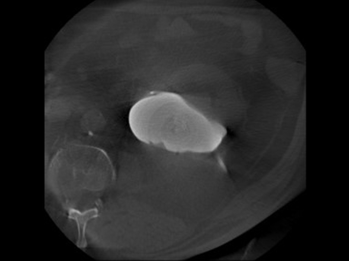
내원 후 시행한 CT 요로조영술에서 좌측 신우주위 낭종으로 인한 수신증이 확인되며 (Fig. 1A), 경화치료 시 시행한 cone beam CT에서 신우주위 낭종과 신우신배와의 communication은 없는 것을 확인하였다 (Fig. 1B). 경화치료 후 요로와의 communication이 의심되어 시행한 관조영술에서 신배로 조영제가 채워지는 것이 보이고 (Fig. 2A), cone beam CT에서 communication 부위로 생각되는 곳이 확인되었다 (Fig. 2B).
시술방법 및 재료
Cone beam CT에서 확인되었던 communication으로 생각되는 부분을 통한 내배액술을 진행하기 위해 삽입된 8.5-F pigtail catheter (Sung Won medical Corp, Cheongju, Korea)를 제거하고, 해당 경로를 통해 7-F Radiofocus introducer (Terumo, Tokyo, Japan)를 삽입하여 0.035-inch hydrophilic guide wire (Terumo, Tokyo, Japan)과 5-F Kumpe catheter (Angiodynamics, Latham, New York, USA)를 이용하여 communication 부위를 선택하고자 하였다 (Fig. 3A). 목표하였던 communication 부위에 대한 선택에 실패하여 투시 유도 하에 신우주위 낭종을 거쳐 조영제가 채워진 상극 신배를 21G needle (Cook, Bloomington, IN, USA)을 이용하여 직접 천자하여 인위적으로 communication을 형성하였다 (Fig. 3B). 이 communication을 5mm x 40mm sized balloon catheter (Mustang, Boston Scientific, Marlborough, MA, USA)를 이용하여 확장시킨 후 (Fig. 3C) 8.5-F pigtail catheter (Sung Won medical Corp, Cheongju, Korea) 를 거치하였다. 8.5-F pigtail catheter에는 추가로 측면에 구멍을 내어 신우주위 낭종 안의 체액이 내배액 및 외배액 될 수 있도록 하였다 (Fig. 3D). 2주 뒤에는 6mm x 60cm sized balloon catheter (X-force, Bard, Covington, USA)를 이용하여 communication 부위를 확장하고 10.2-F pigtail catheter (Sung Won medical Corp, Cheongju, Korea) 를 거치하였다. Communication 부위의 maturation을 위해 4주간 pigtail catheter 거치하고 maturation을 최종 확인 후 catheter를 제거하였다.
추적관찰
경피적 접근로 및 비뇨기과에서 삽입한 double J stent를 모두 제거 후 한달 뒤에 시행한 복부 CT에서 신우주위 낭종은 거의 없어지고 수신증도 호전된 모습을 보였다.
Fig. 1. A
Fig. 1. (A) CT shows left hydronephrosis due to parapelvic cyst.
Fig. 1. B
(B) There is no communication between parapelvic cyst and renal pelvocalyx on pre-procedural cone beam CT
Fig. 2. A
Fig. 2. (A) The contrast is filling in renal calyx during tubography via previously inserted pigtail catheter.
Fig. 2. B
(B) The communication between parapelvic cyst and renal pelvocalyx was confirmed on cone beam CT.
Fig. 3. A
Fig. 3. (A) A fluoroscopy image shows an attempt to select a communication part using 5-F Kumpe catheter and 0.035-inch hydrophilic guide wire.
Fig. 3. B
(B) Direct puncture of upper pole calyx using 21G needle wasdone through the parapelvic cyst.
Fig. 3. C
(C) Dilatation of the communication between parapelvic cyst andrenal pelvocalyx was done using 5mm x 40mm sized balloon catheter .
Fig. 3. D
(D) Internal and externaldrainage was achieved using 8.5-F pigtail catheter with additional side holes
고찰
신우주위 낭종을 비롯한 신장낭종의 치료에 에탄올을 이용한 경화치료의 유용성과 안전성이 증명된 후(3), 낭종의 치료에 경화치료가 널리 이용되고 있다. 경화치료 시에는 경화제를 주입하기 전에 조영제를 낭종 내에 주입하여 조영제가 주변 조직으로 새어 나가는 부분이 있는지 확인하는 과정이 중요하다. 본 증례에서는 경화치료 과정에서 주변 조직과의 communication이 확인되지 않아 경화제 주입이 이루어졌으나, 치료 2일 후에 신배와의 communication이 확인되었다. 새롭게 communication이 생긴 원인은 명확하지 않으나, 체외충격파쇄석술 이후 신실질의 손상(4, 5)이 발생할 수 있는 점을 고려하면 환자가 경화치료 2주 및 4주 전 2차례의 체외충격파쇄석술을 받은 과거력의 영향을 완전히 배제하기는 어려울 것으로 생각된다. 신우주위 낭종을 요로를 통해 내배액하는 개념은 요관경을 통해 역행적 접근하거나 경피적으로 신장경을 이용하여 접근한 후 신우주위 낭종의 벽을 직접 절개하는 방식으로 치료한 사례들에서 소개되어 왔으나(6-8), 경피적으로 pigtail을 이용하여 communication 부위를 maturation 시킴으로써 신우주위 낭종을 치료한 사례는 문헌상 보고된 바가 없었다. 직접 절개를 하는 방법은 주로 레이저를 이용하기 때문에 maturation 에 필요한 시간이 줄일 수 있다는 장점이 있지만, 내시경을 이용한 육안적 확인만으로 주위 구조물에 대한 정확한 정보 없이 낭종의 벽을 절개해야 한다는 위험이 있다. 경피적 접근을 통한 신우신배로의 내배액술은 투시 및 cone beam CT 유도 하에 주변 구조물에 대한 정보를 확인하면서 시술을 진행할 수 있다는 측면에서 위험도를 낮출 수 있을 것으로 생각되며, 본 증례는 특이 합병증 없이 성공적으로 신우주위 낭종과 수신증이 치료된 것을 보여주었다는 점에서 의의가 있다.
참고문헌
1. Amis Jr E, Cronan J, Pfister R. The spectrum of peripelvic cysts. British journal of urology 1983;55:150-153
2. Hinman F. Obstructive renal cysts. The Journal of urology 1978;119:681-683
3. Bean WJ. Renal cysts: treatment with alcohol. Radiology 1981;138:329-331
4. Evan AP, Willis LR, Connors B, Reed G, McAteer JA, Lingeman JE. Shock wave lithotripsy-induced renal injury. American journal of kidney diseases 1991;17:445-450
5. Smith LH, Drach G, Hall P, Lingeman J, Preminger G, Resnick MI, et al. National High Blood Pressure Education Program (NHBPEP) review paper on complications of shock wave lithotripsy for urinary calculi. The American journal of medicine 1991;91:635-641
6. Luo Q, Zhang X, Chen H, Liu Z, Chen X, Dai Y, et al. Treatment of renal parapelvic cysts with a flexible ureteroscope. International urology and nephrology 2014;46:1903-1908
7. Shao Z-Q, Guo F-F, Yang W-Y, Wang G-J, Tan S-F, Li H-L, et al. Percutaneous intrarenal marsupialization of symptomatic peripelvic renal cysts: a single-centre experience in China. Scandinavian Journal of Urology 2013;47:118-121
8. Zhao Q, Huang S, Li Q, Xu L, Wei X, Huang S, et al. Treatment of parapelvic cyst by internal drainage technology using ureteroscope and holmium laser. The West Indian medical journal 2015;64:230
Citations
Citations to this article as recorded by