중심단어
Hepatocelluar carcinoma (HCC), radiofrequency ablation (RFA), portal vein, bleeding
임상소견
간암에 대해 9차례 경동맥 화학 색전술(Transcatheter arterial chemoembolization)을 시행한 이후에 진행된 두 개의 간암 병변에 대하여 cool tip needle을 이용하여 고주파 열치료(radiofrequency ablation, RFA)를 시행하였다. 고주파 열치료 전 수축기/이완기 혈압이 111/68 mmHg에서 시술 2시간 후 70/50 mmHg로 감소되어 시행한 간 CT에서 동맥 그리고 문맥기에 출혈이 보여 간동맥 조영술과 색전술을 시행하였다(Fig. 1, 2). 간동맥 색전술 직후 수축기 /이완기 혈압이 90/60mmHg로 상승되었으나 4시간 이후 수축기 혈압이 60mmHg로 감소되어 간문맥 조영술을 다시 시행하고 출혈부위에 대해 색전술을 시행하였다 (Fig. 3).
진단명
간암의 고주파 열치료 후 발생한 간문맥 출혈
영상소견
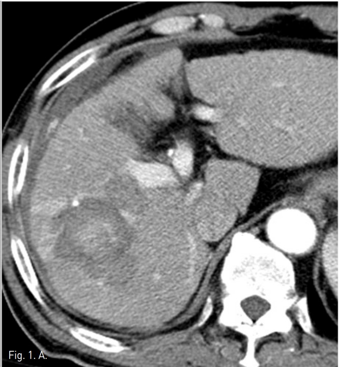
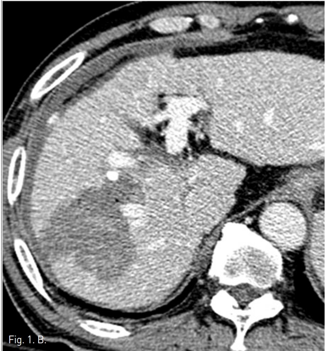
동맥기 간 CT 횡단면 영상에서 간우엽에 두 개의 간암의 크기가 증가되어 고주파 열치료를 시행하였다. 시술후 추적 CT에서 간우엽의 표면을 따라 조영제가 혈관 밖으로 흐르는 것이 발견되었고, 출혈성 복수의 양이 증가되었다 (Fig. 1).
시술방법 및 재료
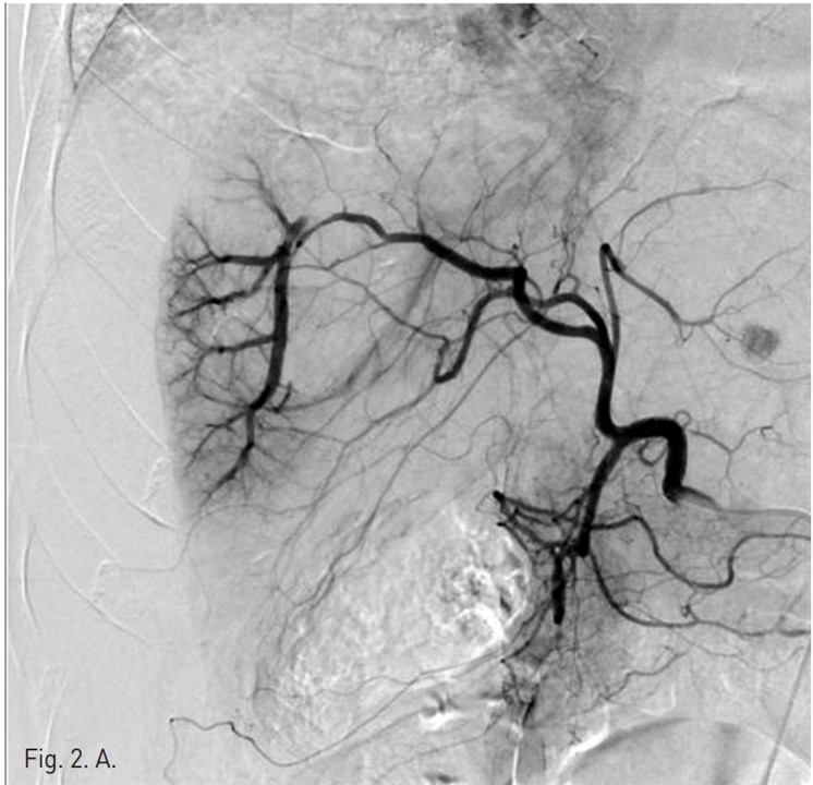
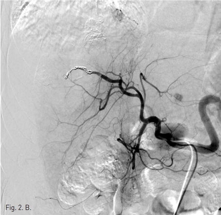
간동맥 혈관 조영을 하기 위해 오른쪽 대퇴동맥 주변을 국소 마취 후, angiocath로 대퇴동맥을 puncture하고 guidewire유도 하에 RH catheter삽입 후 celiac trunk를 선택하여 간동맥 혈관조영을 시행하였다. 간동맥 혈관 조영상에서 분명한 출혈은 보이지 않았지만, right anterior superior hepatic artery에서 arteriovenous shunt가 있어 microcatheter(Sirabe 2.2 Fr)로 간동맥을 선택하여 microcoil (TORNADO microcoil, 4 mm x 2mm, 3 mm x2 mm and 5 mm x 2 mm)과 gelfoam을 이용하여 색전술을 시행하였으나(Fig. 2) 색전술 4시간 후 수축기 혈압이 60 mmHg로 감소되었다. 간동맥 색전술에도 불구하고 계속적인 출혈이 있어서 간문맥 출혈을 의심하였음. Chiba needle로 좌측 간문맥을 puncture하고 guide wire유도 하에 angiography catheter를 삽입하여 간문맥 혈관 조영술을 시행하였다. 간문맥 혈관 조영에서 right anterior inferior portal branch에서 출혈이 관찰되어 microcatheter(Sirabe 2.2 Fr)로 right anterior inferior portal branch를 선택하여 두 개의 microcoil (Nester 35 mm x 4 mm and Tornado 5 mm x 2mm), Glue(Histoacryl 0.5ml) 그리고 Gelfoam을 이용하여 색전술을 시행하였다 (Fig. 3).
고찰
복강내 출혈은 간암의 고주파 열치료 시술중과 이후에 발생되는 중요한 합병증 중 하나로 Korean Study Group of Radiofrequency Ablation(l,139 patients in 11 institutions)의 보고에 따르면,주요한 합병증(2.43 %) 중에서 0.46 %의 빈도로 두 번째를 차지하는 것으로 보고되고 있으며, 출혈과 동반되어 거짓동맥류나 동정맥루가 발생되기도 한다, 고주파 열치료 needle에 의한 직접적인 혈관 손상이 혈관의 열손상보다 주요 원인으로 생각되고 있으며,간경화 환자에서 혈액응고장애도 주된 원인으로 생각된다. 또한 간문맥의 경우 needle에 관통되는 경우 높은 문맥고혈압으로 인하여 출혈의 가능성이 높은 것으로 생각된다. 따라서 간암의 고주파 열치료 후에는 활력 징후와 complete blood cell count, prothrombin time, and partial thromboplastin time을 추적 조사해야 한다. 대부분의 정맥 출혈은 보존적 치료와 수혈로 자가 치유될 수 있는 것으로 보고되고 있지만,본 증례에서와 같이 자가 치유되지 않는 정맥 출혈과 동맥성 출혈은 침습적 치료가 필요하며,최근의 여러 문헌들에 따르면 혈관을 통한 색전술이 간내 출혈의 진단과 치료에 최선의 방법으로 제시되고 있다 (2-4).
참고문헌
1. Rhim H., Yoon K., Lee JM, et al Complications after radio-fiequency thermal ablation of hepatic tumors: spectrum of imaging findings. RadioGraphics 2003; 23:123-136
2. Carrafiello G., Lagana D., lanniello A., et al. Bleeding after percutaneous radiofrequency ablation: successful treatment with transcatheter embolization. Eur J Radiol 2007; 61(2):351-5.
3. Livraghi T, SoIbiati L, Meloni MF, et al Treatment of focal liver tumors with percutaneous radiofrequency ablation: complication encountered in a multicenter study. Radiology 2003; 226(2):441-51.
4. Mulier S, Mulier P, NiY, et al. Complications of radiofrequency coagulation of liver tumors. Br J Surg 2002; 89(10):1206-22.
Fig. 1. A
Fig. 1. Dynamic liver CT arterial (A) and portal (B) phase scans show extravasation (arrows) of contrast materials at the surface of the liver and hemoperitoneum around the liver. The focus of extravasation was not clear.
Fig. 1. B
Fig. 1. Dynamic liver CT arterial (A) and portal (B) phase scans show extravasation (arrows) of contrast materials at the surface of the liver and hemoperitoneum around the liver. The focus of extravasation was not clear.
Fig. 2. A
Fig. 2. A. Hepatic arterial angiography shows AP shunts (arrow) from the right anterior superior hepatic artery.
Fig. 2. B
B. Right anterior superior hepatic artery was embolized with 5 microcoils and gelfoam. Note there is no definite extravasation of contrast media on initial and final hepatic angiography.
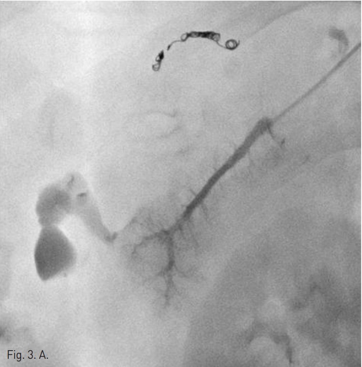
Fig. 3. A
Fig. 3. A. Direct portogram shows active bleeding (arrow) from the right anterior inferior portal branch.
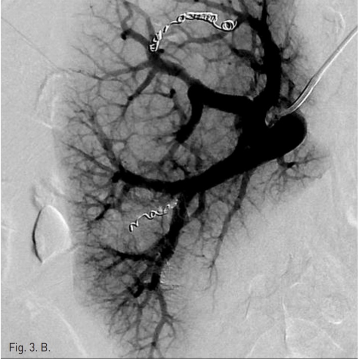
Fig. 3. B
B. The right anterior inferior portal branch was embolized with 2 microcoils, glue and gelfoam. After embolization, there was no further contrast extravasation.
Citations
Citations to this article as recorded by