중심단어
Angioplasty, subintimal angioplasty, retrograde access
임상소견
Diabetic foot 환자로서 2009년 5월 left big toe gangrene으로 인해 toe amputation 시행함. 외래 추적 관찰 중 1주일 전부터 left big toe amputation site에서 discharge가 발생하였으며 양쪽 두번째 발가락의 color change가 발생하여 내원함.
왼쪽 SFA의 협착에 대해서는 2008년 이미 stent를 삽입하였으나 폐색되어 있는 상태로서 왼쪽 다리에 대해서는 입원 후 femoropopliteal bypass수술을 시행하였으며, 수술 시행 후 우측 다리에 대해 혈관 성형술을 시행하기 로 함.
진단명
Critical limb ischemia due to chronic atherosclerotic occlusion
영상소견
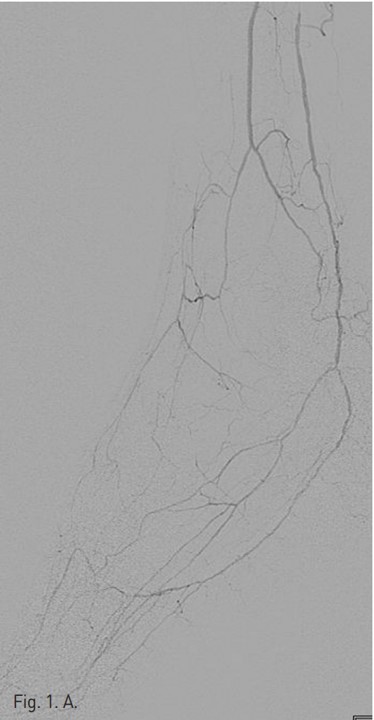
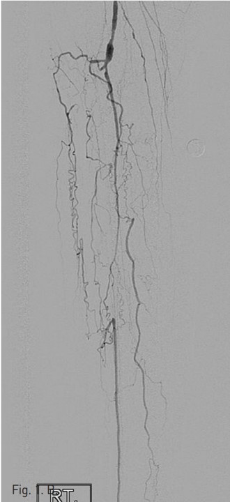
시술 당시 시행한 우측 하지 혈관조영술 (Fig. 1A)에서 right superficial femoral artery(SFA)의 mid-portion에 severe segmental stenosis가 있다. Popliteal artery는 segmental narrowing이 있고, tibio peroneal(TP) trunk 기시부에서부터 완전 폐색을 보이고 있으며, 우측 비골동맥과 후경골 동맥이 기시부에서부터 근위부 2/3 segment가 완전 폐색되어 있다(Fig. 1B).
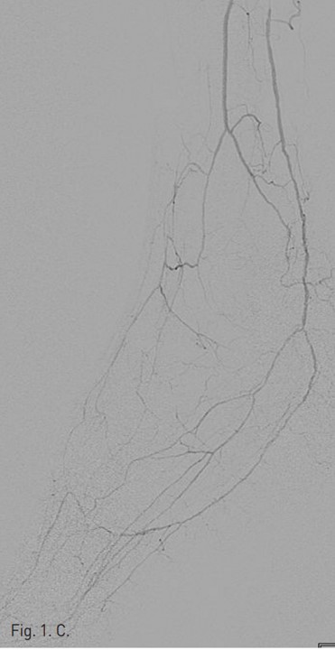
Distal lower leg angiogram에서는 우측 비골동맥의 원위부가 collateral에 의해서 reconstitution되고 있고, 우측 후 경골동맥 역시 calcaneus tip level을 기준으로 상방 약 8 cm 길이에 걸쳐 내강이 확보되어 있고 혈류가 유지되어 있다. Plantar artery는 혈류가 유지되어 있다(Fig. 1C).
시술방법 및 재료
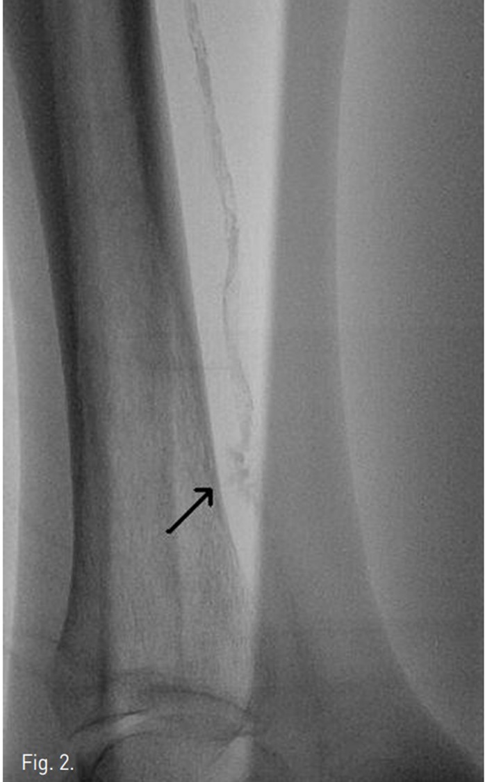
우총대퇴동맥을 antegrade puncture하여 6Fr sheath 를 삽입하였다. 4Fr Berenstein catheter(Glide catheter, Terumo, Japan)를 사용하여 우측superficial femoral artery의 협착 부분과 popliteal artery의 협착 부분을 확인하였다. 4 Fr catheter 와 J-tip guide wire (RADIFOCUS, Terumo, Japan) 로 TP trunk 끝부분의 후경골동맥 시작부위 폐색부분에서 subintimal channel내로 들어간 다음, 후경골동맥의 원위부까지 subintimal channel을 따라 J-tip guide wire를 전진 시켰으나,후경골동맥의 원위부 1/3지점에 위치하는 heavy calcification으로 인해 더 이상 wire가 원위부 후경골동맥의 true lumen까지 전진이 불가능하였다. Wire를 subintimal channel에서 조작하는 동안 heavy calcification부위에서 subintimal space가 perforation되어 wire passage를 일단 중지하였다 (Fig. 2).
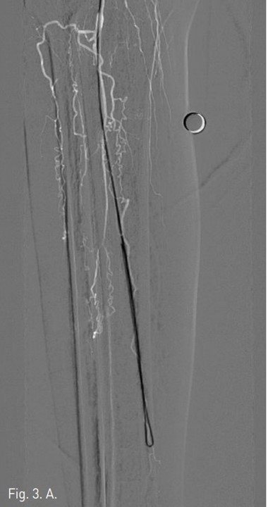
일단 후경골 동맥은 그대로 두고, TP trunk에서 0.016 inch guide wire(GT guide wire, Terumo, Japan)을 사용하여 막혀 있는 비골동맥의 입구에서 subintimal guide wire passage한 다음 (Fig. 3A) 비골동맥의 원위부 true lumen으로 re-entry가 되었다. 3mm/10cm long balloon (SAVVY, CORDIS, Miami, USA))으로 우측 비골동맥 에 대해 subintimal angioplasty를 시행하였으며 우측 비골동맥의 혈류는 재개통 되었다 (Fig. 3B).
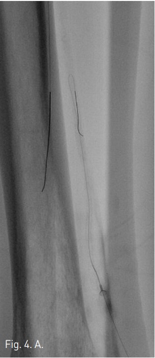
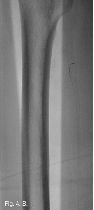
이어서 ankle level보다 약간 위쪽에서 원위부 후경골동맥을 22G needle로 puncture한 다음 0.016 inch GT Guide wire를 retrograde하게 삽입하였으며 이를 따라 4Fr dilator를 안전하게 삽입한 다음, Re-entry에 실패하였던 heavy calcification부분을 retrograde하게 subintimal passage하여 (Fig. 4A), guide wire를 SFA상방까지 전진시켰다 (Fig. 4B).
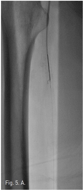
우총대퇴동맥을 통해서 5Fr catheter 내로 goose neck snare를 insertion하여 GT Guide wire를 잡아빼낸 후(Fig. 5A), 이를 다시 SV 5 Guide wire (Cordis, Miami, USA)로 교환하였다 (Fig, 5B). 3mm/10cm balloon을 우총대퇴동맥에서부터 삽입하여 우측 후경골동맥에 대해 subintimal angioplasty를 시행하였고 (Fig. 6), final arteriogram 상 우측 비골동맥과 우측 후경골동맥의 혈류는 완전 재개통되었다 (Fig. 7A 7B). 우측 SFA 협착 부위에 대해서 6mm/12cm SMART stent를 삽입한 후 시술을 마쳤다. 일주일 뒤 시행한 doppler 검사는 시술 이전과 크게 차이가 나지는 않으나, 발의 온도는 시술전보다 따뜻해져서 임상적인 호전을 보였다.
고찰
Chronic atherosclerotic occlusive disease에 의한 critical limb ischemia 환자에서는 infrapopliteal artery occlusion이 빈번하며 특히 diabetic foot환자에서는 extensive calcification을 동반하는 lower leg artery occlusion이 흔하다. 동맥 내강이 완전 폐색된 경우 intraluminal angioplasty를 시행하기는 어려운 경우가 많으며, 대개 subintimal angioplasty가 시도된다. Infrapopliteal arteries에 대한 subintimal angioplasty는 long term patency 자체는 낮지만, subintimal angioplasty의 임상적 추적결과 limb salvage effect는이 시술을 받지 않은 환자들에 비해 분명히 더 나은 것으로 알려져 있다.
Subintimal angioplasty는 폐색 원위부 동맥이 족부관절 혹은 그 상방에서 측부순환에 의해 개통되어 보이는 경우에만 시행하는 것이 원칙이다. Infrapopliteal angioplasty의 경우 technical failure rate는 약 20% 가량으로 실패시 bypass surgery가 권장되며, 일부에서 pedal arch나 communicating branch를 통한 SAFARI(subintimal arterial flossing with antegraderetrograde intervention) technique, 혹은 transpedal access technique등이 보고되고 있다. 이러한 시술들은 아직 그 효과는 명확히 입증되지 않았으나, Spinosa등의 보고에 따르면 SAFARI technique으로 시술한 사람의 6개월 limb salvage rate가 90%가량에 이른다.
Infrapoliteal artery 3개 branch중에서 재개통에 성공하는 혈관의 갯수가 많을수록 보다 limb salvage에 도움이 되는 것으로 보고 있으며, 따라서 본 증례의 경우 원위부 true lumen이 전혀 없는 전경골 동맥은 subintimal angioplasty의 적응증이 되지 못하지만, 나머지 두 개의 비골동맥과 후경골동맥은 원위부 내강이 살아 있으므로, antegrade subintimal angioplasty가 실패하였을 경우 retrograde puncture를 통한 angioplasty를 시도해 볼 수 있다.
본 증례는 popliteal artery 상방의 경우 intraluminal angioplasty및 stent삽입을 통해 혈류를 완전히 회복할 수 있는 상태로서, 비골 동맥에 대해서는 antegrade subintimal angioplasty를 시행할 수 있었으나 재개통된 비골동맥 원위부와 후경골동맥 사이에 SAFARI technique을 적용할만한 communicating branch가 없었으며, 전경골동맥의 원위부 역시 완전 폐색되어 있으므로 pedal arch를 통한 retrograde approach또한 적용할 수 없다.
따라서, 후경골동맥을 통해 retrograde access를 통해 적극적인 angioplasty를 시행하여 혈류의 개선을 얻어 증상이 호전되었으며 나아가, 절단해야 할 부위를 최소화 하는데 도움이 되었을 것으로 판단된다.
이러한 retrograde puncture기법의 경우 시술이 끝난 뒤, 후경골동맥의 천자 부위를 지혈하는 것이 문제가 되며 만약 puncture site에서 출혈이 있을 경우 compartment syndrome의 위험성이 따른다. 본 증례의 경우 retrograde puncture site에 확보된 4Fr dilator는 subintimal angioplasty성공 직후 제거하고 약 10분간의 manual compression만으로 천자부위가 합병증 없이 지혈이 되었으나, 일부 시술자의 경우 subintimal angioplasty시행한 다음, 다시 3mm나 2.5mm직경의 balloon catheter를 천자부위의 동맥내강에서 inflation하여 intraarterial hemostasis를 시행하기도 하며, 천자 부위에 BP cuff를 적용하여 지혈을 하기도 한다.
결론적으로, infrapopliteal arteries의 완전 폐색에 대해서는 subintimal angioplasty의 limb salvage effect가 증명되어 있으므로, 원위부 동맥 내강이 확보되어 있는 경우, antegrade subintimal angioplasty가 실패하더라도 retrograde access를 통해 동맥 혈류 재개통을 시도해 보는 것이 임상적으로 도움이 된다.
참고문헌
1. Miguel MB, Andrej S, Sven B, et al. Retrograde approach for complex popliteal and tibioperoneal occlusion s. J Endovasc Ther, 2008; 15:594-604.
2. Spinosa DJ, Harthun NL, Bissonette EA, et al. Subintimal arterial flossing with antegrade-retrograde intervention (SAFARI) for subintimal recanalization to treat chronic critical limb ischemia. J Vasc Intervent Radiol. 2005; 16:37-44.
3. Vraux H, Hammer F, Verheist R, Goffette P, Vandeleene B, Subintimal angioplasty of tibial vessel occlusions in the treatment of critical limb ischemia: mid-term resuIt. Eur J Vasc Endovasc Surg. 2000; 20:441-446.
Fig. 1. A
Fig. 1. A. An angiography of the right lower extremity shows segmental stenosis of the right superficial femoral artery.
Fig. 1. B
B. The popliteal artery has multifocal luminal narrowing and tibioperoneal trunk is occluded.
Fig. 1. C
C. An angiography at level of ankle shows reconstitution of posterior tibial artery by collateral vessels and also shows patency of plantar artery.
Fig. 2.
Fig. 2. A posterior tibial arteriography shows perforation of subintimal space. Faint extravasation of contrast media is noted (arrow)
Fig. 3. A
Fig. 3. A. A U-looped wire tip is passed along the subintimal channel of occluded left peroneal artery.
Fig. 3. B
B. Completion angiogram of left pffoneal artery after subintimal angioplasty. Complete restoration of arterial flow in peroneal artery.
Fig. 4. A
Fig. 4. A. Through the 22G needle in true lumen of left distal posterior tibial artery, 0.016 inch GT guide wire is passed into subintimal channel of left posterior tibial artery with U-loop end.
Fig. 4. B
B. A wire is passed along the subintimal channel to the superficial femoral artery.
Fig. 5. A
Fig. 5. A. The GT guide wire is captured by goose neck snare.
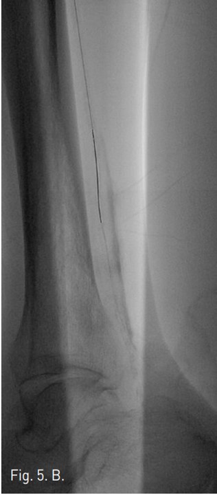
Fig. 5. B
B. The SV 5 Guide wire is passed through antegrade pathway.
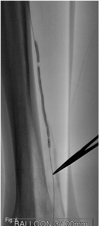
Fig. 6.
Fig. 6. Subintimal balloon angioplasty is performed at posterior tibial artery with a 3mm/10cm balloon. Note the balloon waist at distal portion of the inflated balloon catheter that is compatible to the location of calcification(retrograde wire passage point).
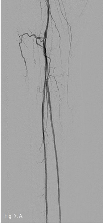
Fig. 7. A
Fig. 7. Final angiography shows complete recanalization of arterial flow in both left posterior and left peroneal arteries.
Fig. 7. B
Fig. 7. Final angiography shows complete recanalization of arterial flow in both left posterior and left peroneal arteries.
Citations
Citations to this article as recorded by