중심단어
Arteries, interventional procedure, therapeutic embolization, aneurysm, omental
임상소견
내원 당일 작업 중 500Kg의 철판이 하복부 위로 떨어져 약 20분 가량 깔려 있다가 구출 된 후 복부통증을 호소하였다. 혈압 및 맥박은 정상범위이었다.
진단명
Traumatic pseudoaneurysm of the omental artery
영상소견
복부 CT상 우측하복부의 장간막 위치에 복강내출혈과 조영제의 유출 소견이 관찰되었고 복강동맥 조영술 상 그물막동맥에 가성동맥류가 형성되어 있었다.
시술방법 및 재료
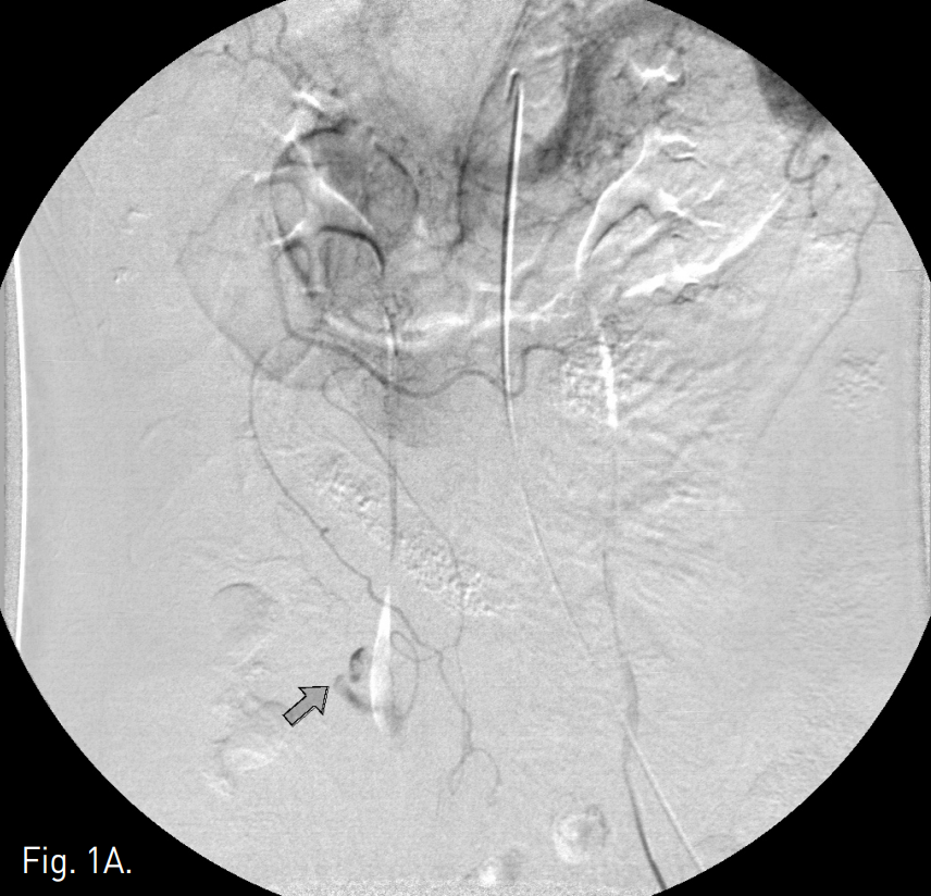
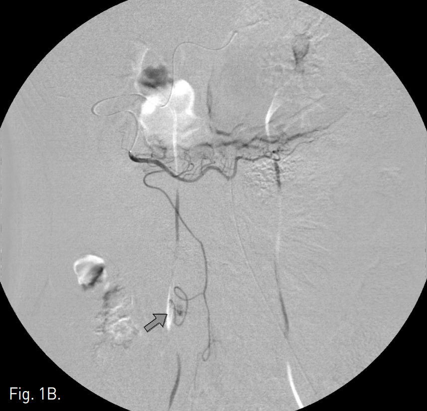
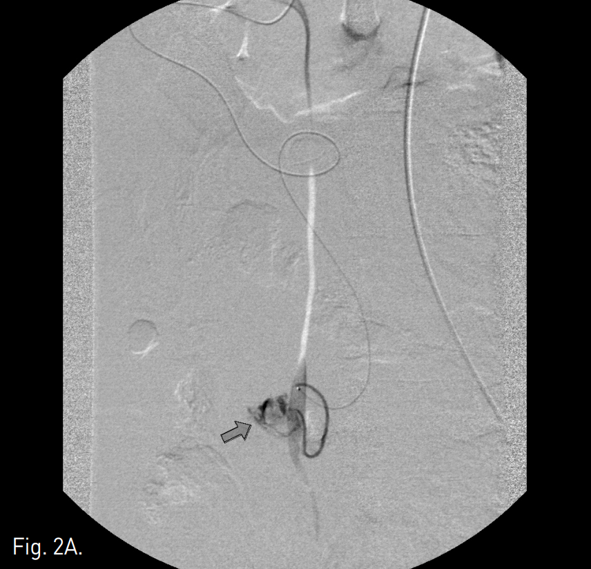
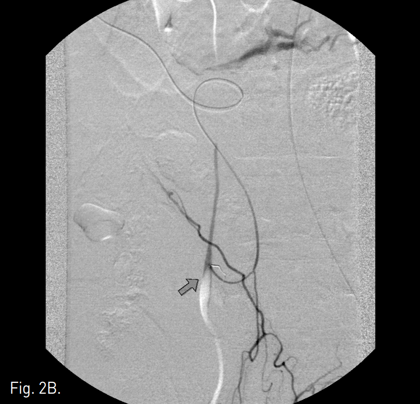
우측 대퇴동맥을 천자하여 5F Cobra 카테타로 양측 총장골동맥 및 장간막동맥 조영술을 시행하였으나 이상 소견은 발견하지 못했으며 이어 시행한 복강동맥 조영술에서 우 위그물막동맥에서 분지하는 그물막동맥에서 조영제의 유출을 보이는 가성동맥류가 발견되었다(Fig. 1A, B). 2.2F Stride 카테타(Asahi intecc, Aichi, Japan)를 이용하여 출혈동맥을 선택한 후 3mm 코일(Cook, Bloomington, IN, USA) 한 개로 색전하였고 동맥조영술상 가성동맥류가 더 이상 조영되지 않는 것을 확인하였다(Fig. 2A, B).
Fig. 1
A, B. Celiac angiogram of delayed phase (A) and selective angiogram of the right gastroepiploic artery (B) show extravasation of contrast media (arrow) from an omental branch of right gastroepiploic artery.
Fig. 2
A. A microcatheter was advanced into the distal portion of the bleeding omental artery (arrow).
B. After successful embolization with a 3-mm microcoil, extravasation of contrast media disappeared on the post-embolization angiogram (arrow).
고찰
좌우 위그물막동맥은 각각 위십이지장동맥과 비장동맥에서 나와 좌우가 서로 만나고 gastric branch와 omental branch로 다수의 분지를 내는 동맥이다.
그 중 그물막동맥은 큰그물막(greater omentum)을 담당하며 중결장동맥과 서로 만나 접합을 한다.
이 동맥은 간세포암의 extrahepatic collateral vessel 중 두 번째로 많은 빈도를 차지하고 있기 때문에 간의 주변부에 종양이 발견될 때는 반드시 이 동맥을 선택하여 혈관조영술을 시행해 보아야 한다.
이 동맥에서 출혈이 일어나는 원인질환은 외상, 항응고제 사용, 종양, torsion of the omentum, 혹은 segmental arterial mediolysis 등이 있으나 드물게는 뚜렷한 원인 없이 터지는 경우도 있다.
Greater omentum은 위의 greater curvature에서 시작하여 아래로 길게 뻗어 소장을 덮고 다시 위로 반전하여 횡행결장에 붙기 때문에 하복부의 외상 시 충분히 다칠 수가 있다.
하복부 복강내 출혈이 있을 때는 대개 장간막동맥이나 골반동맥의 조영술을 시행하여 출혈원인을 먼저 찾아 보는데 하복부 출혈이 복벽 앞쪽 가까이에 있을 때는 반드시 이 혈관을 추적하여 출혈 유무를 확인할 필요가 있다.
참고문헌
1. Tsuchiya R, Takahashi S, Takaoka T, et al. Case of idiopathic omental bleeding treated successfully with transarterial embolization. Nippon Shokakibyo Gakkai Zasshi 2009; 106(4):554-9.
2. Matsumoto T,Yamagami T, Morishita H, et al. Transcatheter arterial embdlization for spontaneous rupture of the omental artery. Cardiovasc Intervent Radiol 2011; 34(suppl 2):S142-5.
3. Kim HC, Chung JW, Lee W, Jae HJ, Park JH. Recognizing extrahepatic collateral vessels that supply hepatocellular carcinoma to avoid complications transcatheter arterial chemoembolization. Radiographics 2005; 25(suppl 1):S25-39.
4. 오현준, 장만규, 김재규. 간암의 경동맥화학색전술: 그물막동맥으로부터의 간외 곁혈관. 대한인터벤션영상의학회지 2009; 16:68-71.
Citations
Citations to this article as recorded by