중심단어
Tracheostomy, bleeding, embolization
임상소견
만성 뇌졸증 병력이 있는 환자로 2009년 10월 심장마비 발생 후 허혈성 뇌손상이 발생하여 2010년 01월부터 기관 절개술을 시행하고 요양원에서 치료해오다 기관 절개부에서 급성 출혈이 하루 전부터 발생하여 외부병원으로부터 전원 되었다. 응급실에서 이비인후과 진료 후 기관내 삽관으로 변경하였으나 출혈 지속되어 CT 촬영하였고 기관-경동맥루가 의심되어 혈관 조영술 및 색전술을 하고자 의뢰되었다. 응급실에서 체크한 혈압은 100/60mmHg, 맥박 96회/min, Hemoglobin 수치는 8.3g/dL에서 4시간 만에 7.1g/dL로 감소하였고, WBC는 16,100/uL이었다.
영상소견
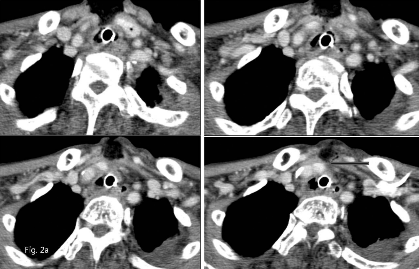
임상적으로 출혈이 발생하는 부위와 비슷하게, Neck CT에서 기관절개부 우측 하방에서 우총경동맥 근위부 좌상방으로 향하는 기관-경동맥루가 의심되었다(Fig. 1, 2A).
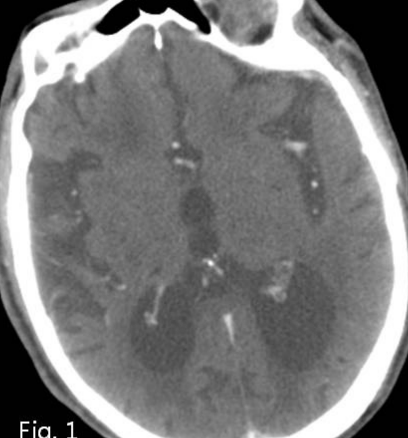
Fig. 1
Contrast enhanced CT scan shows diffuse brain atrophy due to hypoxic brain damage.
시술방법 및 재료
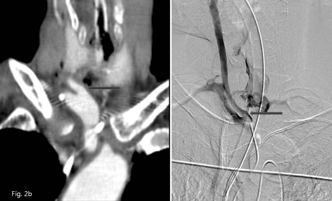
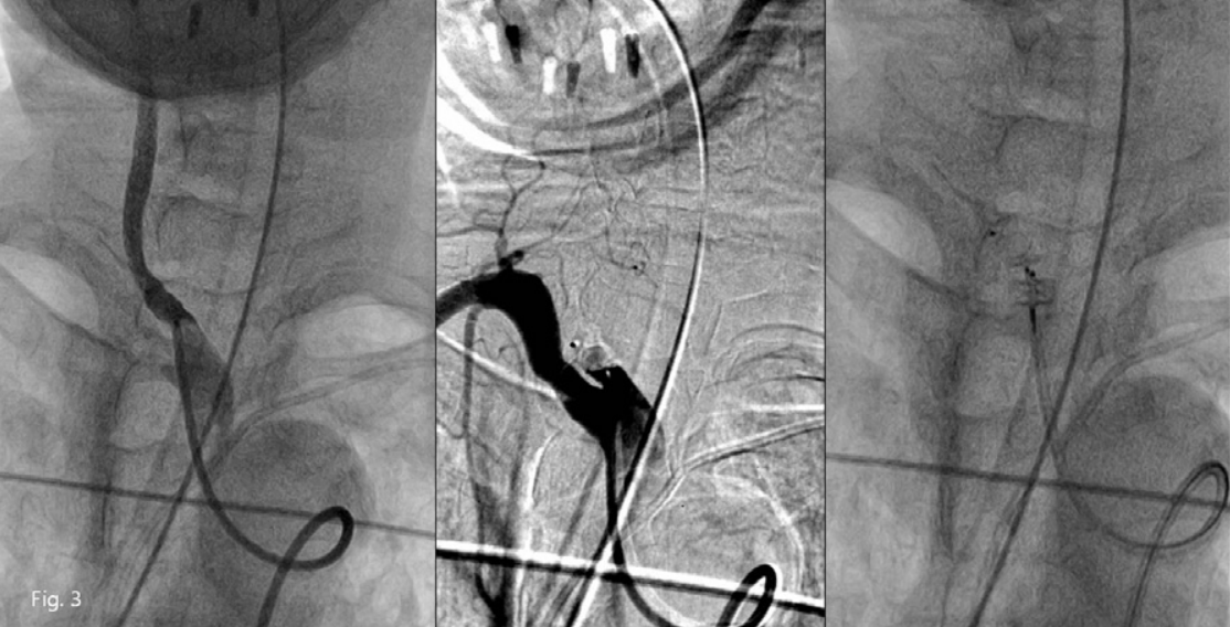
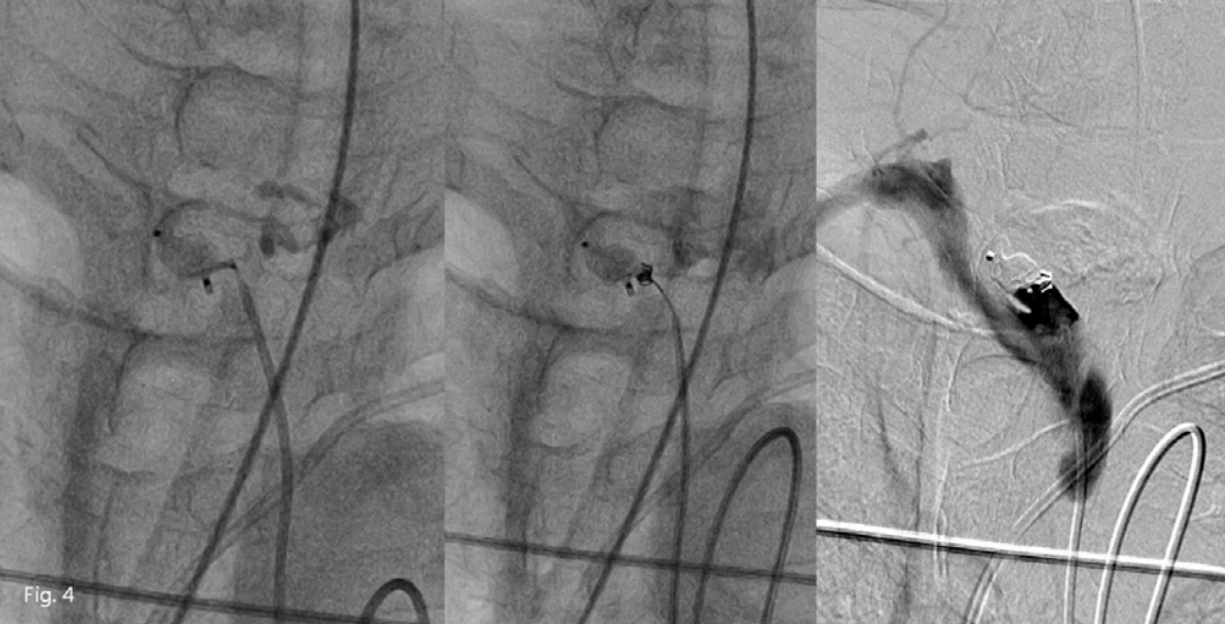
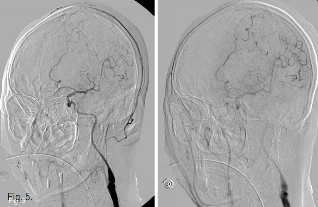
우측 넙다리 동맥을 통해 우측 무명동맥 조영술을 시행하였다. 우측 총경동맥 기시부에서 약 1.0cm 이내인 총경동맥 근위부 좌상방으로 국소적인 돌출부위가 있고 이 부위에서 기관 절개부위로 조영제가 새는 소견이 있었다(Fig. 2B). Sim-1 catheter(Cook, Bloomington, USA)로 selection하고 stiff guidewire(Radifocus, Terumo, Japan)로 교체한 뒤, stiff guidewire tip은 외경동맥 분지인 후두동맥에 위치시키고 8F guiding catheter(Cordis, FL, USA)를 우측 총경동맥 중간부위까지 삽입하고, 8mm diameter를 가진 vascular plug device(AMPLATZER® Vascular Plug II, AGA Medical Corp., Golden Valley, MN)를 총경동맥 기시부에서 무명동맥으로 빠져 나오지 않도록 guiding catheter를 빼면서 총경동맥의 최대한 하방에 설치하였다. 처음 10여 분 동안에는 출혈이 멈추었으나 동맥 박동에 의해 조금씩 plug device가 상방으로 이동하면서 조영제가 조금 새는 모양을 보여서 추가적인 plug device 또는 8mm 직경의 Micronester coil을 설치하려 하였으나 공간이 충분치 않았다(Fig. 3). Plug device의 하내방에 있는 출혈부위를 microcatheter와 microwire(Renegade™, Transend, Boston scientific, Watertown, MA)로 selection 하여 3-2mm 크기의 microcoil(VortX, Boston scientific, Watertown, MA) 두 개를 fistula tract 내부에서 총경동맥 내강으로 튀어 나올 정도의 위치에 정확히 떨어지도록 설치하였고, 이후에는 더 이상 출혈이 발생하지 않았다(Fig. 4). 약 10분 후에 시행한 좌측 경동맥 조영술상 출혈은 없고, 곁동맥들을 통해 상방의 우측 경동맥이 조영되는 것을 확인하고 시술을 마쳤다(Fig. 5). 시술 일주일 후까지 출혈은 없어 퇴원하였으며 퇴원시 혈압은 100/70mmHg, 맥박 92회/min, Hemoglobin 수치는 11.4g/dL, WBC는 10,600/uL이었다.
Fig. 2
A. Contrast enhanced axial CT scans show suspicious focal fistula from the upper medial wall of right proximal common carotid artery to the tracheostomy lumen (arrow).
B. Coronal reformatted CT image right innominate ar teriography show focal bulging of the upper medial wall of right proximal common carotid artery and contrast leakage into tracheostomy site (arrows).
Fig. 3
Vascular plug device was applied into right proximal common carotid artery but the bleeding point seems not to be covered completely. So, placement of an additional plug device was tried to cover the residual lumen of common carotid artery, but failed.
Fig. 4
After selection of fistula itself near the mesh of plug device with a microcatheter, microcoils wer e inserted into the fistula itself and well fixed on completion angiography.
Fig. 5
Left common carotid arteriography shows good contrast filling of right carotid arteries through extracranial and intracranial collaterals.
고찰
기관-경동맥루는 기관절개 수술 후의 치명적인 합병증이며, 72 % 에서 수술 후 3주 이내에 생기는 것으로 보고되었다(1). 수술적 치료 후의 생존율은 약 25 %로 알려져 있고, 주로 median sternotomy를 통해 치료해 왔으나 기관절개부위의 심한 감염 상태로 인한 수술 후 종격동염을 비롯한 감염의 가능성, 수술적 어려움 등의 애로 사항이 있어 최근에는 코일 색전술, 스텐트 그라프트 삽입술 등으로 치료한 보고들이 증가하고 있다(2, 3, 4).
기관-무명동맥루, 기관-경동맥루는 같은 기전으로 발생하며 기관절개부위의 출혈은 기관절개 수술 전후의 혈액 응고 기능에 영향을 주는 다양한 변수에 의해 영향을 받는다는 보고가 있지만, 수술적 요인과 tracheostomy tube의 overinflated cuff, 또는 tube 자체에 의한 기관 연골 미란, 또는 무명동맥 자체의 혈관 기형 등의 원인들로 생긴다(2, 3, 5, 6).
경동맥 파열이나 기관 경동맥루의 질환에서 혈관내 스텐트 그라프트, 코일 색전술 등의 시술은 응급 출혈 상황에서 환자가 가지고 있는 기저 질환으로 인한 낮은 장기 생존율을 고려한다면 보존적인 방법으로 매우 유용하며 다만 출혈의 원인 기전으로 작용한 tracheostomy tube의 존재가 계속 남아있다는 점에서 좀더 넓은 부분의 경동맥을 치료 대상으로 해야 한다는 보고도 있다(4).
본 증례는 vascular plug device를 사용하였으나 우측 쇄골하 동맥에 영향을 주지 않고 치료하려고 노력하다 plug device가 좀더 상방으로 이동한 경우로, plug device의 아래쪽 mesh와 기관-경동맥루 자체의 인접성을 확인하고 microcoil 두 개로 성공적으로 지혈 시킨 경우이다. Vascular plug device의 색전 효과가 생기는 동안 뇌혈관으로 색전이 발생할 가능성은 기존 환자가 허혈성 뇌 손상을 가지고 있는 환자라는 점에서 코일 색전술에 서와 비슷할 것으로 보이며, 코일을 이용할 때 출혈부위 근처에 고정시킬 수 있는 방법이 많지 않은 상황에서는 vascular plug device가 색전술에 더 용이할 것으로 판단된다.
참고문헌
1. Jones JW, Reynolds M, Hewitt RL, Drapanas T. Tracheo innominate artery erosion: Successful surgical management of a devastating complication. Ann Surg 1976; 184:194-204.
2. Allan JS, Wright CD. Tracheoinnominate fistula: diagnosis and management. Chest Surg Clin N Am 2003; 13:331-41.
3. Kenji Takasaki, Kaori Enatsu, Masahiko Nakayama, Takatoshi Uchida, Haruo Takahashi. A case with tracheo-innominate artery fistula: Successful management of endovascular embolization of innominate artery. Auris Nasus Larynx 2005; 32:195-198.
4. Sorial E, Valentino J, Given CA, Eric D. Endean, David J. Minion, Lexington, K. The emergency use of endografts in the carotid circulation to control hemorrhage in potentially contaminated fields. J Vasc Surg 2007;46:792-798.
5. Schaefer OP, Irwin RS. Tracheoarterial fstula: an unusual complication of tracheostomy. J Intensive Care Med 1995;10:64-75.
6. Beiderlinden M, Eikermann M, Lehmann N, Adamzik M, Peters J. Risk factors associated with bleeding during and after percutaneous dilational tracheostomy. Anaesthesia 2007;62:342-346.
Citations
Citations to this article as recorded by