중심단어
Intestinal varix, Liver cirrhosis, Coil embolization
임상소견
2년 전부터 C형 바이러스성 간경화로 치료받던 환자로 내원 20일 전부터 흑색변이 지속되고 5일전부터 전신위약감이 생겨 내원하였다. Capsule endoscopy에서 공장(jejunum) 출혈로 확인되어 탐색개복술을 시행하였으나 소장에 특별한 출혈소견 보이지 않아 영상의학과에 의뢰되었다.
진단명
Liver cirrhosis with jejunal variceal bleeding
영상소견
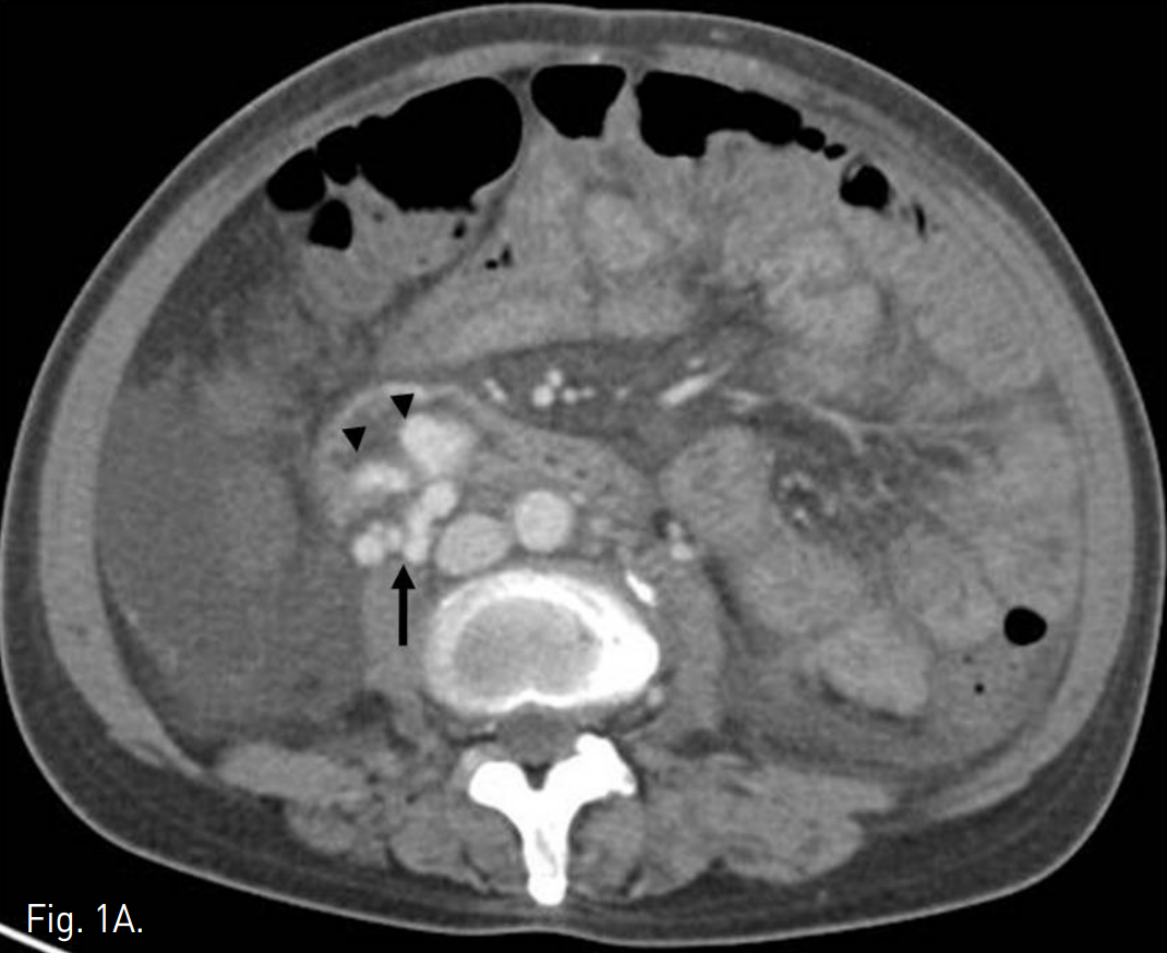
복부 CT에서 정맥류로 생각되는 확장되고 구불구불한 모양의 이상 혈관구조가 주문맥 윈위부에서 기시하여 우하복부 장간막을 따라 있으며, 일부 혈관구조가 소장 벽에 노출되어 있는 모습을 보인다. 그 외에 위, 식도에도 정맥류가 관찰되고 복수와 간 표면의 불규칙화, 경한 비장종대, 소장의 점막하부종이 동반되어 있다(Fig. 1A, B).
Fig. 1
A. Contrast-enhanced abdominal CT shows a tortuous and dilated vasculature (arrow) suggesting varix is located in mesentery of right lower quadrant area and some branching vessels from varix are protruding into adjacent intestinal wall (arrowheads).
B. Coronal scans show entire course of the varix supplied from main portal vein.
시술방법 및 재료
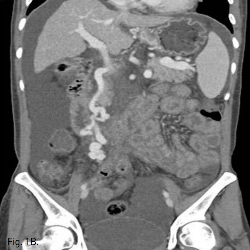
우후간문맥(right posterior portal vein)을 경피적으로 천자하여 6F sheath를 삽입하고 5F curved diagnostic cahteter를 상장간막정맥(superior mesenteric vein, SMV)에 위치시킨 뒤 시행한 direct portal venogram에서 주문맥(main portal vein)의 원위부에서 기시하는 비후된 혈관이 관찰되고 jejunal varix와 연결되어 있으며, 주문맥으로부터 다량의 혈류가 이 혈관으로 역류하고 있음(Fig. 2A). Varix의 nidus로부터 기시하는 drainage veins가 다수 관찰되고, 그 중 varix의 기시부에서 하대정맥으로 직접 유출되는 다량의 혈류가 있음(Fig. 2B).
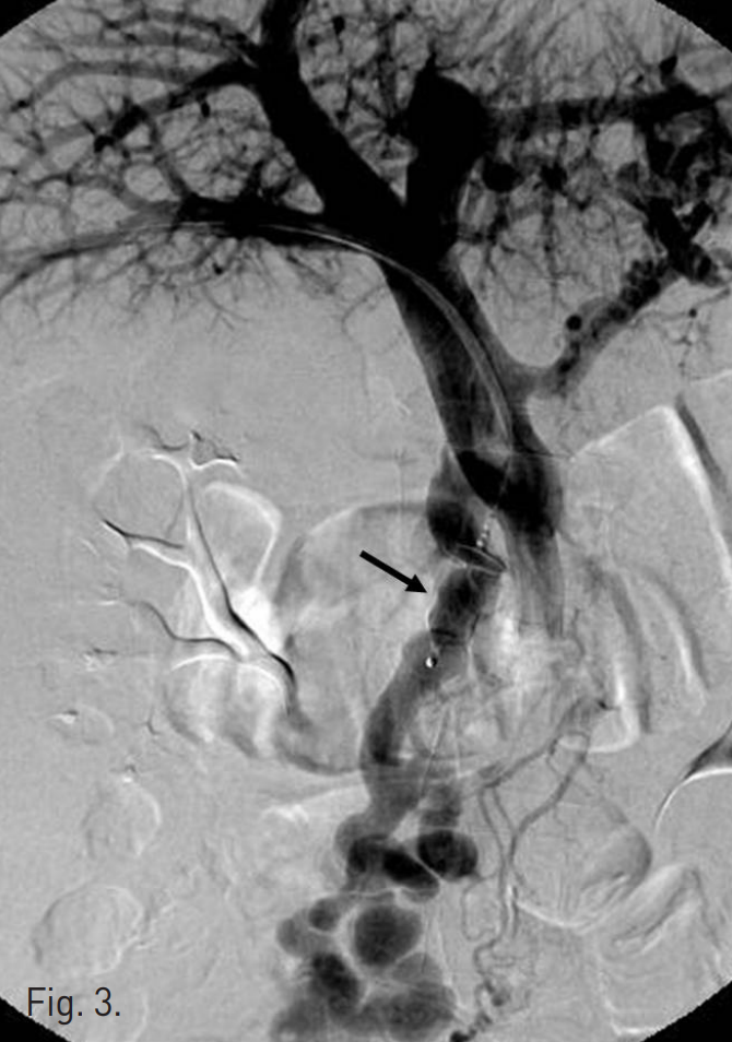
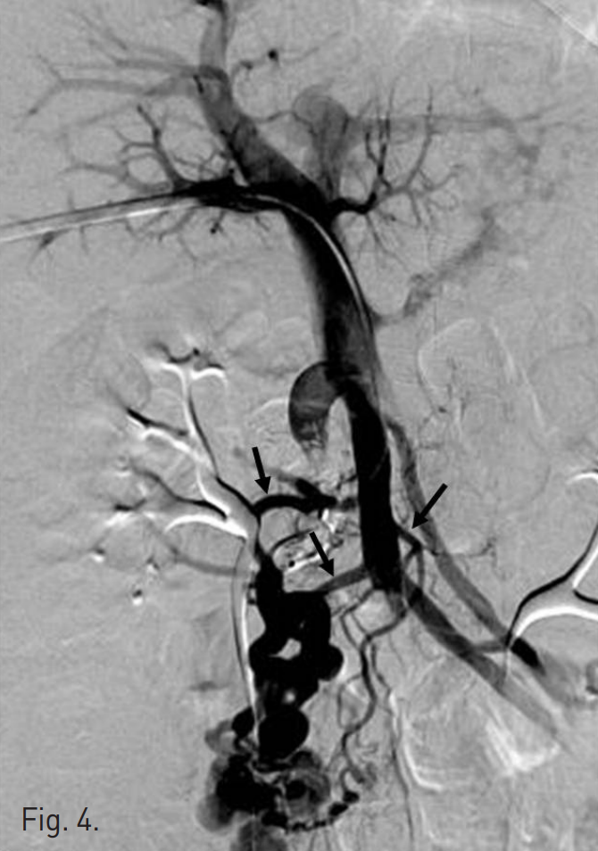
12mm AMPLATZER® Vascular Plug(AVP; AGA Medical, Golden Valley, MN)를 이용하여 주문맥의 원위부에서 기시하는 비후된 afferent vein에 대한 색전술을 시행함(Fig.3). 이후 시행한 direct portal venogram에서 상장간막 정맥으로부터 기시하여 varix nidus를 supply하는 3개의 측부혈관(collateral vessels)이 관찰되고 이로부터 varix의 nidus가 retrograde filling되는 소견이 지속적으로 관찰됨(Fig.4). 이에 대하여 2.4F microcatheter(Progreat®; Terumo, Tokyo, Japan)를 이용하여 각각의 측부혈관을 선택한 후 다수의 microcoils(Tornado®; Cook, Bloonminton, USA)을 이용하여 색전술을 시행함.
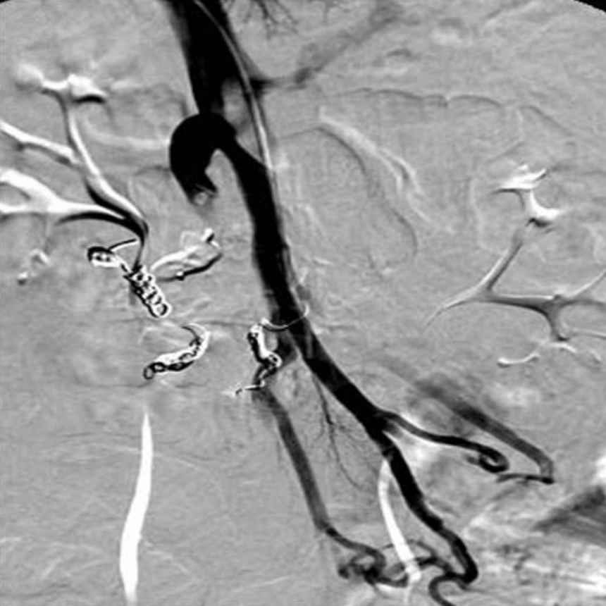
최종적인 direct portal venogram에서 더 이상의 varix가 관찰되지 않음(Fig. 5). 이후 portal vein puncture tract을 microcoils로 폐쇄 후 시술 종결함. 환자는 2주 후 퇴원시까지 더 이상의 장출혈 소견 보이지 않아 퇴원함.
Fig. 2
A. Direct portal venogram shows large amount of portal flow refluxed into hypertrophied vein (arrows) originated from distal portion of main portal vein, which supplies huge jejunal varix (arrowhead) without definitive con trast extravasation.
B. Multiple draining veins (arrows) from nidus of varix are found and one of them is hypertrophied and drained directly into IVC.
Fig. 3
The main trunk of jejunal varix is embolized with 12mm in diameter vascular plug (arrow).
Fig. 4
Follow-up portal venography after the main trunk embolization shows continuous opacification of the varix due to three small colla ter al vessels (arrows) from SMV.
Fig. 5
All of small collateral veins are successfully embolized using 2.4F microcatheter and multiple microcoils and completion portal venogram shows the varix and its afferent veins are not opacified.
고찰
문맥고혈압은 보통 측부순환의 발달을 촉진시키며 그 중 위장관에 생기는 정맥류는 생명을 위협하는 대량출혈을 일으킬 수 있으므로 빠른 진단과 치료가 필요하다. 이소성 정맥류(ectopic varix)는 간경화 환자의 1~5%에서 나타나게 되며, 그 중 소장에서 정맥류가 발달하는 것은 극히 드문 것으로 알려져 있다. 위식도 정맥류가 정맥류 출혈(variceal bleeding)의 가장 흔한 원인이며, 이소성 정맥류에 의한 장출혈은 매우 드물기 때문에 특히 위식도 정맥류가 동반되어 있는 경우 조기진단이 어려울 수 있다.
지금까지 이소성 정맥류의 치료로는 수술적 절제가 시행되어 왔으나, 최근 endovascular embolization으로 variceal flbw를 차단하는 방법이 시도되고 있다. 하지만 gastric varix가 보통 splenorenal shunt를 통해 drainage되는 것과는 달리, 소장 정맥류(intestinal varix)의 경우 대부분 상장간막 정맥이나 하장간막 정맥에서 나와서 하대정맥으로 직접 유입되기 때문에 기존의 BRTO에서와 같이 drainage vein을 통해 shunt와 varices 자체를 색전하는 것은 iatrogenic IVC embolism을 일으킬 위험성이 크다. 이런 경우 afferent vein인 portal vein으로 접근하여 최대한 varix기시부를 막을 수 있도록 selection하여야 하며 하대정맥으로 색전물질이 떨어져 나가는 것을 방지하기 위해 coil embolization을 하는 것이 좋다. 또한 afferent vein의 직경이 굵은 경우 완전한 정맥류의 폐쇄를 위해 본 증례와 같이 vascular plug을 사용할 수 있다.
참고문헌
1. Joo YE, Kim HS, Choi SK, et al. Massive gastrointestinal bleeding from jejuna varices. J Gastroenterol 2000; 35:775-778.
2. Watanabe N, Toyonaga A, Kojima S, et al. Current status of ectopic varices in Japan: results of a survey by the Japan Society for Portal Hypertension. Hepatol Res 2010;40:763-776.
3. Sato T, Yasui O, Kurokawa T, et al. Jejunal varix with extrahepatic portal obstruction treated by embolization using interventional radidlogy: report of a case. Surg Today 2003; 33: 131-134.
Citations
Citations to this article as recorded by