중심단어
Varicocle, Embolization
임상소견
좌측에 생긴 grade II (visible through scrotal skin)의 정계정맥류에 대해 6개월 전 정계정맥류 절제술 (varicocelectomy; surgical ligation)을 받았으나 정계정맥류의 크기에 변화가 없어서 인터벤션 치료가 의뢰됨.
진단명
Recurrent varicocele after surgical varicocelectomy
영상소견
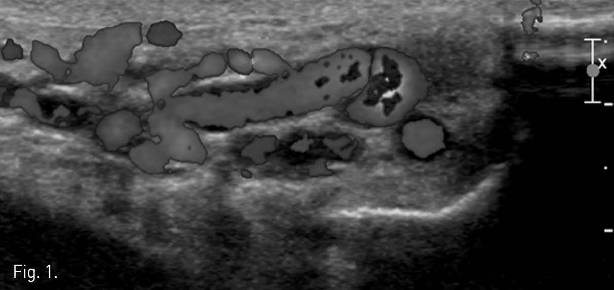
신체검진에서 좌측 고환 상부에 grade ⅡI의 정계정맥류가 있으며 좌측 서혜부에 수술흉터가 있음. 고환 도플러초음파에서 좌측 고환에 pampiniform plexus가 확장되어 있으며 Valsalva maneuver에서 더 두드러지는 소견을 보이고 최대 직경 약 4.5mm로 측정됨 (Fig. 1).
Fig. 1
Doppler US shows dilated veins within the left pampiniform plexus. The maximal diameter of the vein was about 4.4mm. Dilation became more prominent with Valsal va man euver.
시술방법 및 재료
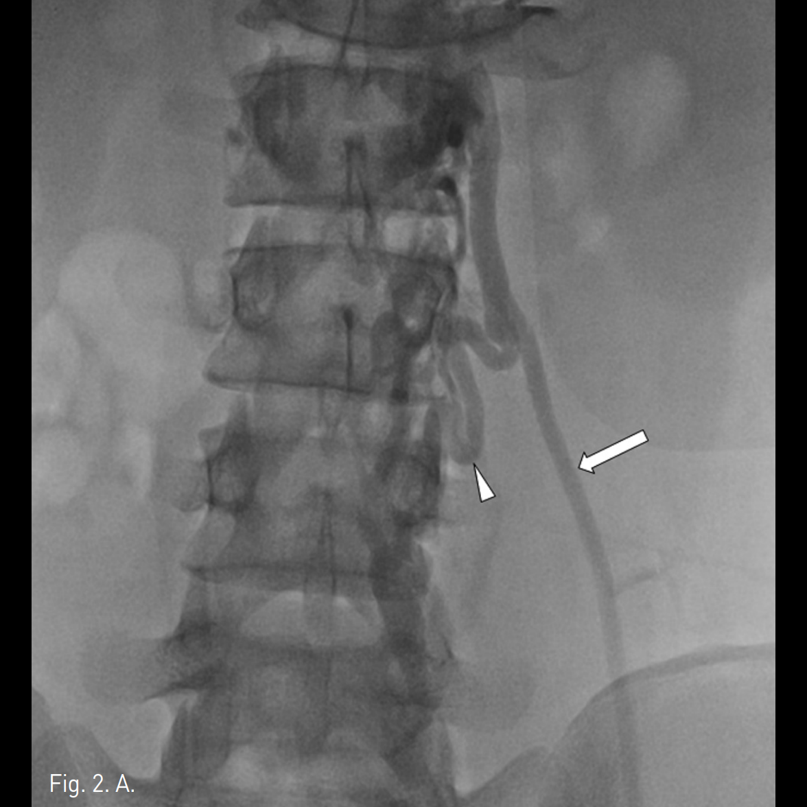
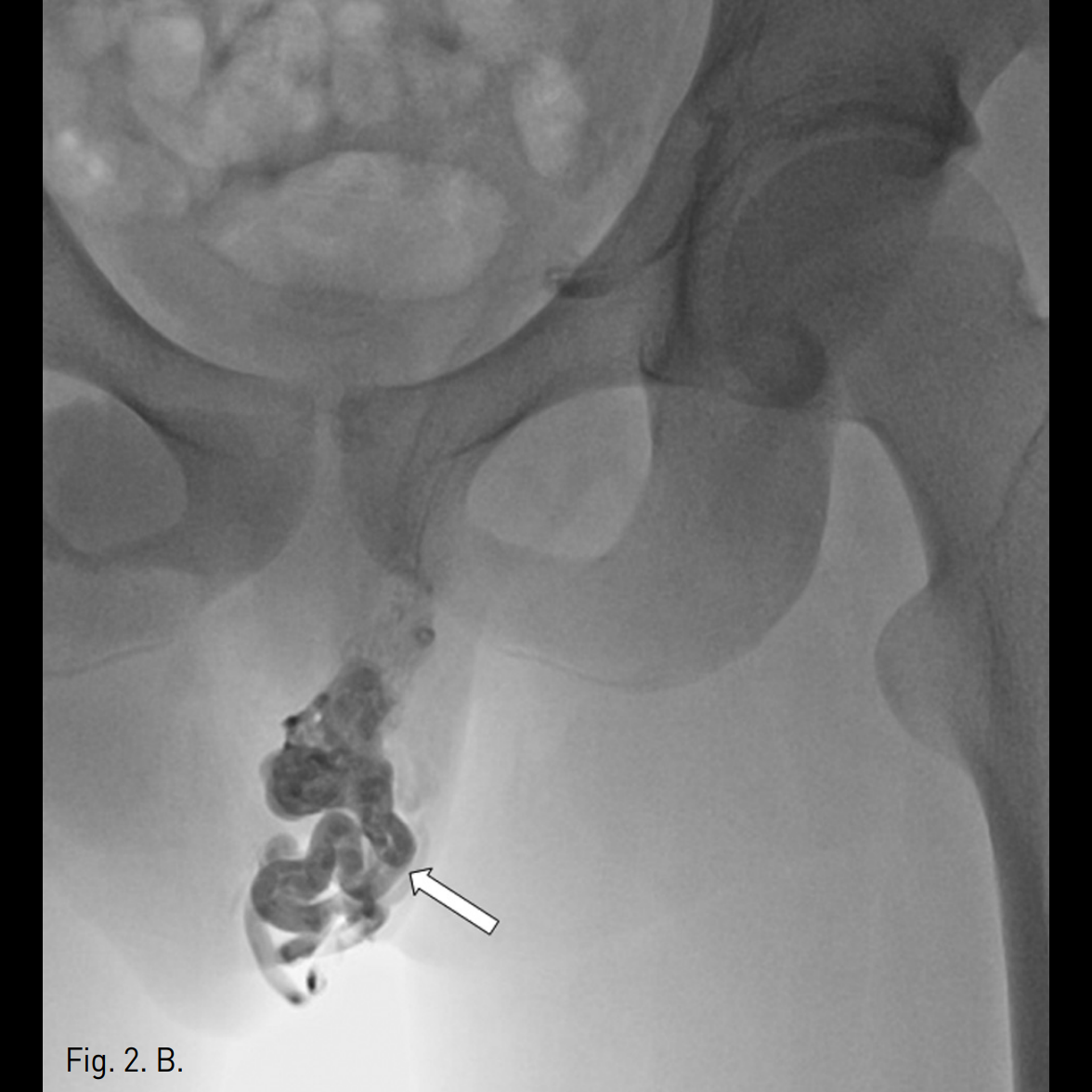
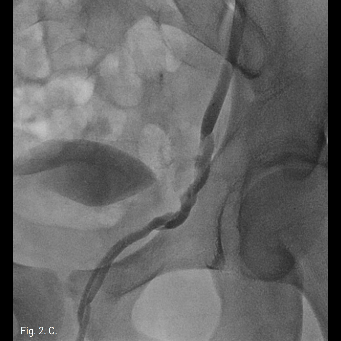
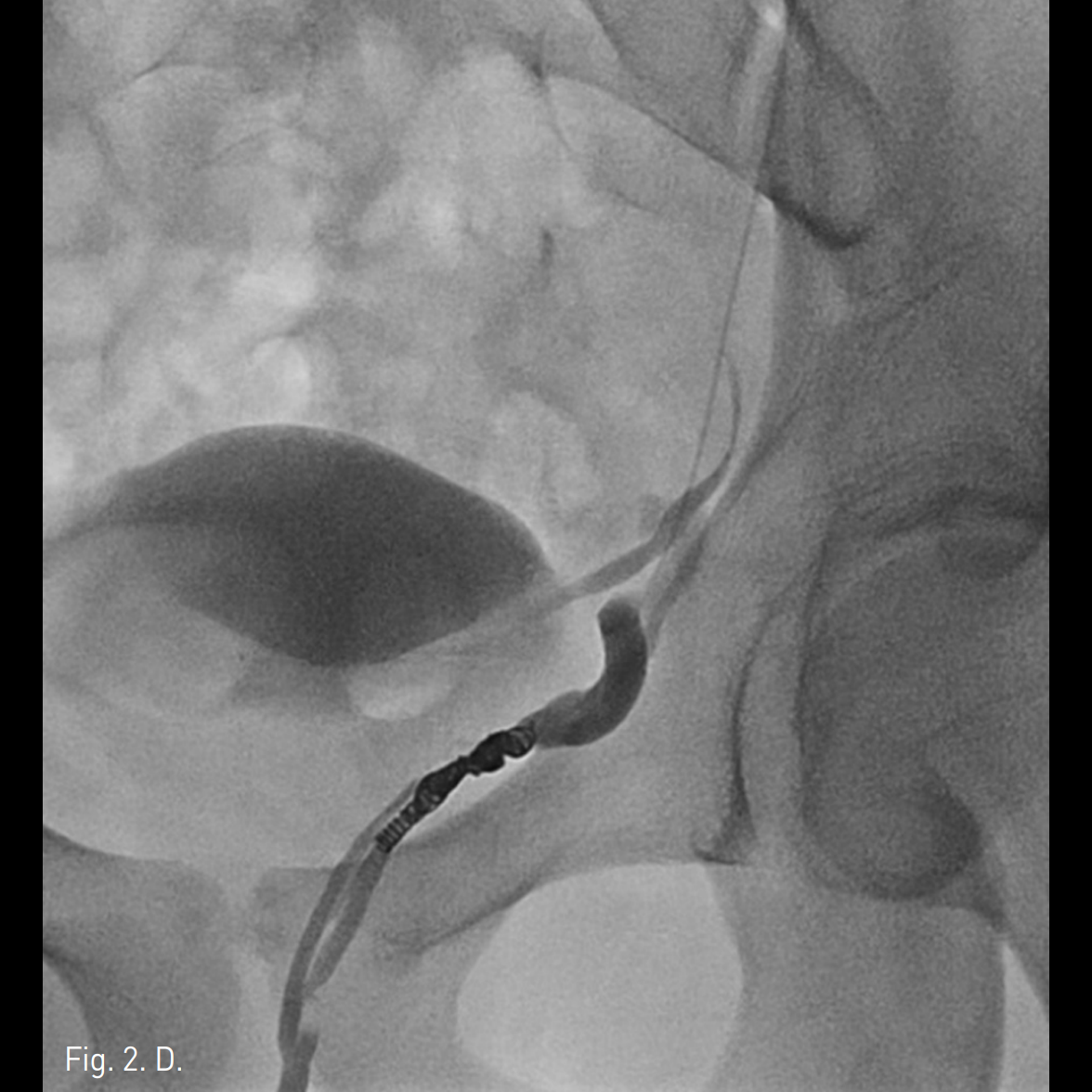
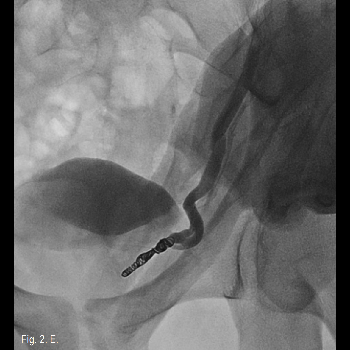
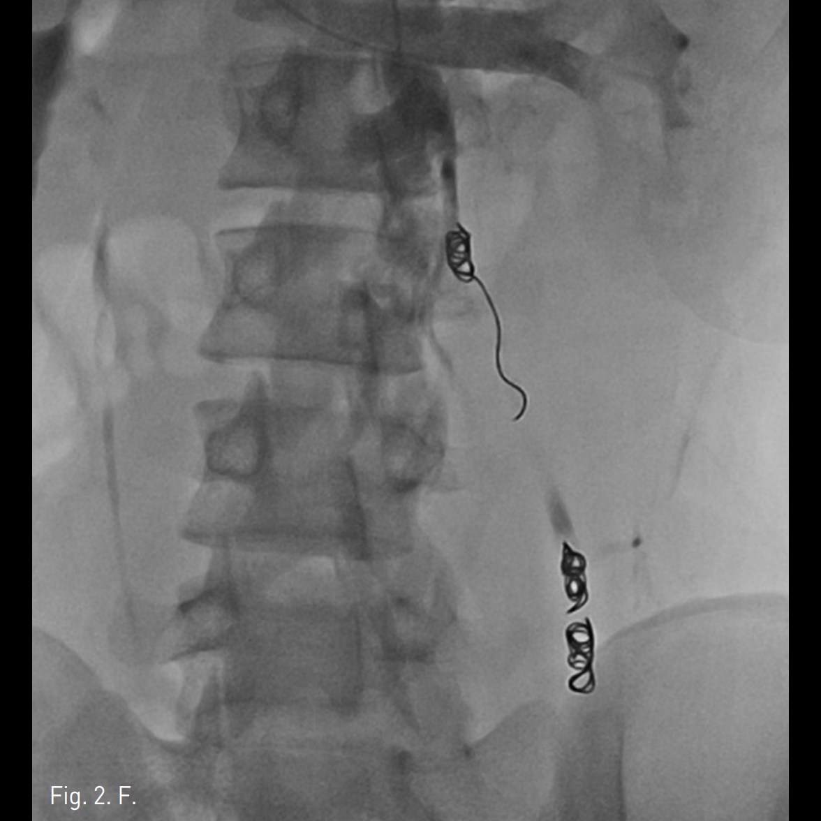
우측 상박을 tourniquet으로 묶은 후 초음파 유도 하에 우측 basilic vein을 minipuncture set을 이용해 천자하여 4Fr Cobra catheter (100cm)를 좌측 신정맥에 위치시킨 후 table tilting을 하여 조영제를 넣었을 때 좌측 spermatic vein.으로의 역류가 관찰되며 L3 level에서 retroperitoneum으로 통하는 venous tributary가 있으며 main tributary는 좌측 pampiniform plexus로 역류되는 소견이 보임(Fig. 2A, 2B). Microcatheter (Renegade, Boston Scientific, Watertown, MA)를 이용하여 보다 자세한 정맥조영을 얻었을 때 inguinal canal level에서 두 개의 정맥이 관찰됨(Fig. 2C). Subinguinal level에서 3개의 microcils (2~3mm ; micronester, Cook, Bloomington, IN)를 이용하여 색전술을 시행하였음 (Fig. 2D). 이어서 환자의 왼쪽 손가락을 이용해 inguinal level을 압박하게 하고 조영제를 주입했을 때 고환으로의 조영제 흐름이 차단된 것을 확인한 후(Fig. 2E), foam sclerotherapy (2mL of 3% STS [sodium tetradecyl sulfate, Thrombojet] + 3mL of air) 5mL를 Cobra catheter를 통해 시행함. 근위부 spermatic vein에 3개의 coils (6mm Nester coils)로 색전술을 시행함. 최종 좌측 신정맥조영술에서 spermatic vein이 조영되지 않음(Fig. 2F).
Fig. 2
A, B. Left renal venograms (via right brachial approach) show confluence of the left spermatic vein (arrow in 2A) presenting an incompetent valve and reflux of contrast medium into an ectatic pampiniform plexus (arrow in 2B). Another tributary (arrowhead in 2A) to the retroperitoneum is seen.
C, D. Selective spermatic venograms show two spermatic veins at spermatic cord levd. Three microcoils were deployed in one of the two veins.
E. Seetive spermatic venogram during manual compression of spermatic cord shows no contrast medium reflux into pampiniform plexus, which indicates the efficacy of the clamping.
F. Completion left renal venogram after sclerotherapy and coil embolization for the proximal part of the left spermatic vein shows no reflux of the contrast into the spermatic vein.
고찰
Varicocele은 spermatic venous insufficiency에 의해 pampiniform plexus가 비정상적으로 확장된 질환을 말하며 9~15%의 높은 유병율을 보이며 저류된 정맥혈의 온도효과로 인해 남성 불임의 주요 원인으로 알려져 있다.
이에 대한 치료로 수술적 결찰술이 inguinal, subinguinal, 또는 internal inguinal ring 위치에서 행해지고 있으나 전신마취가 요구되기도 하며 수술 후 혈종, hydrocele, 고환 위축의 합병증 발생이 문제가 된다. 반면 색전술은 좌측 신정맥을 통해 좌측 spermatic vein으로 catheter를 위치시키고 늘어나 있는 pampiniform plexus의 직상방인 subinguinal level부터 inguinal canal 및 그 상방의 spermatic vein까지 cail이나 경화제 치료를 병행하면서 치료를 할 수 있다는 장점이 있다. 또한 측부순환 정맥들을 함께 치료할 수 있어서 보다 높은 치료 성공률을 기대할 수 있다. 본 증례에서는 일종의 샌드위치 방법으로 coil을 쓰고 경화요법을 쓰고 다시 상방에 coil로 색전술을 하여 색전을 완벽하게 시행할 수 있다.
거품 경화요법 (foam sclerotherapy)은 본 증례와 같이 3% STS 2mL와 air 2mL를 섞어서 약 5mL의 foam을 얻을 수 있는데 이러한 foam을 씀으로써 경화제의 양을 줄일 수 있으며 또한 경화제의 표면적을 늘릴 수 있는 장점이 있다. 섞는 비율은 일부 연구자들은 STS : air를 1 : 4로 하는 경우도 있다.
본 증례처럼 table tilting과 Valsalva maneuver를 병행하면 정맥내 역류를 보다 쉽게 관찰할 수 있고 상완의 basilic vein이나 cephalic vein을 이용하면 외래에서도 시술이 가능하고 지혈도 손쉽게 된다. 또한 우측 대퇴정맥에서보다 좌측 spermatic vein으로 들어가는 각도도 보다 유리할 수 있다.
이와 같은 경피적 색전술의 기술적 성공율은 90~97%로 알려져 있고 정액검사 소견도 27~78%에서 향상되는 것으로 알려져 있어서 수술과 비슷한 소견을 보인다. 재발율은 4~11% 정도이며 최근 보고에서는 3.6%로 알려져 매우 고무적이라고 할 수 있다.
합병증으로는 pampiniform plexus의 thrombophlebitis가 1~4% 정도에서 생길 수 있으나 환자의 손가락으로 spermatic cord를 압박하여 역류를 방지함으로 thrombo phlebitis를 최대한 예방할 수 있다.
References
1. Gandini R, Konda D, Reale CA, et al. Male varicocele; Tran scatheter foam sclerotherapy with sodium tetradecyl sulfate - outcome in 244 patients. Radiology 2008; 246:612-618.
Citations
Citations to this article as recorded by