중심단어
Iliac vein aneurysm, stent-graft, gastrointe stinal stromal tumor
임상소견
2년전 갑자기 생긴 야뇨증을 주소로 내원함. 조영증강 CT상 방광을 누르고 있는 6cm 크기의 종괴소견 보여 소장기원의 위장관 간질종양(gastrointestinal stromal tumor) 의증 진단하에 진단적 복강경 수술 시행함(Fig. 1). 복강경상 장골정맥류 의심되어 수술 종료 후 인조혈관 스텐트설치술 의뢰됨.
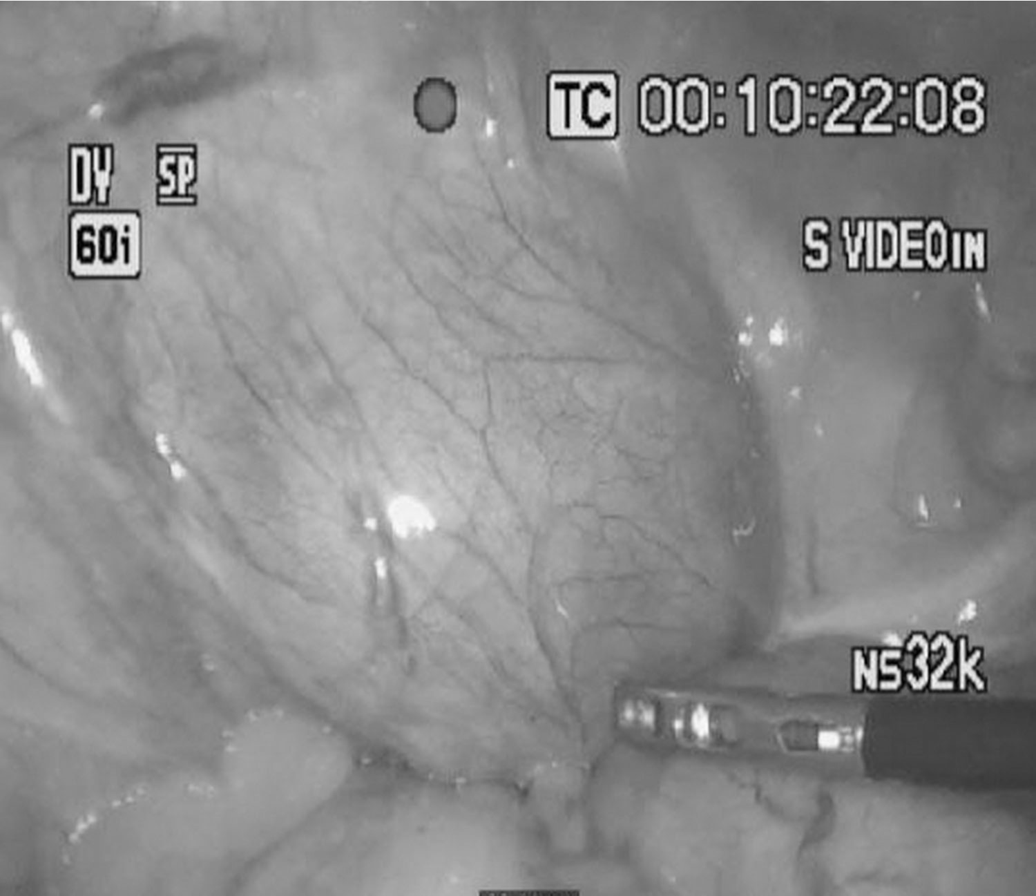
Fig. 1.
Fig. 1. Diagnostic laparoscopy revealed a pulsating extraperitoneal mass which presumed to have vascular origin.
영상소견
조영증강 CT에서 좌측 골반에 66mm x 56mm 크기의 종괴가 보이며, 이는 지연기에서 정맥과 비슷한 정도로 조영증강 정도를 보임(Fig. 2a). 하지 정맥 조영술상 좌측 외장골정맥에서 기시하는 커다란 주머니모양 정맥류(saccular aneurysm) 관찰됨(Fig. 2b).
Fig. 2.
Contrast enhanced CT revealed 66mm x 56mm sized mass lesion in left pelvic cavity (a, arrow). It showed similar enhancement with veins in delayed phase. On venography, there was a huge saccular aneurysm originating from the left external iliac vein (b, arrow)
시술방법 및 재료
좌측 오금 정맥을 초음파 유도 하에 천자하여 7F long sheath (Terumo, Tokyo, Japan)를 삽입 후 5Fr angled tip catheter (Cook, Bloomington, IN, USA)로 좌측 하지 정맥 조영술을 시행하였고 좌측 외장골정맥에 주머니모양 정맥류가 관찰되었음(Fig. 3a). 12Fr long sheath (Terumo, Tokyo, Japan)를 삽입 후 sheath 내로 2개의 인조혈관 스텐트(Excluder, 16mm-14.5mm x 100mm, 16mm-14.5mm x 70mm, W. L. Gore & Associates, Flagstaff, AZ, USA)를 좌측 총장골정맥 원위부부터 좌측 총대퇴정맥까지 삽입하고(Fig. 3a), 14 x 40mm balloon catheter (Foxcross, Abbott Vascular, Abbott Park, IL, USA)를 이용하여 인조혈관 스텐트를 혈관벽에 부착시켜 주었음. 인조혈관 스텐트설치술 및 혈관성형술 후 좌측 외장골 정맥에 위치한 주머니모양 정맥류로의 혈류는 완전히 배제된 소견을 보였음(Fig. 3b).
시술 후 2개월째 시행한 조영증강 CT상 정맥류의 크기는 73mm x 68mm로 측정되었으며, 8개월째와 13개월째 시행한 조영증강 CT에서는 각각 35mm x 34mm와 28mm x 25mm로 측정되었음(Fig. 4). 치료 전 환자가 호소하였던 야뇨증은 호전되었으며, 특이 합병증 및 새로운 증상 호소 없이 외래 추적 관찰 중임.
Fig. 3.
Before stent-graft placement, venography revealed a huge saccular aneurysm originating from the left external iliac vein (a, arrow), and two pieces of stent-grafts (b, arrows; Excluder, 16mm-14.5mm x 100mm, 16mm-14.5mm x 70mm, W. L. Gore & Associates, Flagstaff, AZ, USA) were inserted from distal part of the left common iliac vein to the common femoral vein. After stentgraft placement, venography confirmed complete aneurysm exclusion without endoleak (b).
Fig. 4.
Follow up CT after 2 months (a, arrow) revealed thrombosed iliac vein aneurysm, size of 73mm x 68mm. The aneurysm showed progressive shrinkage with size of 35mm x 34mm on 8 months (b, arrow), and 28mm x 25mm on 13 months (c, arrow).
고찰
정맥류는 드문 혈관 질환으로 신체 여러 부분에서 나타날 수 있으며, 장골정맥을 침범하는 예는 드물다고 알려져 있다. 정맥류의 발생 원인은 뚜렷하게 알려져 있지 않다. 보통 외상 후에 이차적으로 발생하나 외상없이 나타나는 경우도 있다. 외상없이 나타나는 경우에는 결합조직 질환이나 감염 등에 의한 국소적 염증 반응에 의해 정맥의 벽이 약해져 발생할 수 있다고 알려져 있으나 아직 뚜렷한 원인은 밝혀져 있지 않다(1). 임상 증상은 병변의 크기와 위치에 따라 달라져 증상이 나타나지 않을 수도 있으며, 주변 장기의 압박, 정맥류 파열, 혈전생성, 폐색전 등으로 다양하게 나타날 수 있다(2). 영상 소견에서 본 환자와 같이 연조직 종양이나 탈장 등으로 오인되는 경우도 보고되어 있다(3).
정맥류의 치료 적응증은 위에서 언급한 임상 증상이 나타나거나 크기가 점점 커지거나 모양에 변형이 있는 경우에 치료가 필요하다. 그러나 증상이 나타나지 않는 경우, 정맥류의 위치에 따라 치료의 적응증이 달라진다(4). 흉부 정맥류는 수술의 이환율과 사망률이 높기 때문에 증상이 없는 경우 수술 없이 추적 관찰할 수 있다(5, 6). 복부 정맥류는 크기가 작아도 후에 합병증이 나타날 가능성이 높기 때문에 저위험 환자군에서는 치료를 적극 고려한다. 하지의 심부 정맥류는 치명적인 폐색전 합병증을 유발할 위험성이 많기 때문에 무증상이더라도 치료해야 한다고 알려져 있다(7, 8). 정맥류의 치료 방법에 대해서는 현재까지는 주로 수술을 통해 정맥류를 제거하거나, 혈관결찰술, 혈관 우회술 등으로 치료되어 왔으며, 혈관내 치료법을 이용한 치료는 최근 증례로 보고되는 정도이다(9, 10). 혈관내 치료법으로 정맥류를 치료하는 방법으로는 코일, glue, thrombin 등을 이용한 색전술, 스텐트 혹은 인조혈관 스텐트설치술 등을 단독으로 혹은 복합적으로 이용할 수 있다. 그 중 스텐트를 이용한 정맥류의 혈관내 치료법은 크게 두 가지 방법으로 나눌 수 있는데, 첫번째는 피복이 없는 스텐트를 설치 후 기타 색전 물질을 이용하여 색전술을 시행하는 방법이고, 두번째는 인조혈관 스텐트를 이용하여 정맥류로의 혈류를 배제시키는 방법이다.
본 환자는 크기가 큰 좌측 외장골 정맥류의 방광 압박 증상으로 내원하였던 환자로, 처음 시행한 조영증강 CT상 소장 기원의 위장관 간질 종양으로 오인되었던 환자이다. 외장골 정맥에서 기시하는 정맥류 확인 후 인조혈관 스텐트를 통해 정맥류를 치료하였고, 시술 후 13개월까지 합병증 없이 정맥류의 크기 감소를 보여 만족스러운 결과를 보여주었다. 본 증례는 장골정맥류를 혈관내 치료법을 통해 치료한 드문 경우로, 정맥류의 혈관내 치료법의 실행 가능성과 만족스러운 결과를 제시하는 증례이다.
참고문헌
1. Friedman SG, Krishnasastry KV, Doscher W, Deckoff SL. Primary venous aneurysms. Surgery 1990;108:92-95
2. Gillespie DL, Villavicencio JL, Gallagher C, Chang A, Hamelink JK, Fiala LA, et al. Presentation and management of venous aneurysms. J Vasc Surg 1997;26:845-852
3. Petrunic M, Kruzic Z, Tonkovic I, Augustin V, Fiolic Z, Protrka N. Large iliac venous aneurysm simulating a retroperitoneal soft tissue tumour. Eur J Vasc Endovasc Surg 1997;13:221-222
4. Calligaro KD, Ahmad S, Dandora R, Dougherty MJ, Savarese RP, Doerr KJ, et al. Venous aneurysms: surgical indications and review of the literature. Surgery 1995;117:1-6
5. Modry DL, Hidvegi RS, LaFleche LR. Congenital saccular aneurysm of the superior vena cava. Ann Thorac Surg 1980;29:258-262
6. Taira A, Akita H. Ruptured venous aneurysm of the persistent left superior vena cava. Angiology 1981;32:656-659
7. Donald IP, Edwards RC. Fatal outcome from popliteal venous aneurysm associated with pulmonary embolism. Br J Radiol 1982;55:930-931
8. Greenwood LH, Yrizarry JM, Hallett JW, Jr. Peripheral venous aneurysms with recurrent pulmonary embolism: report of a case and review of the literature. Cardiovasc Intervent Radiol 1982;5:43-45
9. San Norberto EM, Gutierrez VM, Revilla A, Vaquero C. Subclavian venous aneurysm: endovascular treatment. J Vasc Interv Radiol 2010;21:1306-1308
10. D'Souza ES, Williams DM, Deeb GM, Cwikiel W. Resolution of large azygos vein aneurysm following stent-graft shunt placement in a patient with Ehlers-Danlos syndrome type IV. Cardiovasc Intervent Radiol 2006;29:915-919
Citations
Citations to this article as recorded by