중심단어
Uterus, rupture, embolization, postpartum hemorrhage
임상소견
내원 2시간 전 외부병원 산부인과에서 자연분만(39주6일, 남아, 3.66kg)후 발생한 출혈로 refer됨. 응급실 내원 당시 RBC 2 pack 수혈중인 상태였으며 분만 전 Hb이 12.8g/dL, transfusion후 11.9g/dL, embolization 후에도 9.9g/dL로 감소하는 추세였음. CT angiography에서 uterus rupture 확인되었고 환자 혈압 감소되면서(113/73mmHg --> 78/55mmHg) 맥박 빨라져(88회/분 --> 122회/분) 응급 자궁적출술을 시행받았음.
진단명
자궁파열(rupture of uterus)
영상소견
시술 직후 시행한 CT angiography에서 자궁내에 다량의 혈종이 있고 시술 시 사용한 조영제가 남아있음. 자궁내막의 조영증강이 끊긴 형태를 보이고 있으며 이 부위를 통해 자궁외공간으로 조영제가 흐르고 있어 자궁파열에 합당한 소견을 보임.
시술방법 및 재료
초음파 유도 하에 우총대퇴동맥을 천자하고 5F sheath(Terumo, Tokyo, Japan)를 우총대퇴동맥에 삽입함. Pigtail catheter(Cook, Bloomington, USA)를 복부 대동맥에 위치시키고 대동맥조영술을 시행하였으나 다소 커진 양측 자궁동맥만 관찰되며 active bleeding소견은 보이지 않음(Fig. 1A). R.U.H catheter(Cook, Bloomington, USA)를 이용하여 우측 내장골동맥을 선택하고 Right anterior oblique projection 동맥조영술을 시행하였을 때 우측 자궁동맥에서 다량의 조영제 유출이 보임. 미세도관(Renegade, Boston Scientific, Natick, USA)과 미세유도철사(Transend, Boston Scientific, Natick, USA)를 이용하여 우측 자궁동맥을 선택하고 동맥조영술을 시행하였을 때, 자궁내강 및 자궁주위공간으로 조영제 유출이 보여 uterus rupture 소견으로 판단하였음(Fig. 1B). 자궁동맥 원위부의 선택이 불가능하여 N-butyl-2-cyanoacrylate(Histoacryl, B. Braun, Melsungen, Germany)와 리피오돌의 1:3 혼합액을 이용하여 색전술을 시행함(Fig. 2). 좌측 자궁동맥 및 양측 내장골동맥의 전방분지에 대해서 gelfoam 색전술을 시행하였음. 색전술 후에도 혈색소 수치 감소 지속되었고, CT angiography에서 자궁파열에 합당한 소견을 보이면서(Fig. 3) 환자 신체징후가 악화되어 수술을 권유하였으며, 응급 자궁적출술을 시행하였음.
Fig. 1.
A. Pelvic arteriogram shows slightly prominent both uterine arteries (white arrows) and right ovarian artery (black arrow). But active bleeding focus is not demonstrated.
B. Selective angiogram of the right uterine artery shows active contrast leakage to the uterine cavity (white arrow) and extrauterine space (black arrow), which means uterine rupture.
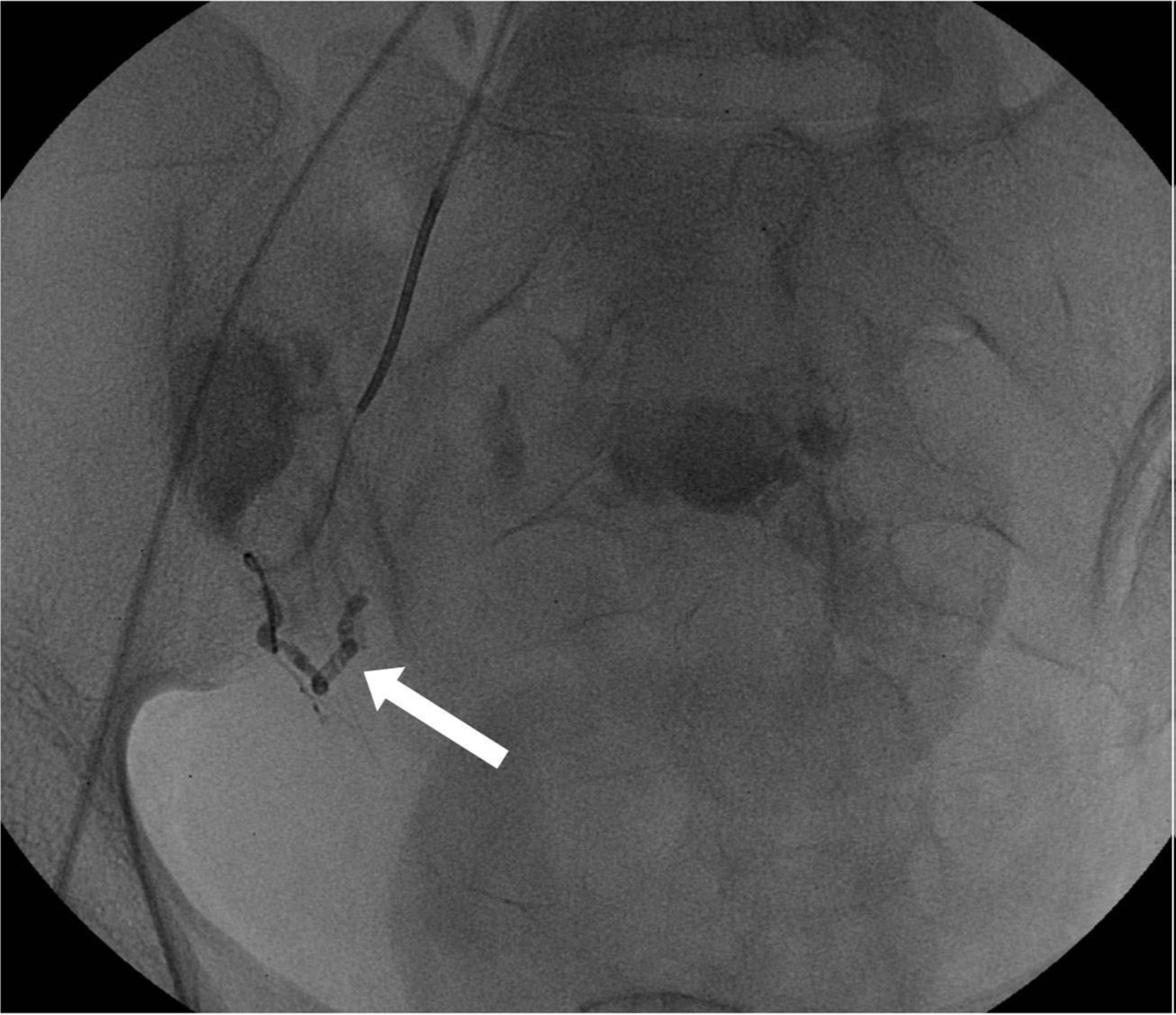
Fig. 2.
The mixture of N-butyl-2-cyanoacrylate and lipiodol with a ratio of 1:3 was injected at the tip of a microcatheter, but it did not reach to the rupture point. The proximal part of the right uterine artery was only embolized (arrow).
Fig. 3.
A. The axial image of CT angiography shows intrauterine (white arrow) and extrauterine (black arrow) contrast leakage, suggestive of uterine rupture.
B. On the coronal image of CT angiography, endometrial disruption is demonstrated in the lower anterior wall of the uterus (arrows).
고찰
자궁파열은 산후출혈과 소화기관파열 다음으로 흔한산모의 사망원인이며 0.03% 정도의 빈도를 보인다(1,2). 자궁파열의 경우 수술적 치료를 요하지만, 3차 병원의 경우 산후출혈을 주소로 의뢰된 환자를 치료하는 경우가 많으며 정확한 information을 얻지 못하는 경우가 있어 그 진단이 지연되는 경우가 있다. 산후출혈을 보이는 산모의 치료는 보존적 약물치료가 먼저 시도되며 심한 경우 자궁동맥결찰술이나 자궁적출술 등의 수술적 치료도 고려하게 된다(3). 하지만 자궁동맥 색전술로 치료하는 경우가 점차 흔해지고 있으며(4) 이는 수술적 치료보다 더 쉽게 접근이 가능하기 때문이다. 따라서 자궁파열이 의심되는 자궁출혈환자의 경우라 할지라도 자궁동맥 색전술을 먼저 시행하여 자궁파열의 소견을 확인할 수 있으며 색전술을 통해 산모의 활력징후가 안정화되면 수술적 치료를 시행하는 것이 환자에게 도움이 되겠다. 상기 환자의 경우도 자궁동맥색전술을 통해 활력징후가 안정화 되었고 이후 복부 CT로 자궁파열을 확인하였으며 자궁적출술로 치료되었다
참고문헌
1. Dwyer R. Post partum deaths of mares. Equine Dis Qtly, UK Dept Vet Sci 1993; 2:104.
2. Waterstone M, Bewley S, Wolfe C. Incidence and predictors of severe obstetric morbidity: casecontrol study. BMJ 2001; 322:1089-1094.
3. Pinto A, Niola R, Brunese L, Pinto F, Losco M, Romano L. Postpartum hemorrhage: what every radiologist needs to know. Curr Probl Diagn Radiol 2012; 41:102-110.
4. Banovac F, Lin R, Shah D, et al. Angiographic and interventional options in obstetric and gynecologic emergencies. Obstet Gynecol Clin North Am 2007; 34:599-616
Citations
Citations to this article as recorded by