중심단어
Arteriovenous malformation, renal vascular malformation, embolization
임상소견
갑자기 발생한 육안적 혈뇨 및 통증으로 내원
진단명
Arteriovenous malformation in the right kidney
영상소견
조영증강 동맥기 CT에서 우측 신장 신우 하측에 구불구불하고 불규칙하게 늘어난 동맥 및 정맥 분지들이 복잡하게 엉켜 종괴 양상으로 보이며(Fig. 1A), MIP영상에서 tortuous vascular structure로 구성된 약 2cm x 2.5cm 크기의 덩어리로 보였다(Fig. 1B). 우측 신동맥조영술에서 이 병변은 불규칙하게 늘어난 혈관이 엉켜있는 과혈관성 동정맥기형으로 보였다(Fig. 2).
Fig. 1.
Early arterial phase image of axial CT scan (A) shows tortuous and dilated vascular structures (arrows) at the inferior aspect of the right renal pelvis, suggestive of vascular malformation. The MIP image of the right Kidney(B) shows well demarcated and conglomerated hypervascular arteriovenous malformation (arrow).
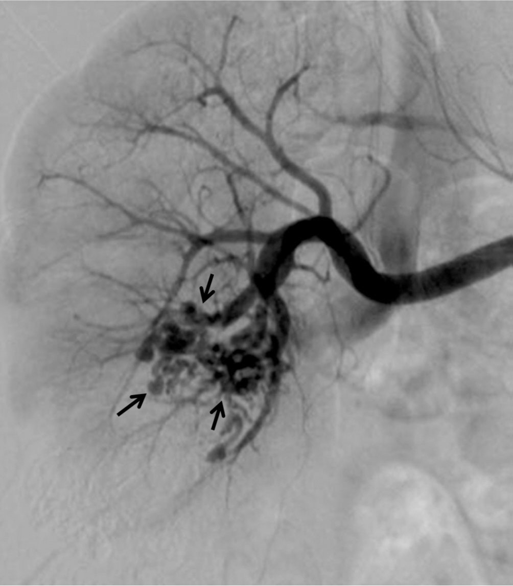
Fig. 2.
Fig. 2. Right renal arteriogram shows a network of tangled hypervascular structures (arrows) with early venous drainage.
시술방법 및 재료
5F Cobra-catheter로 우측 신동맥조영술을 시행하였고 신우 하측에 early arterial opacification을 보이는 약 2cm x 2.5cm 크기의 tangled hypervascular mass를 확인하였다. 이후 6F renal guiding catheter를 이용하여 우측 신동맥을 선택하고 microcatheter를 이용하여 초선택적 동맥조영술(superselective arteriography)을 시행하여 우측 신장 동정맥기형의 nidus 및 영양동맥을 확인하였다(Fig. 3A). N-butyl 2-cyanoacrylate(Histoacryl, B. Braun, Melsungen, Germany)를 이용한 색전술을 계획하고 우선 1:3 비율의 glue/lipiodol 혼합액을 injection하였으나 다량의 glue material이 동정맥단락을 통하여 체정맥으로 배출되는 양상이 관찰되었다. Glue와 lipiodol을 1:1 비율로 혼합하여 mixture의 점도를 높인 혼합액을 nidus 및 동정맥기형의 영양동맥에 대하여 serial embolization을 시행하였다(Fig. 3B, C). 최종 동맥조영술에서 우측 신동맥의 하엽분지까지 색전되어 상엽의 실질만 contrast staining이 관찰되었으며 더 이상의 동정맥기형은 관찰되지 않아 시술을 종료하였다(Fig. 3D). 시술 후 3개월째 시행한 조영증강 전 CT영상에서 우측 신장에 보이던 동정맥기형의 nidus 및 영양동맥들에 high attenuation의 glue materials가 남아 있으며(Fig. 4A) 조영증강 CT영상에서 비정상적으로 늘어나거나 구불구불한 혈관은 보이지 않고 우측 신장의 상엽 및 하엽 실질은 대부분 보존되었으며 glue로 색전된 부위 주변 mid pole 신실질에 mild contraction이 관찰되었다(Fig. 4B-D).
Fig. 3
A. Superselective angiogram shows well demarcated AVM and its feeding arteries.
B. Renal angiogram after one session of intraarterial glue embolization, demonstrates decreased extent of AVM.
C. Spot radiograph shows large amount of glue/lipiodol casting within the AVM.
D. Final angiogram demonstrates non-visualization of the AVM and preserved parenchymal staining in the upper portion of the right kidney.
Fig. 4.
The precontrast image (A) of CT scan obtained 3 months after embolization shows hyperdense glue/lipiodol mixtures in the nidus of the AVM at the mid portion of the right kidney. Postcontrast CT images (B-D) show preserved parenchymal perfusion in the upper and lower poles of the right kidney with partial parenchymal contraction of the mid portion.
고찰
신장의 동정맥기형은 유병율이 0.04% 정도로 보고된 매우 드문 질병으로 대부분 30대까지 증상이 없고, 40대 이후 임상 양상이 발현될 시 다량의 육안적 혈뇨를 동반하는 경우가 많아 신세포암종과 감별이 필요하다. 이에 도움이 될 수 있는 진단적 방법으로는 CT angiography및 신동맥조영술이 대표적이며, 특히 신동맥조영술은 동정맥기형의 감별뿐만 아니라 효과적인 치료를 동시에 시행할 수 있다는 이점이 있다. 선택적 동맥색전술은 과거의 개복식 수술을 대체하게 되면서 근래에 와서는 치료의 gold standard가 되었다. 동정맥기형 동맥색전술의 원칙은 모든 영양동맥을 차단하되 유출정맥과 parent artery는 보존하여 신실질의 경색을 방지하는 것이다. 이러한 효과를 위한 색전물질로서 gelfoam, coil, alcohol, N-butyl 2-cyanoacrylate 등이 이용되고 있는데, gelfoam particle 및 coil의 경우 약 절반의 환자에서 재개통되거나 측부혈관이 발달하여 동정맥기형이 재발했다는 보고가 있다. N-butyl 2-cyanoacrylate의 경우 동정맥기형의 nidus를 완벽하게 색전시켜 영구적인 효과를 기대할 수 있는 것으로 여겨지고 있다. 이번 증례의 경우 N-butyl 2-cyanoacrylate를 이용하여 성공적인 초선택적 동맥색전술을 시행하였고 시행 후 특별한 임상 증상의 재발 없었으며 추척관찰 CT에서도 동정맥기형의 재발이나 신실질의 경색을 의심할 만한 소견은 보이지 않았다.
참고문헌
1. Defreyne L, Govaere F, Vanlangenhove P, Derie A, Kunnen M. Cirsoid renal arteriovenous malformation treated by endovascular embolization with n-butyl 2-cyanoacrylate. Eur Radiology 2000; 10:772-775.
2. Vasavada SP, Manion S, Flanigan RC, Novick AC. Renal arteriovenous malformations masquerading as renal cell carcinoma. Urology 1995; 46:716-721.
3. Sountoulides P, Zachos I, Paschalidis K, et al. Massive hematuria due to a congenital renal arteriovenous malformation mimicking a renal pelvis tumor: a case report. J Med Case Rep 2008; 2:144.
Citations
Citations to this article as recorded by