중심단어
Uterine artery embolization, uterine necrosis, gelfoam, postpartum hemorrhage
임상소견
임신 33주 4일인 쌍둥이 임산부로 만산 전 조기파막으로 내원하여 입원 관찰 중 태반조기박리로 응급 제왕 절개술을 시행하였다. 수술 후 질출혈 지속되며 보존적인 치료에도 혈색소 8.3g/dL로 감소하여 응급으로 자궁동맥 색전술 의뢰되었다. 시술 후 한 달 여간 지속된 복통과 출혈 있어 자궁절제술 시행하였다.
영상소견
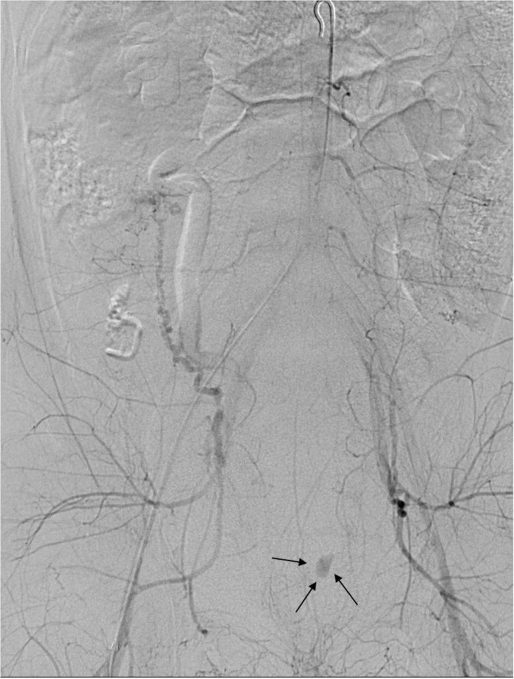
자궁동맥조영술상 비대해진 양측 자궁동맥이 관찰되었으나(Fig. 1), 혈류 유출은 없었다. 양측 자궁동맥을 gelfoam을 이용하여 색전술을 시행하였으며, 시행 후 골반동맥조영술에서 양측 자궁동맥의 혈류는 모두 소실되었으나 좌측 골반강에 조영제 혈관외 유출이 관찰되었다(Fig. 2).
Fig. 1.
Left (A) and right (B) internal iliac angiograms show bilateral hypertrophied uterine arteries.
Fig. 2.
Pelvic angiogram shows extravasation of contrast media in the left pelvic cavity(arrows).
시술방법 및 재료
초음파 유도 하에 우측 대퇴동맥을 천자하여 5F Accu-Sheath Introducer System(Sungwon Medical Co, Cheongju, Korea)을 삽입하였다. 0.035-inch 유도철사(Terumo, Tokyo, Japan)와 5F pigtail catheter(Cook, Bloomington, USA)를 이용하여 복부 대동맥에서 골반동맥조영술을 시행하였으며 비대해진 양측 자궁동맥을 확인하였다. 5F Omni-Flush Catheter(Angiodynamics, Queensbury, USA)를 이용하여 양측 내장골동맥을 선택하여 혈관조영술을 시행하였고, 3F Renegade catheter(Boston Scientific, Natick, USA)와 0.016-inch Fathom guidewire(Boston Scientific, Natick, USA)로 자궁 동맥을 선택하였다. 자궁동맥은 1-2mm 크기의 gelatin sponge(Cutanplast, Mascia Brunelli, Milan, Italy)를 이용하여 색전하였고, 자궁동맥 내에조영제의 정체가 보일 때까지 색전술을 시행하였다. 색전술 후 시행한 골반동맥조영술에서 양측 혈류는 모두 소실되었으나 좌측 골반강에 조영제 혈관 외 유출이 관찰되었다. 좌측 외장골동맥조영술시 아래배벽동맥 분지에서 가성동맥류가 있었으며(Fig. 3), N-butyl-2-cyanoacrylate(Histoacryl, B. Braun, Melsungen,Germany) 와 리피오돌 1:4 혼합액으로 색전술을 시행하였다. 다시 시행한 혈관조영술에서 가성동맥류가 완전히 폐색된 것을 확인하였다(Fig. 4). 시술 12시간 후 환자는 다시 질출혈 발생하였으며 수액공급 및 수혈 시행 후에도 혈압 80/55mmHg, 맥박 98회/min, 혈색소 6.4g/dL로 감소하여 응급 자궁동맥 색전술 다시 시행하였다. 골반동맥조영술 시행 시 우측자궁동맥에서 조영제 혈관외 유출이 관찰되었으나(Fig. 5) 혈관연축으로 인하여 자궁동맥 선택이 어려워 자궁동맥 입구에서 300-500μm polyvinyl alcohol particles(Cook, Bloomington, USA)과 gelatin sponge를 이용하여 색전술 시행하였다. 시술 후 시행한 혈관조영술에서 우측 자궁동맥 폐색과 조영제 혈관외 유출이 사라진 것을 확인하였다(Fig. 6). 색전술 후 환자는 소량의 질출혈외 특별한 증상 없었으며 한달 뒤 하복부 통증과 다량의 질출혈로 내원하였다. 조영증강 복부 CT상 자궁내막과 자궁근층에 조영 증강이 거의 보이지 않고 테두리 조영증강을 보였다(Fig. 7). 보존적 치료에도 증상 계속되었으며 지속적 출혈, 감염의 위험 등 있어 자궁절제술 시행하였다. 병리 소견상 자근근층에 괴사와 출혈이 있었다.
Fig. 3.
Left external iliac (A) and inferior epigastric (B) angiograms show a pseudoaneurysm (arrows) from the left inferior epigastric artery.
Fig. 4.
Left external iliac angiogram after embolization shows no opacification of the pseudoaneurysm with obstruction of the supplying artery.
Fig. 5.
Pelvic angiogram obtained during the second embolization procedure shows extravasation of contrast media from the right uterine artery (arrows).
Fig. 6.
Pelvic angiogram after the second embolization shows no blood flow to the right uterine artery with no extravasation of contrast media.
Fig. 7.
Axial (A) and sagittal (B) enhanced CT images show uterine necrosis with peripheral enhancement. Air infiltration in the necrotic myometrium is also noted.
고찰
자궁동맥 색전술을 통한 산후출혈의 치료는 높은 성공률(85-95%)과 낮은 합병률(6-7%)을 보인다. 시술과 관련된 합병증은 드물며 6-7%에서 발생한다고 알려져 있다. 가장 흔한 합병증은 통증과 일시적인 발열 등이며 이들은 3-5일내 대부분 회복된다. 드물게는 자궁 및 방광의 괴사, 신경손상, 방광-질 누공 형성, 혈관 천공 또는 폐쇄 등이 발생할 수 있다. 자궁괴사의 원인으로 알려져 있는 것은 너무 작은 색전물질을 사용하여 측부 혈관이 막혀버리거나 출산 후 팽창된 자궁에 난소동맥과 자궁동맥 사이의 연결이 불충분하여 원위부 혈관에 혈류공급이 부족해지는 경우이다. Sone 등의 연구에 의하면 막힌 혈관의 diameter가 자궁괴사와 중요한 연관성을 보였다. 자궁괴사는 대부분의 비흡수성 색전물질과 관련하여 보고가 많으나 흡수성 색전물질을 사용한 경우에서도 알려져 있으며, 색전물질의 종류보다는 사용한 물질의 크기가 자궁괴사와 더욱 연관이 있다고 생각되고 있다. 그 밖에 예방적 항생제의 사용유무, 패혈증과 같은 환자의 상태도 자궁괴사와 관련이 있다. 본 환자에서 발생한 자궁괴사는 원인을 정확히 알 수 없는 자궁으로의 불완전한 곁순환에 의한 것으로 생각된다. 시술 시 비교적 큰 PVA particle(>300μm)과 gelatin sponge를 사용하여 색전술을 시행하였으며 시술 전 환자는 다른 위험인자를 가지고 있지 않았다. 보존적 치료에도 멈추지 않는 산후출혈 치료에 있어자궁동맥 색전술은 수술적 치료에 비해 시술과정이 덜 침습적이고 자궁과 생식능력을 보존할 수 있으며 다른 시술을 시행한 이후에도 사용할 수 있다는 장점이 있다. 최근에는 자궁동맥 색전술이 자궁근종의 대체적인 치료방법으로도 많이 사용되고 있다. 자궁괴사는 자궁동맥 색전술에 아주 드물게 발생하는 합병증 이지만 예방하기 위해서는 선택적 색전술과 함께 크기가 작은 색전물질의 사용을 피해야 할 것이다. 또한 색전술 전에 불완전한 곁순환이 있을 수 있는 과거 병력에 대한 조사가 필요할 것이다.
참고문헌
1. Tseng JJ, Ho JY, Wen MC, Hwang JI. Uterine necrosis associated with acute suppurative myometritis after angiographic selective embolization for refractory postpartum hemorrhage. Am J Obstet Gynecol 2011; 204:4-6.
2. Coulange L, Butori N, Loffroy R, et al. Uterine necrosis following selective embolization for postpartum hemorrhage using absorbable material. Acta Obstet Gynecol Scand 2009; 88:238-240.2.
3. Courbiere B, Jauffret C, Provansal M, et al. Failure of conservative management in postpartum haemorrhage: uterine necrosis and hysterectomy after angiographic selective embolization with gelfoam. Eur J Obstet Gynecol Reprod Biol 2008;140:291-293.
4. Porcu G, Roger V, Jacquier A, et al. Uterus and bladder necrosis after uterine artery embolization for postpartum hemorrhage. BJOG 2005; 112:122-123.4.
5. Cottier JP, Fignon A, Tranquart F, Herbreteau D. Uterine necrosis after arterial embolization for postpartum hemorrhage. Obstet Gynecol 2002; 100:1074-1077.
6. Sone M, Osuga K, Shimazu K, et al. Porous gelatin particles for uterine artery embolization: an experimental study of intra-arterial distribution, uterine necrosis, and inflammation in a porcine model. Cardiovasc Intervent Radiol 2010; 33:1001-1008.
Citations
Citations to this article as recorded by