중심단어
Iatrogenic pseudoaneurysm, post-catheterization pseudoaneurysm, thrombin injection
임상소견
공사현장에서 커다란 돌이 환자의 골반에 부딪혀 내원함. 내원 당시 다발성 골반뼈 골절과 동반된 혈복강이 있고 활력징후가 불안정하여 내장골동맥조영술을 시행하였으나 출혈은 없었고 gelfoam을 이용한 예방적 색전술을 시행함. 이후 catheter를 제거하고 나서 시행한 CT상 puncture site였던 우측 표재성 대퇴동맥(right superficial femoral artery, SFA)에 가성동맥류가 발견됨.
진단명
Post-catheterization pseudoaneurysm
영상소견
CT상 우측 표재성 대퇴동맥의 catheterization을 시행한 부위에 1.2cm 크기의 가성동맥류가 있음(Fig. 1). 일단 초음파 유도 하에 약 한 시간 정도 compression을 하였으나 이틀 뒤 시행한 초음파 상에서도 역시 SFA와 communication을 하고 있는 가성동맥류를 확인할 수 있음(Fig. 2). 이때 가성동맥류의 장경이 약 1.6cm로 크기가 증가하였고 다시 이틀 뒤 시행한 혈관조영상에서 남아 있는 가성동맥류를 확인함(Fig. 3).
시술방법 및 재료
21 Gauge needle(Chiba needle, Cook, Bloomington, USA)을 초음파 유도 하에 가성동맥류내로 진입시킨 후 angiography를 시행하였다. 혈관조영술상에서 경계가 명확한 가성동맥류를 확인하였으며 SFA로의 flow는 분명하지 않은 것으로 확인되었다. 이 부위에 0.3mL(300 U)의 thrombin을 주입하였으며 이때 혈압이 150mmHg에서 120mmHg로 잠시 하강하였으나 몇 분 뒤 다시 정상화되었다. 이후 시행한 도플러 초음파 상에서 가성동맥류 내로의 flow는 확인되지 않음(Fig. 4A). 다시 이틀 후 시행한 CT상에서도 더 이상의 가성동맥류는 확인되지 않음(Fig. 4B).
고찰
대퇴동맥의 의인성 가성동맥류는 catheter-based vascular procedure의 합병증으로서 잘 알려져 있다. 발생 빈도는 약 0.1%에서 6%까지 보고되고 있으며 이는 시술의 복잡성, 환자의 응고능력 등에 따라 달라진다.
치료방법으로는 초음파 유도하 압박(US-guided compression), 외과적 복원술(surgical repair), covered stent 설치, coil을 이용한 색전술 그리고 thrombin 주입 등이 있다.
가성동맥류가 발생한지 2주 이내의 long accessible neck을 가진 경우에는 초음파 유도 하 압박을 시행할 수 있다. 이는 비교적 손쉬운 방법이나 환자가 느끼는 통증이 크며 시간이 오래 걸린다는 단점이 있고 overlying skin이 잘 보존되어 있지 않은 경우, 비만, 주위의 vascular structure를 막는 커다란 크기의 가성동맥류, arteriovenous fistulous component와 연관되어 있는 경우 등에는 시행할 수 없다. 한편, thrombin 주입은 다른 시술들에 비해 비교적 덜 침습적이며 소요 시간도 적고 환자가 느끼는 통증도 크지 않은 안전하고 효과적인 방법이다. 또한 성공률이 높고 합병증이 적다는 장점이 있다.
Thrombin 주입 시에는 가성동맥류의 neck이나 neck과 sac 사이에 insertion 했을 때는 thrombin을 femoral artery로 흘려 보낼 가능성이 높기 때문에 가성동맥류의 fundus쪽으로 needle을 insertion하는 것이 가장 보편화되어 있으며 현재 가장 선호되고 있는 방법이다.
Complex post-catheterization pseudoaneurysm과 같이 여러 개의 lobe을 가진 가성동맥류의 경우에 한 개 이상의 lobe에 thrombin을 주입하는 것에 대해 서는 아직 논란이 있으며, thrombin 주입 시 thrombin을 femoral artery로 흘리지 않도록 하기 위해 ballooning이나 external compression하는 방법 등이 소개되고 있다.
초음파 유도 하 압박의 경우는 성공률이 74~98%로 알려져 있고 가장 흔한 실패의 요인은 항혈전치료, pain intolerance without sedation 등이다. 또한 thrombin 주입과 비교하여 성공률의 wide range를 보인다. Thrombin 주입의 성공률은 약 86~100%로서, 초음파 유도 하 압박과 비교하여 contraindication의 범주가 작고 더 높은 patient candidacy를 보인다. 또한 항혈전치료와 상관이 없으며 대부분의 환자가 sedation없이도 치료를 받을 수 있다.
Thrombin 주입의 합병증은 3.5%에서 나타나며, 원위 동맥 색전(2.6%), 알레르기 반응(0.4%), 감염(0.9%), 가성동맥류 파열(0.8%), 저혈압과 서맥증(0.2%) 등이 있다.
본 증례에서는 초음파 유도 하 압박을 시행하였음에도 불구하고 크기가 증가하는 표재성 대퇴동맥의 의인성 가성동맥류를 thrombin 주입을 통해 효과적으로 치료하였다.
참고문헌
1. Krueger K, Zaehringer M, Strohe D, et al. Postcatheterization Pseudoaneurysm: Results of USguided Percutaneous Thrombin Injection in 240 Patients. Radiology 2005; 236:1104-1110.
2. Brophy DP, Sheiman RG, Amatulle P, et al. Iatrogenic femoral pseudoaneurysm: thrombin injection after failed Us-guided compression. Radiology 2000; 214:278-282.
3. Samal AK, White CJ, Collins TJ, et al. Treatment of Femoral Artery Pseudoaneurysm with Percutaneous Thrombin Injection. Catheterization and Cardiovascular Interventions 2001; 53:259-263.
4. Saad WEA, Waldman DL. Management of postcatheterization pseudoaneurysms. In Mauro MA, Murphy KPJ, Thomson KR, et al. Image-guided interventions. 1st ed. Philadelphia:Saunders, 2008:525-536.
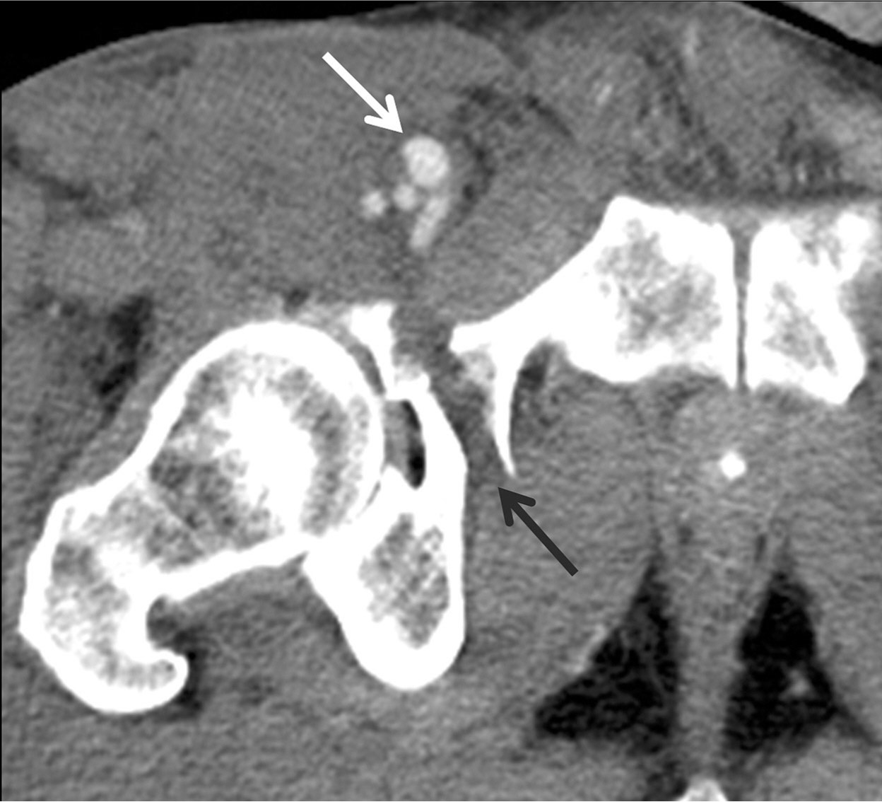
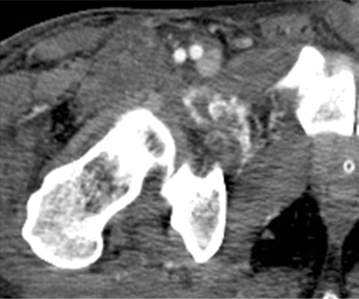
Fig. 1.
Fig. 1. The axial CT image shows a 1.2 cm-sized pseudoaneurysm at the right superficial femoral artery (white arrow). Note the fracture in the right anterior acetabulum (black arrow).
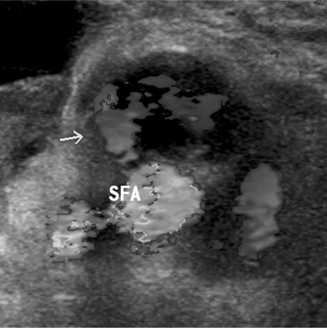
Fig. 2.
Fig. 2. Doppler US 2 days after CT scans shows enlargement in the size of the pseudoaneurysm(arrow), communicating with the right superficial femoral artery.
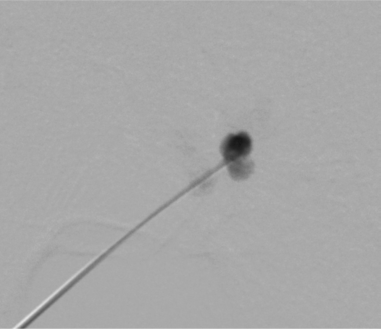
Fig. 3.
Fig. 3. Digital subtraction angiogram obtained after the needle positioning into the pseudoaneurysm shows the sac of the pseudoaneurysm at the proximal portion of the right superficial femoral artery. Note that there is no definite communication between the pseudoaneurysm and right superficial femoral artery. Subsequently thrombin injection was performed under US guidance (not shown).
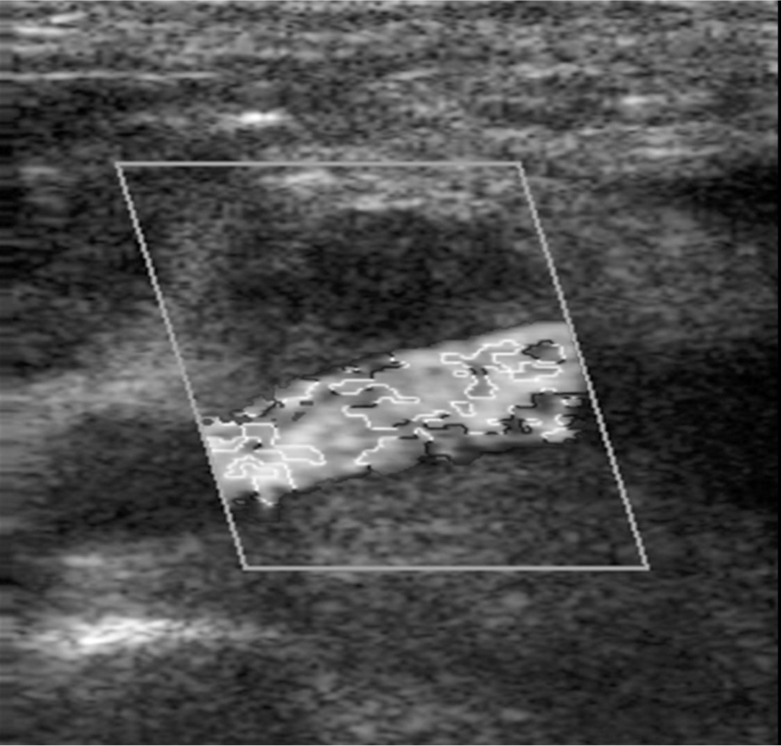
Fig. 4. A
Fig. 4A, B. On Doppler US (A) immediately after thrombin injection, there is no color swirl, indicating disappearance of pseudoaneurysm. On CT scan (B) obtained two days after thrombin injection, there is no visible pseudoaneurysm.
Fig. 4. B
Fig. 4A, B. On Doppler US (A) immediately after thrombin injection, there is no color swirl, indicating disappearance of pseudoaneurysm. On CT scan (B) obtained two days after thrombin injection, there is no visible pseudoaneurysm.
Citations
Citations to this article as recorded by