중심단어
Pulmonary artery pseudoaneurysm, embolization, vascular plug
임상소견
3년전 유방암으로 우측 유방 부분절제술 및 방사선/항암치료를, 2년전 폐전이로 우상엽절제술을 받은 환자로 1년전부터 폐전이가 악화되어 항암치료를 받으며 경과 관찰하던 중 흉부 CT에서 우하엽에 1cm의 가성동맥류가 발견되었고 1개월 후 크기가 3.4cm로 커졌음. 증상은 없었지만 빠른 크기 증가로 인한 massive bleeding 및 sudden death의 위험성이 높아 치료 의뢰되었음
진단명
Pseudoaneurysm in the right lower pulmonary artery
영상소견
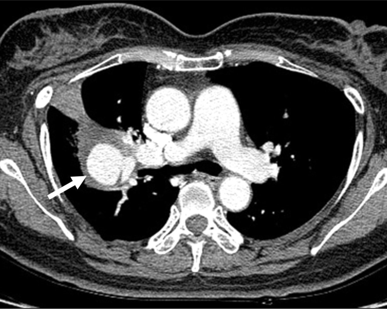
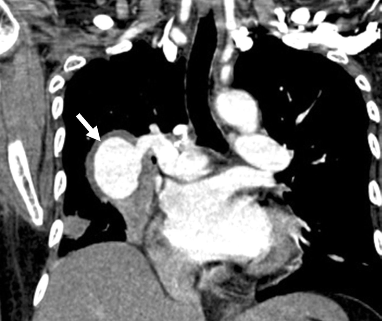
근위부와 연결되는 34mm 크기의 가성동맥류가 관찰되었고, 이 가성동맥류는 과거에 존재하던 cavitary metastatic mass내부를 가득 채우면서 주변으로 혈종으로 추정되는 soft tissue attenuation에 의해 둘러싸여 있었음(Fig. 1).
시술방법 및 재료
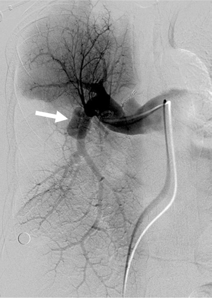
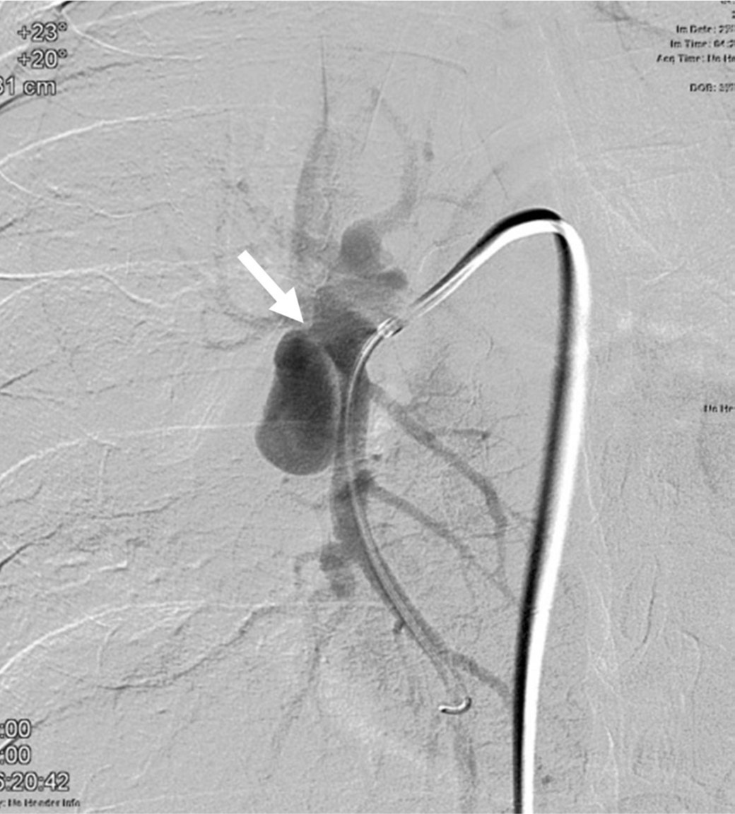
우대퇴정맥을 천자하여 8F sheath를 삽입한 후 5F Tempo 카테타(Cordis, Miami, USA)로 시행한 우폐동맥조영술에서 우하폐동맥 근위부에 가성동맥류가 보였고(Fig. 2), 3차원 우폐동맥조영술을 시행하여 우하폐동맥과 연결되는 가성동맥류 neck의 정확한 위치 및 가장 잘 보이는 projection을 확인하였다. 가성동맥류 neck부위 우하폐동맥에서 우엽간폐동맥까지의 여유 길이가 너무 짧아 코일은 사용하기 어려웠고, plug device를 사용하기 위해 0.035-인치 유도철사(Rosen, Cook, Bloomington, USA)를 따라 8F Envoy guiding catheter (Cordis, Miami, USA)를 우하폐동맥 근위부까지 전진시켰다. 우하폐동맥조영술을 시행하여 가성동맥류 neck의 정확한 위치를 확인하였고(Fig. 3), 우하폐동맥 근위부의 직경이 10mm로 측정되어 직경 16mm의 Amplatzer vascular plug(AGA Medical, MN, USA)를 가성동맥류 neck부위가 포함되도록 우하폐동맥에 deploy하였다. 약 60분 기다린 후 시행한 우폐동맥조영술에서 가성동맥류와 우하폐동맥의 혈류가 완전히 차단되었고 우중엽의 혈류는 잘 유지되었다(Fig. 4).
3주 후 시행한 흉부 CT에서 가성동맥류는 완전 소실되었고, 색전 원위부의 우하엽은 폐동맥 혈류가 차단되었음에도 aeration이 잘 유지되었다(Fig. 5).
고찰
폐동맥류 혹은 폐가성동맥류는 매우 드물지만 치료가 필요한 중요질환이며, 결핵을 포함한 감염, 심장기형, 혈관염, 만성폐색전증, 의인성, 및 외상성 등이 흔한 원인이다. 과거에는 주로 수술적 치료를 했지만 최근에는 인터벤션 치료가 선호되며 병변의 해부학적 위치, 모양, 개수에 따라 코일, vascular plug, stentgraft, NBCA, balloon 등을 선택할 수 있다.
본 증례에서는 우하폐동맥 근위부에 가성동맥류가 존재했기 때문에 최대한 많은 범위의 폐동맥 혈류를 유지하면서 가성동맥류 혈류를 완벽하게 차단해야 된다는 딜레마가 있었다. 이의 해결을 위해서 stent-graft 사용이 가장 이상적이었지만 우엽간폐동맥과 우하폐동맥의 직경차이가 심해 혈류차단에 실패할 가능성이 높은 깔대기 형태의 모양을 보인다는 점과 적절한 직경 x 길이의 제품이 국내에 없다는 점 때문에 제외되었다. Vascular plug 혹은 코일을 사용할 수 밖에 없었는데 우하폐동맥의 직경이 굵은 점을 고려한다면 코일의 색전범위가 plug에 비해 훨씬 클 것으로 예상되었고, 코일의 경우 실제 색전범위를 시술 전에 예측하기 어려워 vascular plug을 사용하여 색전술을 시행하였다. 우하폐동맥 근위부를 색전하였기 때문에 색전술 후 우하엽괴사 등의 합병증을 염려했으나 본 증례에서는 별다른 합병증 없이 퇴원하였고, 1달 뒤 추적 검사에서도 우하폐동맥이 색전되었음에도 우하엽의 aeration이 잘 유지되고 있었다.
참고문헌
1. Jagia P, Sharma S, Juneja R, Guleria R. Transcatheter treatment of pulmonary artery pseudoaneurysm using a PDA closure device. Diagn Interv Radiol 2011; 17:92-94.
2. Matsumura Y, Shiono S, Saito K, Sato T. Pulmonary artery pseudoaneurysm after lung resection successfully treated by coil embolization. Interactive CardioVascular and Thoracic Surgery 2010; 11:364-365.
3. Burrel M, Real MI, Barrufet M, et al. Pulmonary artery pseudoaneurysm after Swan-Ganz catheter placement: Embolization with vascular plugs. J Vasc Interv Radiol 2010; 21:577-581.
Fig. 1. A
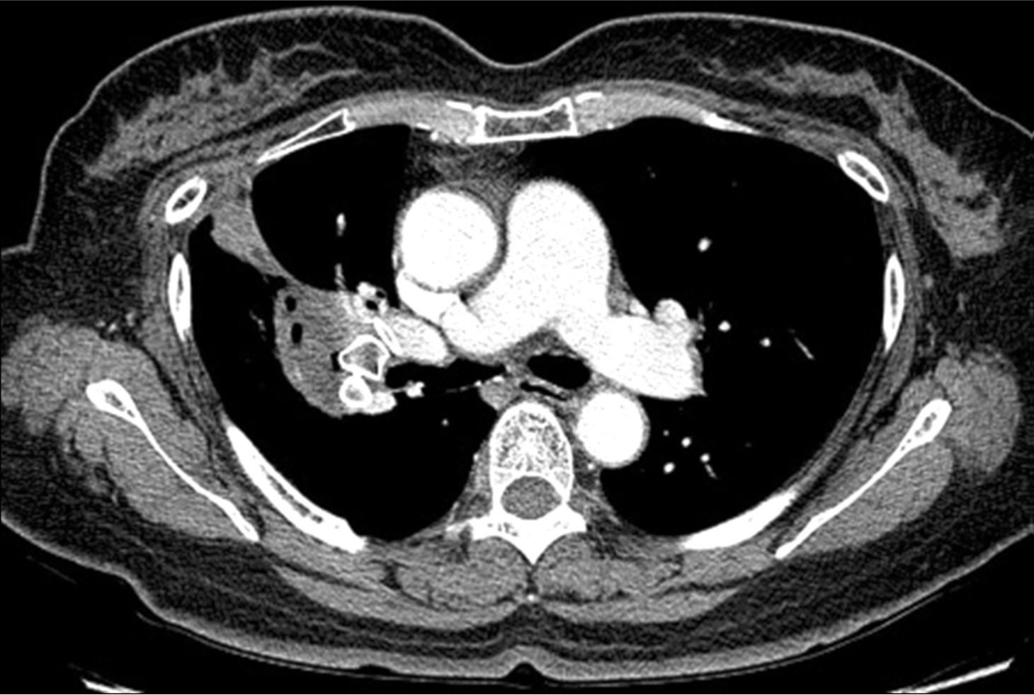
Fig. 1A, B. The axial (A) and coronal (B) images of chest CT scan demonstrate a pseudoaneurysm (arrows) in the right lower lobe, connected to the proximal part of the right lower pulmonary artery.
Fig. 1. B
Fig. 1A, B. The axial (A) and coronal (B) images of chest CT scan demonstrate a pseudoaneurysm (arrows) in the right lower lobe, connected to the proximal part of the right lower pulmonary artery.
Fig. 2.
Fig. 2. Right pulmonary angiogram shows a pseudoaneurysm (arrow) at the proximal part of the right lower pulmonary artery.
Fig. 3.
Fig. 3. On selective angiogram of the right lower pulmonary artery with a properly angulated projection, the neck(arrow) of the pseudoaneurysm is clearly demonstrated.
Fig. 4.
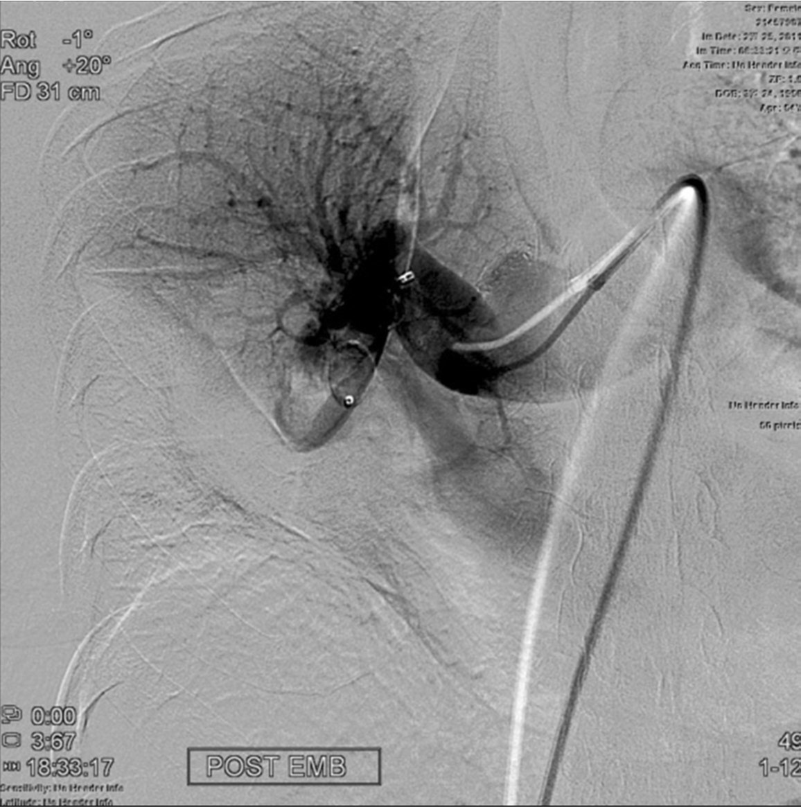
Fig. 4. Right pulmonary angiogram obtained 60 minutes after implanting an Amplatzer vascular plug at the right lower pulmonary artery shows complete exclusion of flow to the pseudoaneurysm.
Fig. 5. A
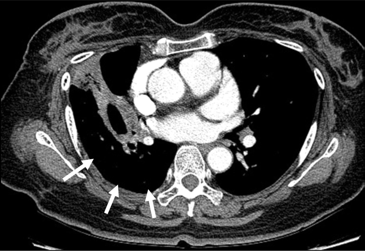
Fig. 5A, B. The axial images (A, B) of Chest CT scan 3 weeks after embolization show disappearance of the pseudoaneurysm and well-preserved aeration (arrows) of the distal right lower lobe.
Fig. 5. B
Fig. 5A, B. The axial images (A, B) of Chest CT scan 3 weeks after embolization show disappearance of the pseudoaneurysm and well-preserved aeration (arrows) of the distal right lower lobe.
Citations
Citations to this article as recorded by