중심단어
Splenic artery aneurysm, embolization, vascular plug
임상소견
1 주일간 지속되는 설사를 주소로 내원한 환자로 염증성 설사 진단 하에 시행한 CT에서 우연히 발견된 비장문 근처의 동맥류.
진단명
Splenic artery aneurysm in hilum of spleen.
영상소견
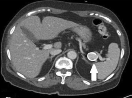
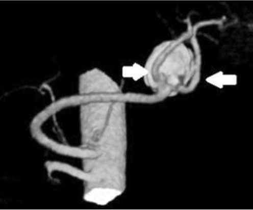
CT상 비장의 hilum의 위치에 2.5 x 2 cm 크기의 타원형의 mass like lesion이 관찰되었고 splenic artery와 연결되고 splenic artery와 동일한 조영증강을 보여 동맥류로 추정되었으며 rim calcification이 동반되어 있었음 (Fig.1). 정확한 해부 구조를 보기 위해 시행한 CT angiography에서 비장의 hilum의 근처 splenic artery 원위부에 동맥류가 관찰되었고 동맥류로부터 두 개의 큰 branching vessels이 기시하고 있었다 (Fig.2).
Fig. 1.
The axial CT image shows 2.5cm sized aneurysm (white arrow) with calcification in the splenic artery at the hilum of spleen.
Fig. 2.
3D reconstruction CT image shows 2.2x2.0cm sized splenic artery aneurysm in which two large branching vessels (white arrows) are originating from the base of the aneurysm.
시술방법 및 재료
우대퇴동맥을 천자하여 6F renal guiding sheath(Ansel, Cook, Bloomington, USA)로 시행한 동맥조영술에서 비장의 hilum 근처에 있는 2.2x2.0cm 크기의 비장동맥류 및 동맥류의 바닥으로부터 기시되는 두 개의 branching vessels을 확인하였다 (Fig.3). 비장 동맥류를 색전술하기로 결정하였고 색전술은 두 개의 branching vessel 근위부와, 동맥류, 동맥류 근위부의 비장동맥을 막기로 하였다. 먼저 두 개의 branching vessels을 5F Head Hunter catheter (Ansel, Cook, Bloomington, USA)를 이용하여 선택하였고 각각 vascular Plug IV (Amplatzer Vascular plug type IV, St. Jude Medical, St. Paul, USA), 5 mm diameter와 6mm diameter를 이용하여 동맥류에 최대한 근접하여 Depoly 하였다 (Fig.4). 이후 Aneurysm의 sac을 Interlock coils (Boston Scientific, Cork, Ireland) (14mmx30cm size, #3)과 Micro Nester coils (Cook, Btoomington, USA) (#6)를 이용하여 색전하였으며 마지막으로 proximal supplying splenic artery 을 Interlock coils (Boston Scientific, Cork, Ireland) (#4)을 이용하여 색전하였다. 색전 후 시행한 비장동맥 혈관촬영술에서 동맥류의 근위부, 원위부, 동맥류 주머니의 조영증강 소실을 확인 후 시술을 종료하였다. (Fig. 6) 1개월 후 시행한 CT에서 동맥류는 조영증강되지 않았고 비장은 조영증강이 잘되어 우회혈관을 통해 혈류가 잘 유지됨을 알 수 있었다 (Fig.6).
Fig. 3.
6Fr renal guiding sheath insertion in splenic artery, and subsequent angiography. 2.2x2.0cm sized splenic artery aneurysm adjacent to spleen hilum and two large branching vessels are originating from the base of the aneurysm.
Fig. 4.
Spot image shows two Amplatzer Vascular plug type IV (5mm diameter : black arrow, 6mm diameter : white arrow) deployed at two large branching vessels originating from splenic artery aneurysm base, respectively.
Fig. 5.
Post embolization splenic artery angiography shows no contrast filling of splenic artery aneurysm. Proximal and distal splenic artery to the aneurysm embolized by microcoils and vascular plugs also shows no contrast filling.
Fig. 6.
Follow up CT image one month after embolization of splenic artery aneurysm shows homogeneous enhancement of the splenic without infarction.
고찰
비장동맥류는 복부장기의 가장 흔한 동맥류로 0.8%의 유병률을 보인다. 비장동맥류는 다산여성에게서 호발하는데 이것은 복압의 증가, 임신 시 호르몬의 변화와 혈역학의 변화에 의해 혈관내피의 과증식과 분열이 발생하기 때문이다. 또한 간경화에 의한 간문맥압 상승과 연관성도 있는 것으로 알려져 있다. 대부분 증상이 없으나 27%의 환자에게 복통이 동반되기도 한다. 본 증례에서 처럼 Arteriosclerosis가 흔하게 동반되어 있는데 이는 동맥류의 원인이라기보다는 동맥류에 의해 발생되는 것으로 알려져 있다. 대부분 2cm 미만으로 증상이 없는 경우 치료가 필요하지 않다. 그러나 비장동맥류의 파열은 사망률이 높기 때문에 복통 등의 증상을 유발하거나 동맥류의 크기가 2.5cm 이상이거나 임산부 또는 가임기 여성에게서 발견된 경우, 간문맥고혈압, 간이식시 치료의 적응증이 된다. 비장동맥류는 고전적으로 수술적 치료를 통해 이루어져 왔으나 상대적으로 mortality와 morbidity가 낮은 transcatheter coil embolization, covered sentgraft, injection of thrombin등의 다양한 중재적 기법을 이용하여 비장동맥류를 치료할 수 있다. 중재적 치료에 있어서 중요한 점은 위, 대망, 그리고 이자로부터의 collateral vessels을 잘 보존하여 동맥류를 색전하는 것이다. 본 증례는 무증상 환자에서 2cm이상의 동맥류로 파열의 위험성 증가에 대비하여 색전술을 시행한 경우로 동맥류에서 직접 기시하는 두 개의 큰 branching vessels0] 동반되어 있었다. 2개의 branching vessels, aneurysmal sac, Proximal atery를 색전하는 것이 치료의 주요 관점이다. 특히 색전후 비장경색을 막기 위해서는 collateral vessels을 보존해야 하므로 비장동맥에서 기시하는 두 개의 혈관을 막을 때 가능한 동맥류에 가까이, 비장 hilum에서는 멀리 막는 것이 필요했다. 두 개의 branching vessel은 vascular plug를 aneurysm에 최대한 근접하여 위치시킴으로써 비장동맥의 collateral 혈류의 패쇄 없이 치료를 할 수 있었고 비장의 경색 없이 동맥류를 색전할 수 있었다.
참고문헌
1. Kenningham R, Hershman MJ, McWilliams RG, Campbell F. Incidental splenic artery aneurysm, J R Soc Med. 2002; 95(9): 460-461.
2. ABADA HT, CAPASSO P, GOLzARIAN J. Endovascular options for splenic artery aneurysms. Endovascular Today 2012; 53-55.
3. Madoff DC, Denys A, Wallace MJ, et al. Splenic Arterial Interventions: Anatomy, Indications, Technical Considerations, and Potential Complications. RadioGraphics 2005; 25:S191-S211
4. Agrawal GA, Johnson PT, Fishman EK. Splenic Artery Aneurysms and Pseudoaneurysms: Clinical Distinctions and CT Appearances, AJR 2007; 188: 992-999.
Citations
Citations to this article as recorded by