중심단어
Renal artery, Aneurysm, Detachable coil, embolization
임상소견
우연하게 발견된 우측 신장동맥의 동맥류를 주소로 내원하였다. 과거력상 5년 전에 미세혈뇨가 있었으나 추가검사는 시행하지 않았다.
진단명
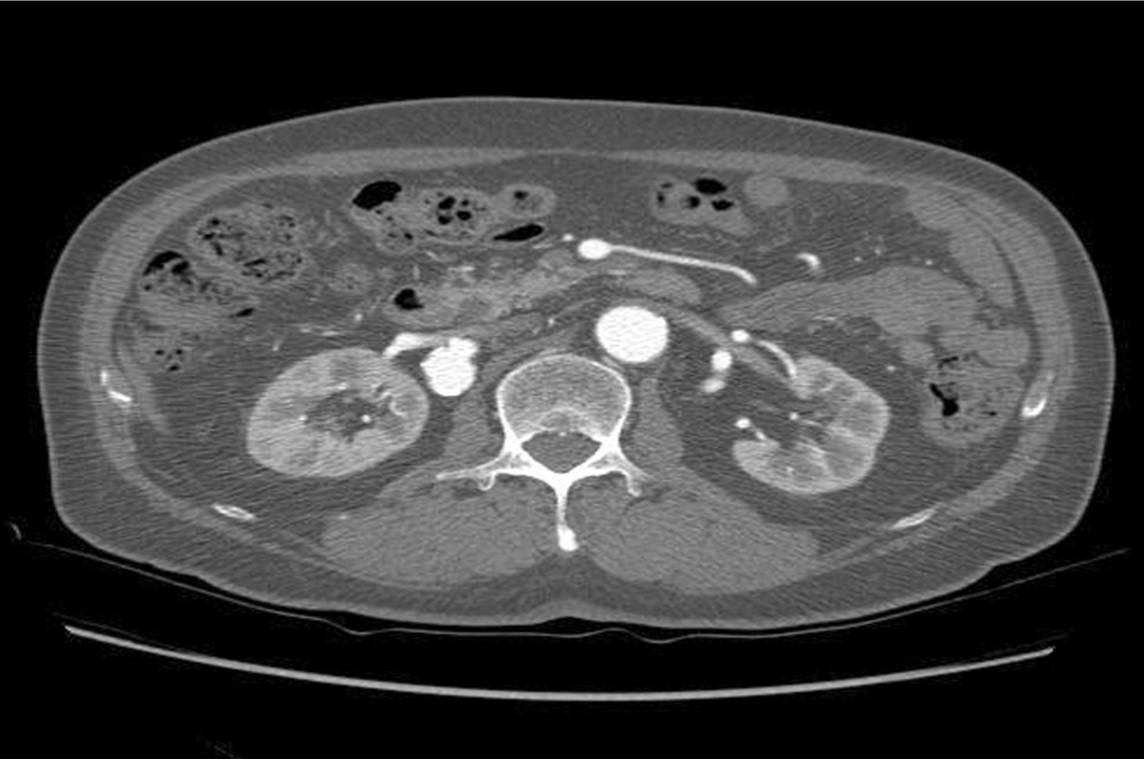
우측 신장동맥의 하부분절이 기시하는 부위의 2cm 크기의 동맥류.
영상소견
전산화단층혈관촬영술에서 우측 신장동맥의 하부분절에 기시하는 부위의 2cm 크기의 동맥류가 확인되었다 (Fig 1-2).
Fig. 1.
Abdominal CT shows aneurysm of right lower segmental renal artery.
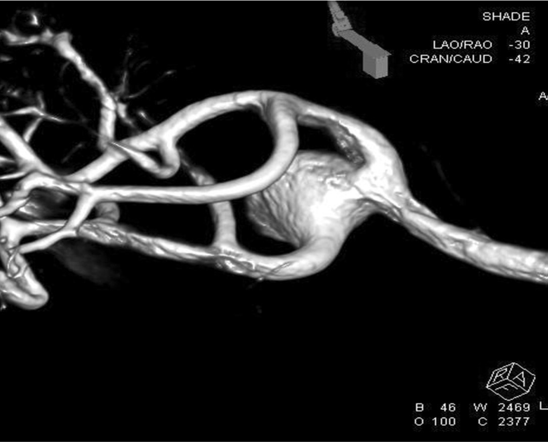
Fig. 2.
3-D reconstructed angiography show wide neck of aneurysm of lower segmental renal artery
시술방법 및 재료
오른 총넙다리동맥을 천자하여 5 Fr 크기의 도관을 삽입하였다. Cobra catheter (C2, Cook, Bjaeverskov, Denmark)를 이용하여 우측신장동맥을 선택하여 시행한 혈관조영술에서 신장동맥의 하부분절 동맥의 이분절이 시작되는 위치에서 조영제가 차는 원형 모양의 동맥류가 관찰됨 , 6 Fr 크기의 long sheath로 교체하고, 6 Fr 안내카테터(RDC, Cordis, Warren, NJ, USA)를 우측 신장동맥에 위치시켰다. 0.014 inch 미세유도철사를 이용하여 동맥류의 하부 분절동맥을 선택하였으며(Fig 3). 그 후 풍선카테터 도움(balloon assisted)에 의한 동맥류의 미세코일 색전을 시행하려 하였으나 풍선카테터의 풍선확장이 여의치 않았음. 미세카테터를 동맥류의 내강에 위치시키고 platinum detachable 미세코일 (Interlock IDC, Boston Scientific, USA) 14mm x 30cm, 14mm x 20 cm, 12mm x 30cm, 12mm x 20cm 들을 이용하여 동맥류 색전을 시행함 (Fig 4).
그 후 시행한 혈관조영술에서 동맥류는 코일로 색전되었고, 신장 동맥의 혈류는 이상 없음을 확인하였다. (Fig 5)
Fig. 3.
Selection of right lower segmental artery of aneurysm by microcatheter.
Fig. 4.
Embolization of aneurysm by multiple detachable microcoils.
Fig. 5.
Completion angiography shows coil embolization of aneurysm and no disturbance of renal arterial flow.
고찰
신장동맥에 생긴 동맥류는 약 0.3%에서 0.7%정도의 발생률을 보인다고 알려져 있다. 환자에게 고혈압이나 섬유근성증식증이 있을 경우에는 최대 9.2%까지 발생하는 것으로 보고되고 있다.[1] 대부분의 경우 증상이 없어서 우연히 발견되는 경우가 많으나 고혈압, 혈뇨, 복통, 신장기능저하 등의 증상이 있을 수 있다. 또한 드물긴 하지만 신장경색, 혈전증, 신장동맥박리 등의 합병증이 있을 수 있으며 동맥류 파열의 가능성이 있다. [1]
신장동맥에 동맥류가 있는 경우 증상이 있는 경우 또는 2cm 이상의 크기를 보이는 경우, 연속적인 혈관촬영술시 크기가 증가하거나 동정맥루 또는 신장동맥박리가 동반된 경우 치료의 적응증이 된다. 가임 여성이 임신 시 파열의 위험성이 증가하여 가임 여성에게서 발견 시 또한 치료의 적응증이 된다.[2]
신장동맥의 동맥류는 주로 분기점에서 발생하며 신장실질내 동맥류 또는 복합종류의 동맥류에 대하여 수술적 치료는 힘들며, 비교적 높은 질병률과 사망률을 보여 점차 혈관내 치료를 이용하고 있다.[3-5] 신장동맥의 동맥류에 대한 혈관내 치료는 수술적 치료보다 덜 침습적이며 안전하고 효과적으로 치료할 수 있는 방법이다. 또한 기존의 혈관내 치료의 기술적 발달과 동시에 재료의 발달을 통하여 기존의 뇌혈관내 시술에서 주로 쓰였던 detachable coil을 이용 시 수술적인 치료보다 좀더 안전하게 신장동맥의 동맥류에 대한 완전한 폐색을 기대할 수 있다. [6]
참고문헌
1. Lumsden AB, Salam TA, Walton KG. Renal artery aneurysm: a report of 28 cases. Cardiovasc Surg. 1996;4(2):185-189.
2. Nosher JL, Chung J, Brevetti LS, Graham AM, Siegel RL. Visceral and renal artery aneurysms: a pictorial essay on endovascular therapy. Radiographics. 2006;26:1687-704;
3. Luis Carlos Mendes de Brito, Joao de Toledo Martins, Eliane Passos, Amanda Jardim dos Santos, Rodrigo Assad Diniz da Gama, Gabriela Ximenes Furlani. Endovascular treatment of a renal artery aneurysm by embolization and aneurism neck remodeling technique: case report. J Vase Bras. 2011;10):181-184.
4. Damascelli B, Bartorelli AL, Ticha V, Trabattoni D, Lanocita R. Large renal artery aneurysm treated with Guglielmi detachable coils: procedural and 4-year follow-up results. Cardiovasc Intervent Radiol. 2008;31 Suppl 2:S88-91.
5. Malacrida G, Dalainas I, Medda M, Nano G, Inglese L.. Endovascular treatment of a renal artery branchaneurysm. Cardiovasc Intervent Radiol. 2007;30:118-120.
6. Klein GE, Szolar DH, Breinl E, Raith J, Schreyer HH. Endovascular treatment of renal artery aneurysm with conventional non-detachable microcoils and Guglielmi detachable coils. Br J Urol. 1997;79:852-860.
Citations
Citations to this article as recorded by