중심단어
Percutaneous transsplenic approach, gastric varix, embolization
임상소견
B형 간염 바이러스로 인한 간경화로 추적관찰 중이던 환자가 하루 전부터 시작된 melena로 응급실 방문하여 전산화단층촬영을 시행하였고 위정맥류출혈이 의심되었다.
진단명
Gastric varix bleeding
영상소견
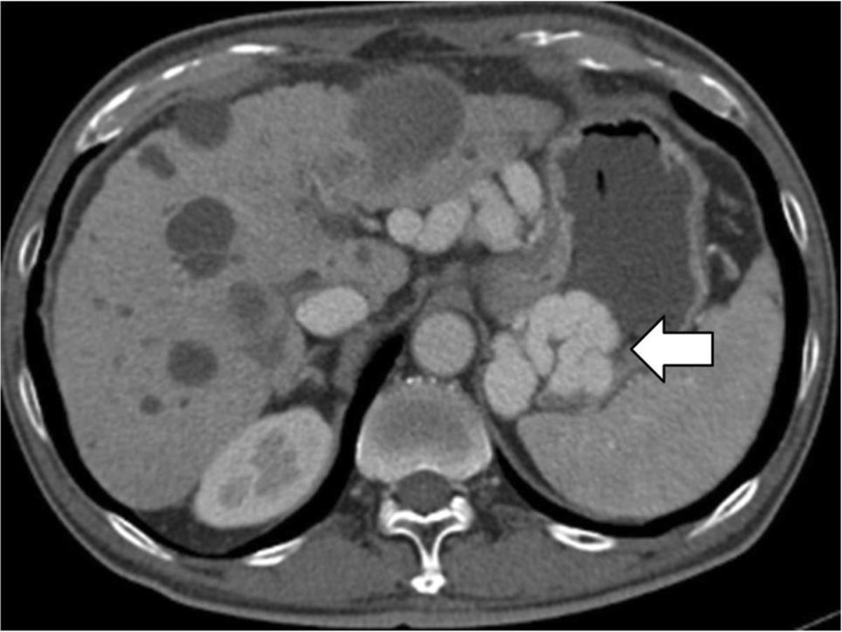
조영증강 복부전산화단층촬영에서 위기저부의 정맥류가 보이지만, 간내부의 문맥은 보이지 않고, 주문맥도 막혀 있음(Fig 1).
Fig. 1.
Fig. 1. Contrast enhanced CT scan of the abdomen demonstrates that the gastric fundal varies(arrow), nonvisulization of the intrahepatic portal vein and main portal vein occlusion.
시술방법 및 재료
초음파유도하에 chiba needle을 이용하여 percutaneous transsplenic approach로 비장정맥을 천자하고 5Fr sheath를 삽입하였다 (Fig 2A). 4Fr Davis catheter & terumo guide wire를 이용하여 후위장정맥을 선택하고 시행한 혈관조영술상 구불구불하게 늘어나 있는 위정맥류가 조영되었고, 조영제는gastrorenal shunt를 통하여 하대정맥으로 배출되고 있었다 (Fig 2B). 이에 위정맥류를 interlock coil 14mmx30cm, nester coil 14cmx10mm(x4), glue와 lipiodol B2 mixture (x3) & 1:3 mixture (x2)를 이용하여 색전술을 시행하였다 (Fig 2C). 색전술후 시행한 비장정맥 조영술상 위정맥류는 막히고 조영제는 gastrorenal shunt를 통하여 하대정맥으로 배출됨을 확인하였다 (Fig 2D). 이후 비장정맥과 percutaneous transsplenic tract을 standard coil 5cmx3mm (x2)와 glue와 lipiodol 1:2 mixture를 이용하여 색전술을 하고 시술을 종료하였다(Fig 2E). 시술 하루 뒤 시행한 조영증강 복부전산화단층촬영에서 위기저부 정맥류가 완전히 폐색됨을 확인하였다 (Fig 3).
Fig. 2
A. Under ultrasonic and fluoroscopic guidance perihilar splenic vein puncture was performed.
B. Initial venography of the posterior gastric vein demonstrates the tortuous and dilated varices(arrow) and drainage into the inferior vena cava through gastrorenal shunt. S=Gastrorenal shunt, R=Renal vein, V=Inferior vena cava.
C. Gastric varies was embolized using multiple coils and mixture of glue and lipiodol.
D. Splenic veiography, after embolization of the gastric varies, shows near totally occlusion of the gastric varices and contrast medium drainage into the gastrorenal shunt.
E. Percutaneous transsplenic tract was embolized by glue & lipiodol mixture.
Fig. 3.
CT scan obtained one day after the procedure, shows complete occlusion of the gastric fundal varices(arrow).
고찰
위정맥류출혈은 만성간질환환자의 morbidity &mortality의 주요원인으로, 위정맥류출혈이 발생할 경우 사망률이 25%-55%에 이르는 것으로 알려져 있다. 위정맥류출혈은 내시경적 지혈술과 경경정맥간내문맥정맥단락술(TIPS), 경피경간색전술, Balloon-occluded retrograde transvenous obliteration (BRTO), 경피경비장색전술과 같은 다양한 경도관혈관내접근법을 이용하여 치료를 할 수 있다. 고식적인 경도관 혈관 내 접근법으로 경경정맥간내문맥정맥단락술(TIPS)와 경피경간접근법이 흔히 이용되지만, 간내문맥의 쇠약, 주문맥의 혈전 또는 폐쇄, 간주변공간의 복수와 혈종 등의 상황에서는 이러한 경도관혈관내치료법의 사용이 불가능하다. 또한 BRTO의 경우 위정맥류와 소통하는 위콩팥회로가 존재해야만 시술이 가능하다.
문맥체계로의 또 다른 접근법으로 경피경비장 접근법이 있다. 경피경비장 접근법은 문맥체계로 곧장 통하는 길로서 문맥의 분지나 다양한 정맥류로 통하는 길을 제공한다. 하지만 심하게 구불구불한 비장정맥의 경우, 도관의 조작이 힘들 수 있다. 또한 비장은 과혈관성 장기로 시술과 관련된 출혈의 가능성이 있는데, 특히 문맥고혈압, 비장비대, 저혈소판증에서는 출혈의 위험이 높아 유의해야 한다. 최근에는 다양한 색전물질의 개발과 기법의 발전으로 비교적 안전하게 경도관혈관내치료법으로 이용되고 있다. 그러므로, 고식적인 경피경간접근법과 경경정맥간내문맥정맥단락술이 불가능하거나 어려운 경우에 안전하고 효과적으로 경피경비장접근법을 이용하여 치료할 수 있다.
본 증례에서는 간문맥이 완전히 막혀서 문맥혈류가 gastrorenal shunt로 배출되고 있었으므로 TIPS나 경피경간색전술이 불가능하였다. 또한 Gastrorenal shunt를 통하여 BRTO를 시행할 경우 문맥혈류가 배출될 경로가 없어지므로 심한 문맥고혈압이 발생할 것으로 판단되었다. 따라서, 위정맥류만 치료하기 위해 경피경비장접근으로 위정맥류만 색전하였다.
참고문헌
1. Chu HH, Kim HC, Jae HJ,et al.Percutaneous Transsplenic Access to the Portal Vein for Management of Vascular Complication in Patients with Chronic Liver Disease. Cardiovasclntervent Radiol. 2012;35:1388-1395
2. Harald B, Eva-Doreen P, Thomas B et al Transsplenic endovascular therapy of portal vein stenosis and subsequent complete portal vein thrombosis in a 2-year-old child. J VascIntervRadiol. 2010;21:1760-1764
3. David JT, Ffrrcsi JR, Mervin HD et al. Percutaneous transsplenic access in the management of bleeding varices from chronic portal vein thrombosis. J VascIntervRadiol. 2007;18:1571 -1575
4. Gong GQ, Wang XL, Wang JH et al. Percutaneous transsplenic embolization of esophageal and gastriofundal varices in 18 patients. World J Gastroenterol. 2001;7:880-883
Citations
Citations to this article as recorded by