중심단어
TIPS, Left hepatic vein
임상소견
2 년전 HCC type B with lung metastasis 진단받고 1-9th transhepatic arterial chemoembolization (TACE), 1-7th High dose Chemotherapy, Radiofrequency ablation(REA) 시행 받았으며, 점차 진행하는 복수로 입원하였다.
진단명
HCC type B with lung metastasis.
영상소견
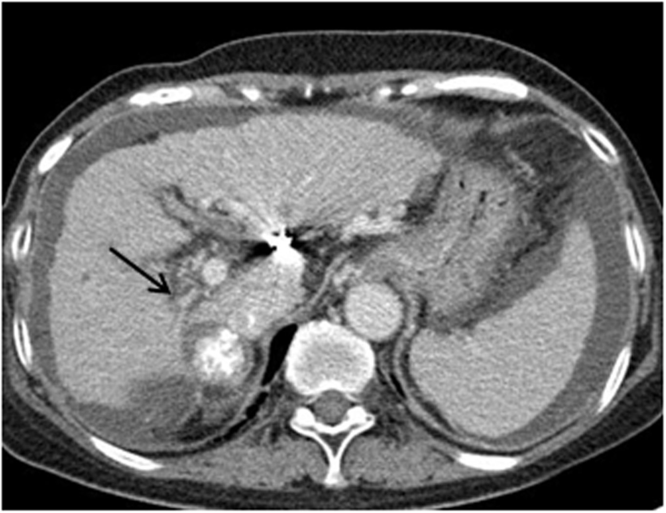
Liver의 Segment 7에 lipiodol uptake된 HCC가 보이고, right hepatic lobe의 atropic change 와 right portal vein의 narrowing이 보인다 (Fig.1).
Fig. 1.
Fig. 1. Portal phase of contrast enhanced CT scan shows atrophic change of the right hepatic lobe and narrowing of the right portal vein(arrow). Lipiodol laden HCC mass is seen near the right portal vein.
시술방법 및 재료
오른쪽 내경정맥을 천자한 후 9 Fr sheath를 삽입하였다. 5 Fr cobra catheter(Cook, Bloomingkn, USA)를 이용하여 왼간정맥을 selection하였다. Venography 시행하였고 왼간정맥에 이상 소견 없는 것을 확인하였다(Fig. 2). 왼간정맥의 중간부위에서 Colapinto needle의 tip을 posterior aspect로 향하게 한 후 왼간문맥을 천자하였다(Fig. 3). 0.035-inch wire를 삽입하고 5 Fr catheter를 상장간정맥에 위치한 후 portal venography를 시행하였다. 이때 측정한 portosystemic pressure gradient 는 19 mmHg 이었다. 8 mm-4 cm balloon(synergy, Boston Scientific, USA)을 이용하여 tract을 확장한 후 tractogram을 시행하여, contrast leakage가 없음을 확인하였다.
10 mm-5 cm self expandabl stent(niti-s vascular stent, Taewoong, Korea)를 삽입한 후 8 mm-4cm balloon(synergy, Boston Scientific, USA)을 이용하여 stent 내부를 확장시켰다. 그 후 측정한 portosystemic pressure gradient는 8mmHg로 감소하였다. Portography를 시행하여 간내문정맥 단락의 flow 가 유지됨을 확인하고 시술을 종료하였다(Fig. 4).
Fig. 2.
Left hepatic venography shows no abnormality.
Fig. 3.
Portal venography after puncture of the left portal vein using the Colapinto needle as posterior direction.
Fig. 4.
Portography after shunt tract dilation between left portal vein and left hepatic vein.
Fig. 5.
Self-expandable stent deployment. Well positioned stent is seen.
고찰
Right side shunt가 이미 존재하는 환자에서 왼간정맥에서 왼간문맥으로 시행된 TIPS는 종종 보고된 적 이 있다. TIPS에서 오른간정맥을 이용하는 이유는 직경이 크고 오른간문맥과 가장 가까이 있고 일정하게 문맥의 상후방에 위치하므로 오른간정맥에서 전하방을 향해 천자하면 문맥 천자를 쉽게 할 수 있기 때문이다. 술후 조기 shunt 폐쇄의 원인인 급성 혈전 형성을 예방하기 위해서 간내 천자 경로는 가급적 직선이고 짧은 것이 좋다. 이에 대부분의 경우 오른간정맥 혹은 중간간정맥에서 간문맥으로 접근하게 되고 이 경우 성공률은 93% 이상이다.
간혹 왼간정맥을 통해 접근이 필요한 경우가 있는데, 이 중 한 가지가 오른간정맥 혹은 중간간정맥이 비전형적 경로를 보이는 경우이다. 오른간정맥과 중간간정 맥에 혈전이 있는 Budd-Chiari syndrome 환자 역시 그러하다. 간문맥의 bifurcation 혹은 오른쪽 분지에 혈전이 있는 경우도 오른간정맥 혹은 중간간정맥으로의 접근이 어렵다. 마지막으로 간우엽절제술를 시행받은 환자 역시 왼간정맥으로의 접근이 불가피하다. 한 randomized controlled trial은 우간정맥을 이용한 간내문맥정맥 단락술과 왼간정맥을 이용한 간내문맥정맥 단락술을 비교하였는데, 왼간정맥을 이용한 간내문맥 정맥 단락술에서 간성 혼수 발생 비율이 현저히 낮았으며, 재출혈이나 reintervention, 복수의 호전에는 두 방법에서 차이가 없었고 생존율도 동일했다. 이번 증례는 왼간정맥을 이용한 간내문맥정맥 단락술의 기술적, 임상적으로의 성공 사례를 보여주고 있고, 오른간정맥 혹은 중간간정맥의 접근이 어려울 때는 왼간정맥을 통한 간내문맥 정맥 단락술의 유용성과 안전성을 보여준다.
참고문헌
1. Hidajat N, Kreuschner M, Rottgen R, et al. Placement of transjugular intrahepatic portosystemic shunt via the left hepatic vein under sonographic guidance in a patient with right hemihepatectomy. Acta Radiol. 2003 Jul;44(4):363-5.
2. Lei Chen, Tianli Xiao, Wensheng Chen, et al. Outcomes of transjugular intrahepatic portosystemic shunt through the left branch vs. the right branch of the portal vein in advanced cirrhosis: a randomized trial. Liver International. 2009 Auguest;29(7);110lli 09.
Citations
Citations to this article as recorded by