중심단어
Carotid artery stenosis, stent placement, radiationtherapy, head and neck tumor
임상소견
35년 전 두경부 림프종으로 타병원에서 방사선 치료받은 병력 있는 자로 내원 3주 전부터 좌안 시력 저하가 있어 좌측 경동맥 협착이 의심됨.
진단명
Radiation-induced carotid artery stenosis
영상소견
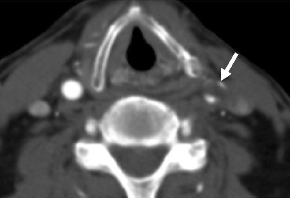
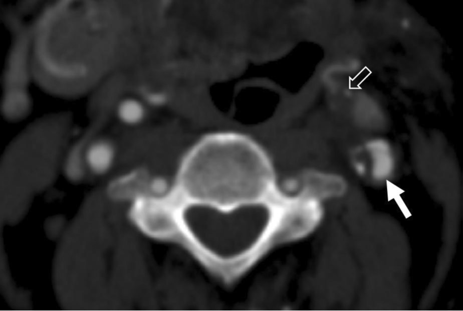
좌측 총경동맥의 원위부에 죽상경화판에 의한 심한 협착이 관찰된다 (Fig. 1). 좌측 총경동맥 분지부의 내경동맥에도 죽상경화판에 의한 심한 협착이 있고 좌측 외경동맥은 막혀있다 (Fig. 2).
Fig. 1.
Axial image of contrast-enhanced CT scan shows significant stenosis (>50%) of left distal common carotid artery (arrow) with atherosclerotic plaque.
Fig. 2.
Axial image of contrast-aihanced CT scan shows significant stenosis of left proximal internal carotid artery (arrow) with eccentric atherosclerotic plaque and occlusion of left external carotid artery (open white arrow).
시술방법 및 재료
좌측 총경동맥에서 원위부 총경동맥에 60% 이상의 협착이 있고 좌측 외경동맥은 막혀 있었다(Fig. 3). 좌측 근위부 내경동맥 에 죽상경화판에 의한 50% 이상의 협착이 있었다(Fig. 3),좌측 내경동맥 조영술에서 대뇌동맥에 심한 협착이나 폐색은 보이지 않았다. 좌측 내경동맥의 근위부에 색전성 폐색을 막기 위해 Filter Wire EZ(Boston Scientific, Natick, MA, USA)를 삽입하여 설치하였다(Fig. 4). 협착이 있는 좌측 총경동맥과 내경동맥에 9mm-50mm Carotid Wallstent Monorail(B oston Scientific, Natick, MA, USA)를 삽입하였다. Stent를 설치한 부위에 6mm-2cm Ultrasoft balloon catheter (Boston Scientific, Natick, MA, USA)로 확장을 시행하여 협착이 소실되었다. 스텐트 내부로 혈류는 잘 관찰되며 남아있는 심한 협착은 없었다(Fig. 4). 좌측 내경동맥 조영술에서 뇌실질로의 혈류가 잘 관찰되며 색전성 폐색은 보이지 않았다.
Fig. 3.
Left common carotid arteriogram shows more than 60% narrowing of common carotid artery (white arrow) and more than 50% narrowing of proximal internal carotid artery (arrowhead) at carotid bifurcation area. Occlusion of left external carotid artery (open arrow) is also noted.
Fig. 4.
Left common carotid arteriogram shows good patency of vascular stent in carotid artery. Filter Wire EZ (white arrow) is placed at distal to stenotic portion of left internal carotid artery to protect from embolic infarction.
고찰
증상이 있는 경동맥 협착에 대한 치료로 surgical endarcterectomy가 효과적임 이 널리 알려져 있다. 유럽과 미국의 carotid endarterectomy trials 에서 70% 이상 협착이 있는 경우 뇌 경색을 예방하데 내과적 치료보다 수술적 치료가 우위에 있다고 보고 하였는데 3년간 신체적 장애나 치명적인 뇌경색을 일으키는 유병률은 수술적 치료를 받은 455명 중에서 6,0%인 반면 비수술적 치료를 받은 323명의 환자들은 11%에 이르렀고 통계학적으로 유의한 차이라고 보고하고 있다. 최근에는 수술 대신 경동맥 혈관 성형을 통해서 경동맥 협착을 치료하는 여러 문헌들이 보고되고 있고 수술과 비교할 때 뇌 합병증이 많지 않다고 제시하고 있다. 특히 방사선 치료를 받은 후 발생하는 경동맥 협착의 경우에는 동맥내 여러층들이 서로 adhesion을 형성하여 endarterectomy로 plaque을 제거하데 어렵기 때문에 혈관 성형술이 유용하다고 보고되고 있다. 또한 혈관 성형술만으로는 재협착률이 높다는 결과가 많아 혈관성형술과 함께 스텐트 삽입을 하는 경향이 있다. 본 증례에서는 추가적으로 Filter Wire EZ를 사용하여 시술 중 발생할 수 있는 embolic occlusion을 예방하고자 하였다. 이러한 filter의 사용의 indication에 대한 consensus는 이루어지지 않았지만 Houdart et al, 에 따르면 long stenosis가 있어서 색전성 폐색의 위험성이 클 경우에 선택적으로 filter를 사용하였다는 보고가 있다. 두경부 암 환자의 방사선 치료 후 발생하는 경동맥 협착은 방사선 치료 후 수 개월에서 수 십년 뒤에 발생할 수 있기 때문에 CT, MRI, 초음파 등을 통해서 경동맥 협착 정도를 주기적으로 관찰하는 것이 필요하며, 협착이 생길 경우 혈관성형술과 스텐트 삽입을 통해서 치료가 가능하겠다.
참고문헌
1. Emmanuel H, Charbel M, Rene C, Jean-Pierre S, Jean-Jacques M. Carotid stenting for radiationinduced stenoses : A Report of 7 Cases. Stroke 2001;32:118-121
2. Dubec JJ, Munk PL, Tsang V, et al.Carotid artery stenosis in patients who have undergone radiation therapy for head and neck malignancy.Br J Radiol 1998;71:872-875
3. Minerva B,Gerhard S, Peter Z, et al.Long-term changes induced by high-dose irradiation of the head and neck region: Imaging findings. Radiographics 1997;17:5-26
4. Paul HK, Yasha K, Colin P, et al.Outcomes of carotid angioplasty and stenting for radiation-associated stenosis. Am J Neuroradiol 2005;26:1781-1788
Citations
Citations to this article as recorded by