중심단어
cystic duct stone, percutaneous stone removal
임상소견
담낭관의 담석을 동반한 담낭염
3일 전부터 복통 있었고 내원일 새벽 통증 악화되어 응급실로 내원. 내원 당시 혈압과 맥박은 정상이었고 혈액검사에서 WBC 13,700, CRP 16.11로 상승되어 있었으며 신체검진에서 Murphy's sign 양성이었음. 이에 시행한 복부 전산화 단층 촬영에서 담낭벽의 비후와 주변 지방조직으로 침윤 소견과 함께 담석이 담낭관에서 발견되었음. 입원 2일째 경피적 쓸개창냄술 시행하였으나 환자 산소포화도 저하되고 빈호흡 보이는 등 septic condition 악화되어 기관 삽관 하였음. 환자의 수술 위험도 크다고 생각되어 담낭절제술은 시행하지 않기로 하였음.
영상소견
CT 소견
담낭벽이 전체적으로 두꺼워져 있고 주변부 지방조직으로 침윤을 보이는 담낭염에 합당한 소견과 함께 담낭관에 담석이 관찰되었음
담낭조영 소견
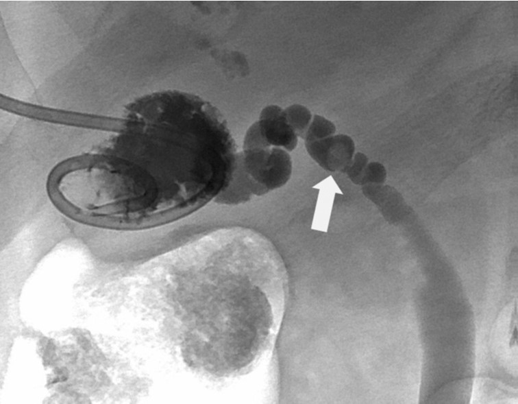
tubogram에서 담낭관에 담석으로 인한 filling defect를 확인할 수 있었음(Fig 1). 담낭 내에는 담석이 뚜렷하게 보이지 않았음.
Fig. 1.
Fig. 1 Tubogram through cholecystostomy tube shows small stone (arrow) in middle portion of cystic duct.
시술방법 및 재료
담낭에 9F sheath를 삽입하고 5F KMP catheter와 microcatheter를 이용해 담낭관을 selection하였다. 5F catheter를 CBD까지 진행시킨 후 0.035” guidewire를 거치시키고 4mmx4cm balloon catheter(Mustang, Boston Scientific, Natick, MA)를 이용해 담낭관을 확장시켰다. 이 과정에서 담낭관-총담관 이행 부위에 waisting이 관찰되었다(Fig 2). 이후 6F Fogarty balloon을 이용해 담낭관에 있는 담석을 총담관으로 밀어내었다(Fig 3). 시술 후 tubogram에서 담낭관과 총담관에 뚜렷한 filling defect는 보이지 않았다.
Fig. 2
Balloon dilatation was performed for cystic duct. There was waisting at the junction of cystic duct and common bile duct (arrowheads).
Fig. 3
The stone was pushed into CBD using Fogarty balloon catheter (arrowheads).
추적관찰 소견
시술 2일 및 4일 뒤 각각 시행한 tubogram에서 담낭, 담낭관 및 총담관에 뚜렷한 filling defect가 보이지 않아 담석이 남아있지 않음을 확인하였고 (Fig 4) 배액관을 제거한 후 별다른 합병증 없이 퇴원하였다.
Fig. 4
Follow-up tubogram reveals no residual stone in the cystic duct and CBD.
고찰
급성 담석성 담낭염은 치료하지 않을 경우 패혈증으로 진행할 수 있으며, 주된 치료는 복강경 담낭절제술이다. 하지만 수술 위험도가 높은 환자에서 응급으로 담낭 감압을 시행해야 하는 경우에 경피적 쓸개창냄술이 대안이 될 수 있다(1-3).
실제로 수술 위험도가 높은 담낭염 환자에서 담낭석을 경피적으로 제거한 사례는 종종 찾아볼 수 있으나(3) 담낭관석을 제거한 사례는 찾아보기 어려웠다. 경피적 담낭관석 제거술은 담낭관 자체의 tortuosity로 인해 시술이 어려울 수 있으나(4,5) balloon을 이용하여 lumen을 확장함으로써 환자에게 덜 침습적인 시술을 시도해 볼 수 있겠다.
참고문헌
1. Lammert F, Miquel JF. Gallstone disease: from genes to evidence-based therapy. J Hepatol. 2008;48:S124-S135.
2. Melin MM, Sarr MG, Bender CE, van Heerden JA. Percutaneous cholecystostomy: a valuable technique in high-risk patients with presumed acute cholecystitis. Br J Surg. 1995;82:1274-1277.
3. Kim YH, Kim YJ, Shin TB. Fluoroscopy-guided percutaneous gallstone removal using a 12-Fr sheath in high-risk surgical patients with acute cholecystitis. Korean J Radiol. 2011 Mar-Apr; 12(2):210-5
5. Shaw MJ, Dorsher PJ, Vennes JA. Cystic duct anatomy: an endoscopic perspective. Am J Gastroenterol. 1993; 88:2102-2106
6. Mary Ann Turner, MD and Ann S. Fulcher, MD. The Cystic Duct: Normal Anatomy and Disease Processes. Radiographics. 2001;21:3-22.
Citations
Citations to this article as recorded by