중심단어
Bronchial artery embolization, hemoptysis, lung cancer
임상소견
20갑년의 흡연력 있는 환자로 기침과 함께 갑자기 500cc정도의 객혈 소견으로 내원함.
진단명
Lung cancer with hemoptysis
영상소견
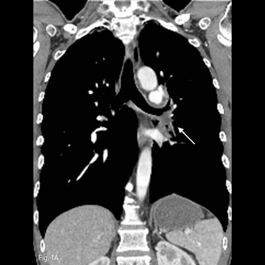
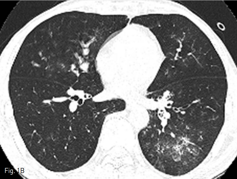
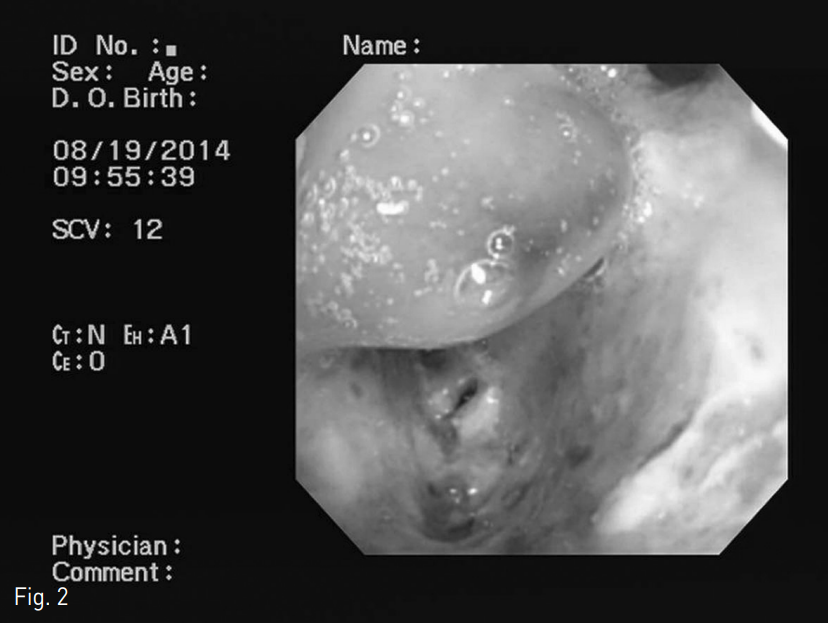
조영 증강 후 chest CT영상에서 좌측폐 주기관지, 좌상엽 및 좌하엽 기관지벽이 두꺼워져 있으며 좌측 엽간 림프절이 커져있음 (Fig. 1A). 우측폐 중엽과 좌측 폐 하엽에 경화가 동반된 간유리음영이 관찰됨 (Fig. 1B). 기관지내시경에서 좌측폐 주기관지에 yellowish exudate가 동반된 mucosal destruction이 있었음 (Fig 2).
Fig. 1
A. The post-contrast chest CT image shows diffuse wall thickening of the left lower bronchus (white arrow).
B. The high resolution chest CTimage reveals multifocal areas of ground glass opacity and consolidation in the right middle lobe and left low er lobe suggesting aspirated blood.
Fig. 2
Bronchoscopy shows mucosal destruction with yellowish exudate in right lower bronchus.
시술방법 및 재료
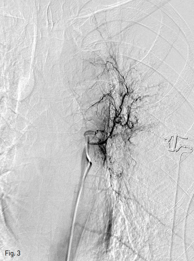
오른쪽 총대퇴동맥을 천자하여 5 Fr sheath (Terumo, Tokyo, Japan)를 삽입한 후, pigtail catheter (Cook, Bloomington, USA)를 흉부 대동맥에 위치시키고 시행한 흉부대동맥조영술에서 좌측에 비대된 기관지동맥이 관찰되었고, 5 Fr catheter를 좌측 기관지동맥에 위치시켜 비대해지고 단락을 보이는 좌측 기관지 동맥을 확인할 있었고(Fig. 3), 이를 미세도관 (Progreat, Terumo, Tokyo, Japan)과 미세유도철사 (GT wire, Terumo, Tokyo, Japan)로 superselection한 후 350-550 microns의 PVA particle 및 gelfoam (Cutanplast, Marscia Brunelli, Milan, Italy)으로 색전함. 경과관찰에서 객혈 보이지 않고 현재 호흡기내과에서 치료중임.
Fig. 3
The left bronchial angiograms show arterial hypertrophy and shunting of pulmonary artery.
고찰
대량 객혈은 24시간동안 100~600ml 이상의 출혈량으로 정의한다. 다량의 객혈을 주소로 내원한 환자들은 폐암보다는 결핵이나 기관지 확장증 등에 의한 경우가 많으나 폐암 환자의 10~30%에서 객혈이 나타나는 것으로 알려져 있다. 대부분 경도 또는 중등도의 출혈을 보이며 10%에서 대량객혈이 생긴다. 폐암에서 객혈은 일반적으로 암세포의 혈관침윤보다는 tumor bed 혈관의 국소적 괴사와 염증반응으로 생긴다. 객혈이 진행하는 양상이거나 소량의 출혈이 지속적으로 보이는 환자에게 색전술을 시행할 수 있는데, 기관지동맥 색전술은 대량 객혈의 치료로서 최소 침습적이 면서도 가장 효과적인 치료법으로 알려져 있다. 시술 후 즉시 객혈의 증상이 조절되는 빈도는 73~99%로 보고되고 있다. 그러나 기관지동맥 색전술 후 1달 내 객혈의 재발률은
10~29%로 비교적 높다고 알려져 있으며 장기적으로도 색전된 혈관의 재개통이나 곁순환의 형성 등으로 재발의 가능성이 있다. 본 증례는 폐암 환자에서 심한 객혈이 있어 기관지 동맥 색전술을 시행하였고 시술 후에 객혈이 호전되는 양상을 보였다.
참고문헌
1. Park HS, Kim YI, Kim HY, et al. Bronchial artery and systemic artery embolization in the management of primary lung cancer patients with hemoptysis. Cardiovasc Intervent Radiol. 2007;30(4):638-643.
2. Kashyap S, Mohapatra PR, Saini V. Endobronchial tuberculosis. Indian J Chest Dis Allied Sci. 2003;45(4):247-256.
3. Sundarakumar DK, Bhalla AS, Sharma R, et al. Multidetector CT evaluation of central airways stenoses: Comparison of virtual bronchoscopy, minimal-intensity projection, and multiplanar reformatted images. Indian J Radiol Imaging. 2011;21(3):191-4.
Citations
Citations to this article as recorded by