중심단어
Liver blunt trauma, embolization, internal mammary artery
임상소견
교통사고 (운전자 TA) 후 발생한 오른쪽 윗배 통증을 주소로 응급실로 내원하였다. 응급실에서 촬영한 CT scan에서 간 손상과 함께 간 내 출혈이 있어 간동맥 혈관조영술과 색전술을 시행하였다.
진단명
Arterial bleeding via internal mammary artery in liver trauma
영상소견
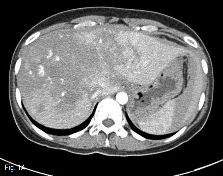
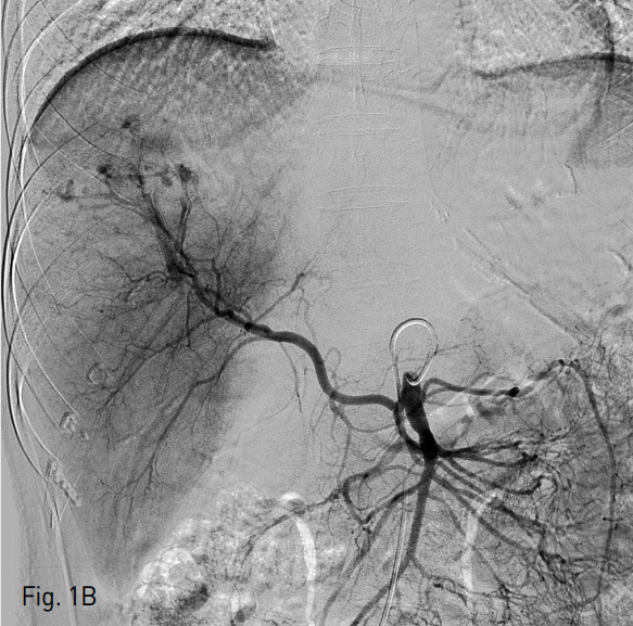
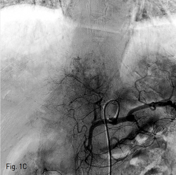
응급실 내원 직후 시행한 복부 CT에서 liver의 4,7,8 segment에 다량의 혈종과 pseudomaneurysm 그리고 multiple active bleeding이 관찰되었다 (Fig.1A). 색전술을 위하여 시행된 혈관조영술에서 replaced right hepatic artery branching off of the SMA를 통한 간 우분절에 다발성의 점상출혈이 있고 (Fig. 1B) 또한 left hepatic artery of celiac trunk를 통한 간 좌분절에도 다발성의 점상출혈이 있어 (Fig. 1C) 색전술을 시행하였다.
시술방법 및 재료
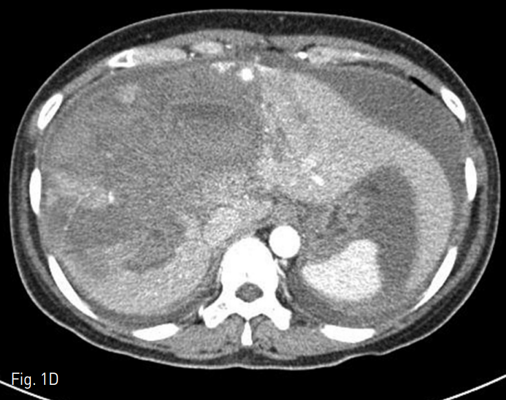
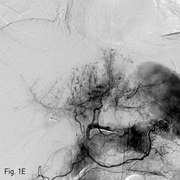
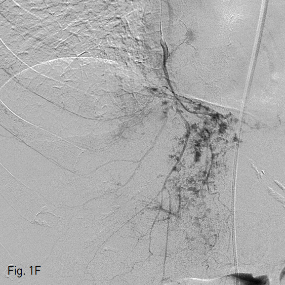
혈관조영술과 색전술을 위하여 우측 총 대퇴동맥을 통해 5 Fr RH catheter를 간동맥에 위치시킨 후 카테터 내에 2.2 Fr 미세도관을 삽입하여 다발성의 점상출혈이 있는 부위에 대하여 gelatin particle을 이용하여 색전술을 시행하였다. 색전술 시행 다음날, 다시 복부팽만과 hemogloin 수치 감소(8.8g/dL → 8.0g/dL)가 발생하여 복부 CT를 시행하였다. 추적 복부 CT상 간 내 혈종의 양이 증가되었고 2, 3번 segment에도 새롭게 다발성의 점상출혈이 보였다 (Fig. 1D). 새로이 보이는 Left lobe의 다발성 점상출혈을 확인하기 위하여 간동맥 혈관조영술을 시행하였으나 명확하게 출혈소견이 보이지 않았기에 간 동맥 외의 다른 동맥을 통한 출혈부위를 찾기 위하여 5 Fr Headhunter angiocatheter를 이용하여 우측 내유동맥의 혈관조영술을 시행하였고 (Fig. 1E, F) 내유동맥 조영술상에서 간 좌분절의 다발성 점상출혈을 확인하고 2.2 Fr 미세도관으로 진입하여 gelatin particle을 이용한 색전술 시행하였다. 이후에 환자는 더 이상의 출혈소견 없이 호전되었다.
Fig. 1
A. A initial contrast enhanced CT scan showed diffuse hepatic contusion of liver with large amount of intrahepatic hematoma and multifocal punctate extravasation of the contrast media, mainly at the segment 4 and 8 of liver. And there is large amount of hemoperitoneum in perihepatic and perisplenic space.
B. SMA angiography revealed multifocal active bleding foci in right hepatic segment through the replaced right hepatic artery originated from the SMA.
C. Celiac angiography revealed faint multifocal small active bleeding at the left hepatic segment of liver through the left hepatic artery.
D. A follow-up contrast enhanced CT scan showed interval increased large amount of intrahepatic hematoma. And newly developed, pseudoaneurysmal active bleeding foci in segment 4 of liver.
E. A celiac angiography revealed still remaining multifocal active bleeding foci at the left hepatic segment of liver.
F. Subsequent right Internal mammary angiography revealed multifocal active bleding foci at the left hepatic segment of liver. Arterial embolization with gelfoam particle was done. And the general condition of patient was improved (not shown).
고찰
내유동맥은 쇄골하동맥에서 분지하여 흉골의 측면으로 길게 내려와 배꼽부위까지 앞복벽을 따라 아래 배벽동맥과 문합을 이루게 된다. 내유동맥의 hepatic supply는 간암환자에서 TACE (Transarterial chemoembolization)를 시행하는 환자에서 중요한 extrahepatic collateral arteries중의 하나로 알려져 있으며 주로 횡경막과 앞복벽에 abutting하는 ventral hepatic area 의 간암에 대하여 feeding artery로 작용할 수 있다는 것이 알려져 있다.
본 증례는 blunt hepatic trauma 환자에서 발생한 간 손상에 의한 출혈 발생시에 간동맥을 통한 색전술을 성공적으로 시행하였음에도 불구하고 지속적인 간 내 출혈이 있는 경우에는 extrahepatic collateral flow에 의한 출혈이 지속될 수 있으므로 필요 시 내유동맥 등의 가능한 other hepatic feeding artery까지도 확인을 하여 적절한 색전술을 시행하여야 한다.
참고문헌
1. Kim HC, Chung JW, Choi SH, et al. Internal mammary arteries supplying hepatocellular carcinoma: vascular anatomy at digital subtraction angiography in 97 patients. Radiology 2007;242(3):925-932.
2. Kim SH, Kim HC, Hur S, et al. Chemoembolization via the left internal mammary artery supplying hepatocellular carcinoma. J Vasc Interv Radiol. 2014;25(9):1389-1397.
3. Kim HC, Chung JW, Lee W, et al. Recognizing extrahepatic collateral vessels that supply hepatocellular carcinoma to avoid complications of transcatheter arterial chemoembolization.Radiographics. 2005;25:S25-39.
Citations
Citations to this article as recorded by