중심단어
Splenic, gastroepiploic, aneurysm, embolization
임상소견
지속적인 피로감과 간헐적 우상복부 동통을 주소로 내원하여 알코올성 간염으로 인한 간경화와 이로 인해 생긴 간세포암을 진단받았다. 초기 혈액 검사상 헤모글로빈치 9.2 g/dl, 백혈구 수치 2.1 x 10^3g/dL, 혈소판 수치 57 x 10^3g/dL로 범혈구 감소증 소견을 보이고 있었고, 프로트롬빈 시간이 52%, INR이 1.60으로 다소 연장되어 있었다. 그러나 간기능 검사를 포함한 그 외 혈액 화학적 검사치는 정상 범위 내에 있었다.
영상소견
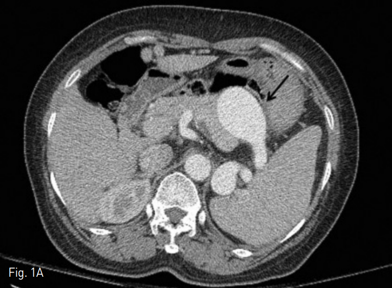
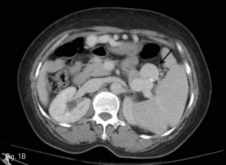
본원 소화기내과 외래에서 시행한 복부단층촬영상 우측 간엽 5번 분절에 약 3.2cm 크기의 간세포암이 있었다. 비장동맥은 매우 구불구불하며, 근위부 1/3 지점부터 비장 문 (hilum)에 이르기 까지 2~4.5 cm 크기의 다발성 동맥류가 있었다 (Fig. 1A-B). 1주일 후, 우측 간엽 5번 분절에 있는 간세포암에 대하여 Doxorubicin 20mg을 이용하여 시행하였고, 4일 이후 비장동맥류에 대한 색전술을 시행하였다.
Fig. 1
A. A contrast enhanced axial CT image on arterial phase shows largest splenic artery aneurysm (arrow) with 4.5cm in diameter.
B. A contrast enhanced axial CT image on venous phase shows multiple splenic artery aneurysms (arrow) around splenic hilum.
시술방법 및 재료
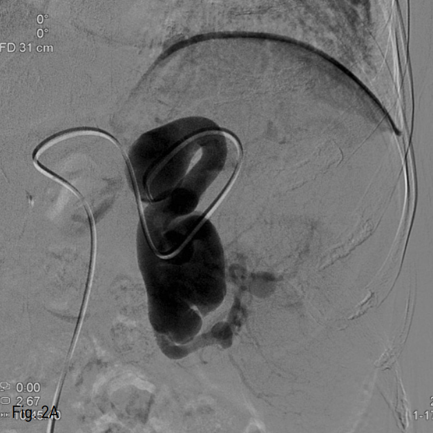
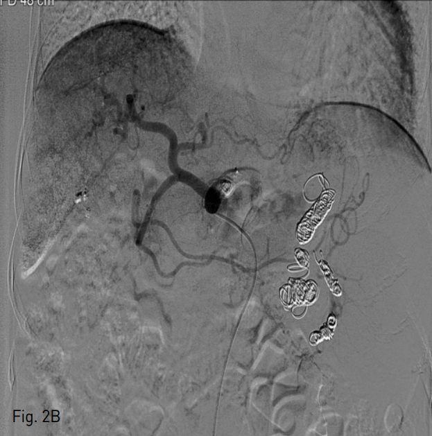
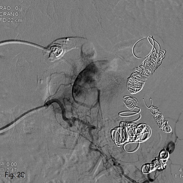
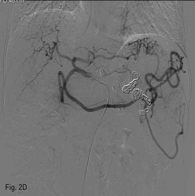
우측 총대퇴동맥을 천자한 후, 5 Fr sheath (Terumo, Tokyo, Japan)를 거치 시켰다. 이후 0.035 inch guide wire (Terumo, Tokyo, Japan)와 5 Fr Roche-Hepatic ca theter (Cook, Bloomington, IN, USA)를 이용하여 비장동맥 조영술을 시행하였으며, 근위부 1/3 지점에 약 4cm 크기의 가장 큰 비장동맥류가 관찰되었다 (Fig. 2A). 2.2 Fr microcatheter (Progreat; Terumo, Tokyo, Japan)을 이용하여 가장 원위부에 있는 동맥류를 지나 비장 문까지 진입한 후 11개의 nester coil (Cook, Bloomington, IN, USA)을 이용하여 색전을 시행하였다. 그 후 microcatheter를 근위부로 옮겨 가장 큰 비장동맥류의 원위부와 근위부에 13개의 nester coils, 1개의 vascular plug, 그리고 histoacryl 1:3 mixture를 이용하여 색전하였다. 색전술 후 복강 동맥 조영술 상 비장동맥은 완전히 색전되어 조영되지 않았으나, 가장 큰 크기의 비장동맥류에 지연기상 조영이 나타났다 (Fig. 2B). 이에 다시 2.2 F microcatheter를 우측 위그물막동맥으로 진입시켜 조영하였고, 많은 측부 곁가지에 의해 비장동맥류가 조영되었다 (Fig. 2C). 다시 histoacryl 1:5 mixture를 이용하여 색전하였으며, 복강동맥 조영술상 더 이상 비장동맥류는 관찰되지 않았다.
Fig. 2
A. Initial splenic arteriogram shows a large splenic artery aneurysm in proximal portion of splenic artery.
B. Post embolization angiogram shows faint visualization of largest splenic artery aneurysm on delayed phase.
C. Right gastroepiploic angiogram shows multiple fine feeders to largest splenic artery aneurysm.
D. Final celiac angiogram shows complete exclusion of splenic artery aneurysm and prominent splenic collateral pathways such as perigastric and gastroepiploic anastomosis.
추적 관찰
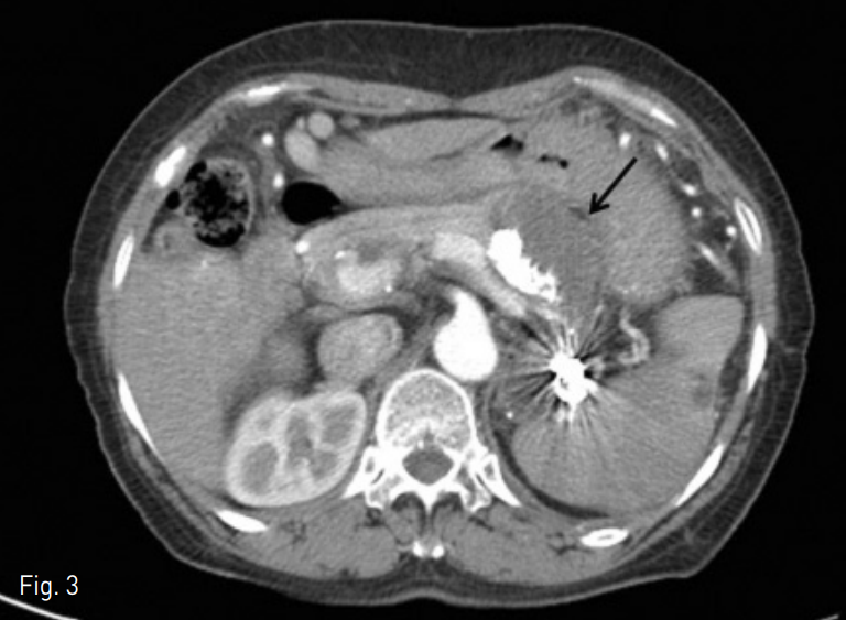
이후 3개월 뒤 재차 간동맥 색전술을 위해 시행한 추적 관찰 복부단층 촬영상 비장동맥류 내에 완전 혈전화가 관찰되었으며, 8개월 뒤 복부 단층 촬영상 가장 큰 비장동맥류가 시술 전 4.5cm에서 3.8cm으로 약간의 크기 감소가 확인되었다.
Fig. 3
A contrast enhanced axial CT image obtained 3 month later shows complete thrombosis of largest splenic artery aneurysm (arrow) with multiple embolic materials.
고찰
비장동맥류는 복부장기의 가장 흔한 동맥류로 0.8%의 유병률을 보인다. 다산 (multiparity), 간경화에 의한 간문맥압 상승과 연관성이 있는 것으로 알려져 있다. 본 증례처럼 대부분 무증상으로 우연히 발견되지만 비장동맥류의 파열은 사망률이 높기 때문에 복통 등 증상이 있거나 동맥류의 크기가 2.5cm 이상이거나 임산부 또는 가임기 여성에게서 발견된 경우, 간문맥고혈압, 간이식시 치료의 적응증이 된다.
비장동맥의 경혈관적 embolization이나 수술적 ligation 후 발생할 수 있는 부작용으로는 감염, 농양, 흉수 그리고 경색 등이 잘 알려져 있다. 이들 중 경색의 발생률을 낮추기 위해서는 main splenic artery embolization보다는 selective 또는 superselective embolization이 선호되나 본 증례와 같이 main splenic artery를 embolization할 경우에도 total splenic infarction은 매우 드문 것으로 알려져 있는데 이는 pancreatic collateral pathway, perigastric collateral pathway, gastroepiploic collateral pathway 등 풍부한 곁순환이 비교적 이른 시간 내에 잘 발달하기 때문이다.
참고문헌
1. Madoff DC, Denys A, Wallace MJ, Murthy R, Gupta S, Pilsbury EP, et al. Splenic arterial interventions: anatomy, indications, technical considerations, and potential complications. Radiographics 2005;25:191-211.
2. Kenningham R, Hershman MJ, McWilliams RG, Campbell F. Incidental splenic artery aneurysm. J R Soc Med. 2002;95:460-461.
3. Keramidas DC, Kelekis D, Dolatzas T, Aivazoglou T, Voyatzis N. The collateral arterial network of the spleen following ligation of the splenic artery in traumatic rupture of the spleen; an arteriographic study. Z Kinderchir. 1984;39:50-51.
4. Cyrany J, Kopacova M, Rejchrt S, Jirkovsky V, AlTashi M, Bures J. Gastric arterial bleeding secondary to chronic occlusion of the splenic artery (with video). Gastrointest Endosc 2010;71:1335.
Citations
Citations to this article as recorded by