중심단어
Angiodysplasia, jejunum, small bowel, embolization
임상소견
4개월 전부터 흑변 및 혈변을 주소로 네 차례 본원 내원하였고 상부, 하부 내시경 및 캡슐 내시경을 포함한 검사들에서 출혈 위치와 원인 찾지 못하였으며, 다시 흑변을 주소로 본원 내원하였다.
영상소견
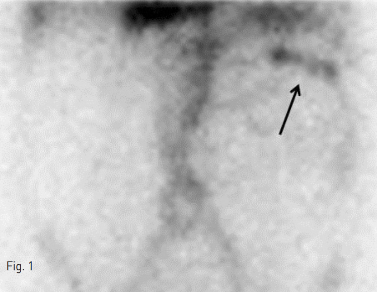
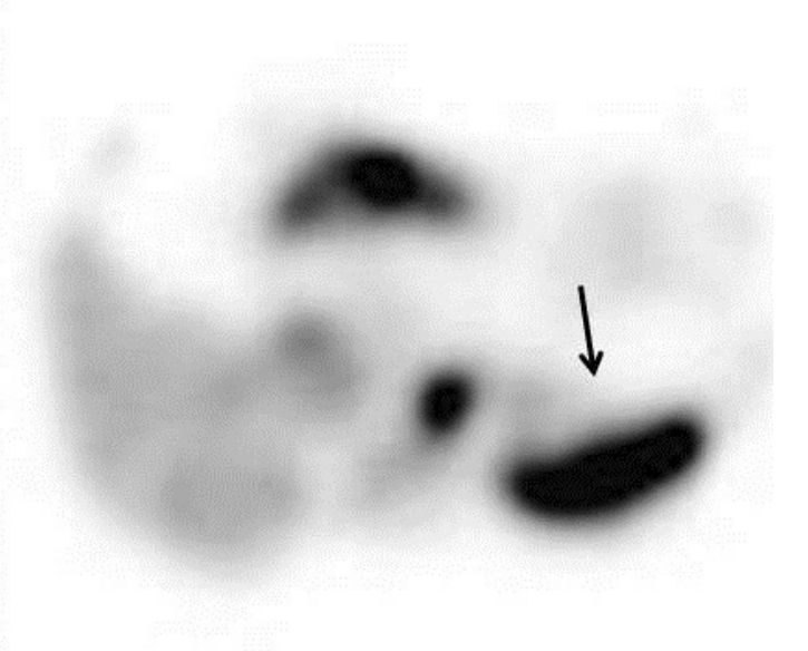
Tc-99m RBC G-I Bleeding scan: 복부 좌상부에 방사성 물질의 섭취가 보이고 함께 시행한 SPECT/CT에서 공장에 섭취 증가 소견이 보여, 공장의 활동성 출혈을 시사하였다 (Fig. 1).
Fig. 1
99m Tc pertechnetate radionuclide scan shows uptake in the left upper quadrant and SPECT/CT image were able to localize radioactivity in the jejunum.
시술방법 및 재료
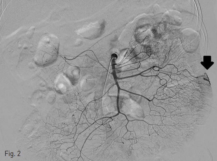
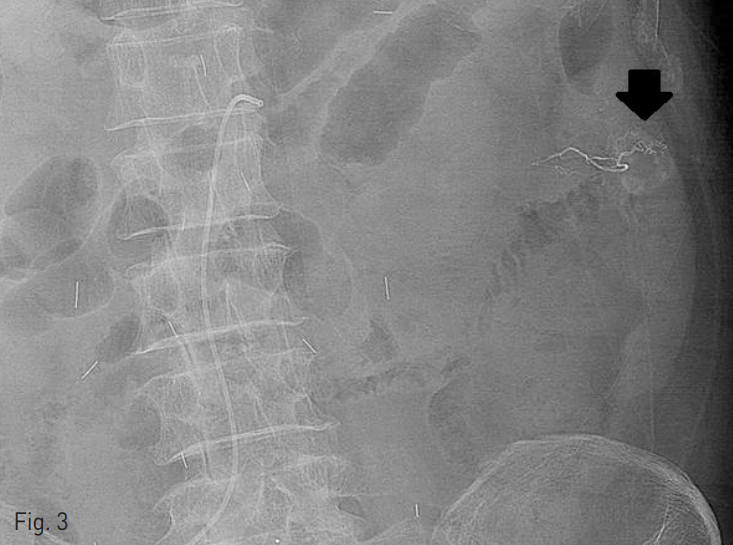
오른쪽 대퇴동맥을 통해 5 Fr 카테터 (RH, Cook, Bloomington, IN)를 삽입하여 상장간동맥 조영술을 시행하였다. 상장간동맥의 공장 가지에 약 1cm 크기의 구불구불한 모양의 늘어난 혈관이 있으면서 조기 배액되는 정맥 병변을 동반하여 혈관형성이상 병변으로 생각하였다 (Fig. 2). 이에 대해 2.0 Fr 미세카테터(Progreat, Terumo, Tokyo, Japan)를 이용하여 공장가지에 접근 하였고, 0.5cc 용량의 glue (Histoacryl, B.Braun, Tuttlingen, Germany) 1 vial을 Lipiodol (리피오돌울트라액, Guerbet, France)과 함께 1:3의 비율로 혼합하여 색전술을 시행하였다 (Fig. 3). 이후 시행한 동맥조영술에서 남아있는 병변이 없는 것을 확인하고 시술을 종료하였다.
Fig. 2
There is an angiodysplasia in the jejunal branch of superior mesentery artery.
Fig. 3
Embolization was done for jejunal angiodysplasia using glue and lipiodol (1:3).
고찰
혈관형성이상은 점막하 혈관이 구불구불한 모양으로 확장된 것으로, 내부의 탄력층이 부족한 얇고 구불구불한 정맥 병변을 동반하기 때문에 쉽게 출혈을 일으키며, 원인이 명확하지 않은 위장관계 출혈의 가장 흔한 원인으로 알려져 있다. 혈관형성이상이 생기는 기전은 노화와 관련하여 퇴행성 과정으로 설명하고 있으며, 구체적으로는 musculovascular unit의 venules, capillary, arteries에 만성적으로 폐쇄가 있어서 생기는 것으로 생각하고 있다. 주로 60~70대 환자에서 발생하며, 다발성 병변이 40~75% 정도로 보고되고 있다.
혈관형성이상 병변은 내시경 검사에서 2-10 mm 크기의 점상형 혹은 반점형의 홍반 병변으로 보인다. 영상의학적으로는, CT 검사에서 5 mm 미만 크기의 조영증강이 되는 점상형 혹은 원반형의 병변으로 보이며, 소장 벽, 특히 공장 벽에서는 점막내 혈관이 둥글게 부어있는 모양으로 보인다. 혈관조영술 검사에서는 혈관 들이 모여있으면서 조기 배액되는 정맥 동반한 형태로 보인다.
혈관형성이상의 치료는 우선적으로 혈관조영술을 시행하여 색전술 및 바소프레신(vasopression) 주입을 시행하게 되고, 그 외 내시경적으로 응고제 및 경화제 주입을 시행할 수 있으며, 경우에 따라 수술적 절제술도 고려해볼 수 있다.
참고문헌
1. Huprich JE, Barlow JM, Hansel SL, et al. Multiphase CT enterography evaluation of small-bowel vascular lesions. AJR Am J Roentgenol. 2013;201:65-72.
2. Huprich JE, Fletcher JG, Fidler JL, et al. Obscure GI bleeding: the role of multiphase CT enterography. Appl Radiol 2011;40:16-20.
3. Krishna K. Peripheral vascular interventions. LWW. 2007.
Citations
Citations to this article as recorded by