중심단어
Hepatocellular carcinoma, portal vein tumor thrombosis, transarterial radioembolization, selective intraarterial radiotherapy
임상소견
환자는 3개월 전부터 시작된 오심, 구토로 시행한 전산화 단층촬영 (CT) 결과 간세포암종으로 진단받았다. Child-Pugh class는 A였으며, ECOG score는 grade 1이었다.
진단명
Hepatocellular carcinoma
영상소견
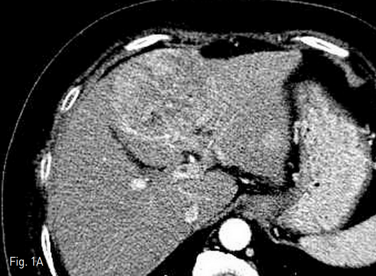
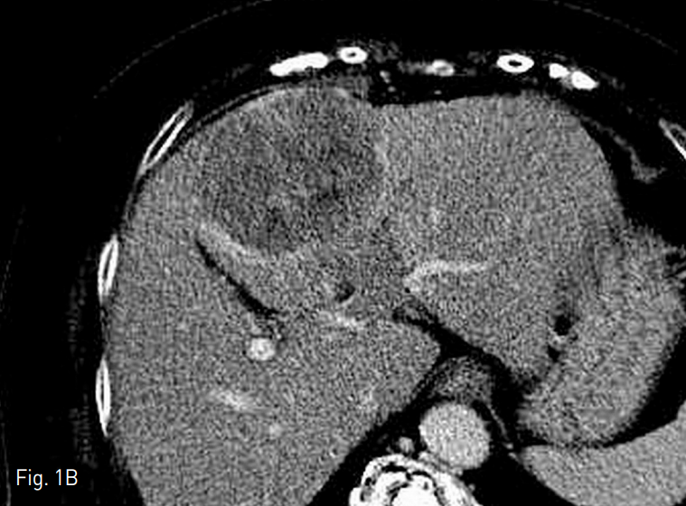
간의 4번 구역에 장경 7.5cm의 간세포암이 있었으며 (Fig. 1A), daughter nodule들이 구역 3, 4, 8번에서 각각 2cm 미만의 크기로 발견되었다. 간문맥의 umbilical portion에서 종양 혈전증이 동반되어 있었다 (Fig. 1B). 이밖에 원격 전이의 소견은 보이지 않았다.
Fig. 1. Computed tomography before transarterial radioembolization
A. There is a 7.5cm sized hepatocellular carcinoma in the liver segment 4 on arterial phase image. Three more daughter nodules are noted (not shown) in segment 3, 4, and 8.
B. There is tumor thrombosis in the um bili cal portion of portal vein on por tal phase image.
시술방법 및 재료
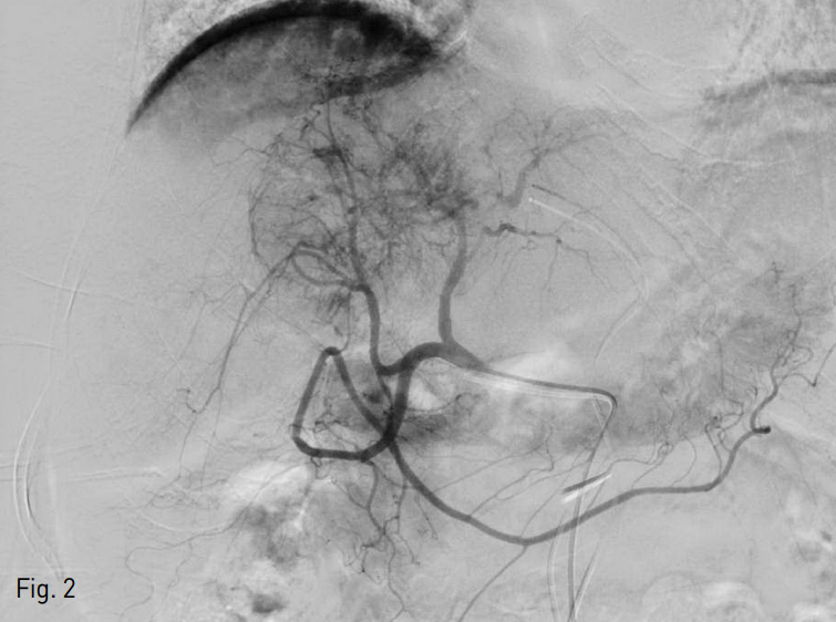
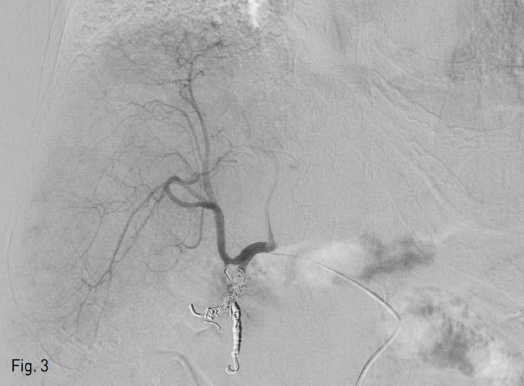
방사선색 전술 시행을 위한 사전 평가를 시행하였다. 우측 총대퇴동맥을 천자하여 4 Fr sheath 3개를 각각 삽입 후 4 Fr Yashiro catheter (Jung Sung Corp., Seoul, Korea) 3개를 사용하여 총간동맥과 좌측 전위 간동맥 (replaced left hepatic artery)이 기시하는 좌측 위동맥 (left gastric artery)에서 혈관 조영술을 시행하였다. 구역 4번의 간동맥이 공급하는 혈관이 풍부한 종양을 확인할 수 있었다 (Fig. 2). 우측 간동맥 근위부의 위이자십이지장동맥 (superior pancreaticoduodenal artery)과 위십이지장동맥 (gastroduodenal artery)을 확인하였고, 방사선색전술 시행시 위장관으로 미세구가 유입되는 것을 방지하기 위해 2.2 Fr microcatheter (Progreat, Terumo, Tokyo, Japan)로 이 동맥들을 미세선택하여 Tornado coil 4개 (3mm/2mm x 1, 4mm/2mm x 3, Cook, Bloomin gton, IN, USA), Nester coil 27H (14cm x 4mm x 2, Bloomington, IN, USA), Interlock detachable coil 27H (8mm x 20cm x 1, 6mm x 20cm x 1, Boston scientific, Natick, MA,USA)를 사용하여 색전술을 시행하였다 (Fig.3).
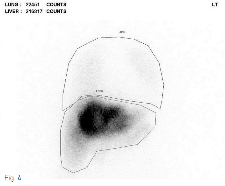
이후 2.8 Fr microcatheter (Progreat, Terumo, Tokyo, Japan)로 종양을 공급하는 동맥을 선택한 뒤 CT 간동맥 조영술을 시행하였고, 의미있는 동정맥 shunt는 확인되지 않았다. 이후 lung shunt fraction을 평가하기 위해 99mTc-MAA scan을 시행하였고, 12.6%로 확인되었다 (Fig. 4).
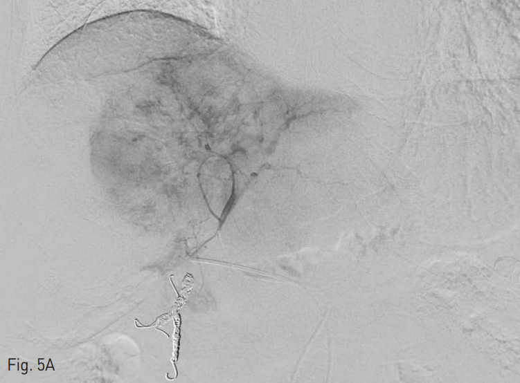
평가 후 1 주 후에 방사선색 전술을 시행하였다. 우측 총대퇴동맥에 4 Fr sheath 2개를 삽입한 뒤 4 Fr Yashiro catheter (Jung Sung Corp., Seoul, Korea) 2개를 이용하여 CT 간동맥 조영술을 시행 하였고 2.0 Fr microcatheter (Progreat, Terumo, Tokyo, Japan)를 이용하여 좌측 간동맥과 우측 간동맥을 각각 미세선택한 뒤 좌측 간동맥에 1GBq, 우측 간동맥에 0.3GBq에 해당하는 SIR-Spheres (Sirtex Medical Limited, Sydney, Australia)를 주입했다 (Fig. 5).
Fig. 2
Angiogram of right hepatic artery. In the segment 4, hypervascular tumor (about 7.5cm) is shown. The superior pancreaticoduodenal artery from right hepatic artery is identified.
Fig. 3
Microembolization of the superior pancreaticoduodenal artery and gastroduodenal artery was done with 8 microcoils.
Fig. 4
Lung scan image using 99mTc-MAA injection through microcatheter.
Fig. 5
90Y microspheres were infused into the tumor through the right hepatic artery (0.3G Bq) (A) and the left hepatic artery (1. 0G Bq) (B).
추적관찰
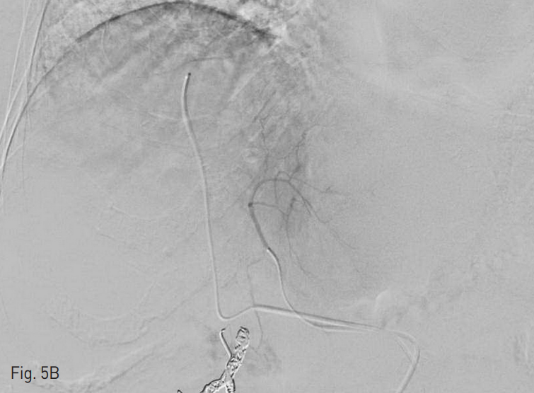
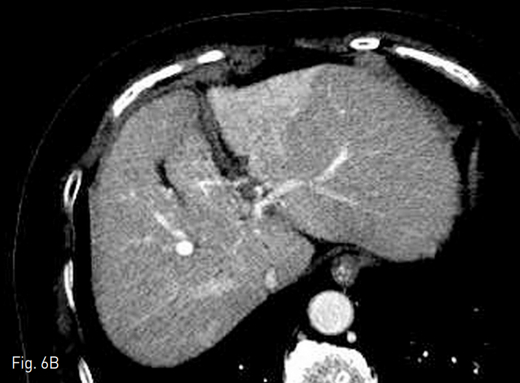
환자는 시술 후 8주 이상 호흡기나 위장관의 증상 발생하지 않았고, 간기능 검사의 큰 변화는 없었으며, 이후 추적 검사에서 지속적인 종양 크기 감소 및 간문맥 종양혈전증의 감소를 보였다 (Fig. 6)
가장 큰 종양은 시술 후 11개월째 장경 7.5cm에서 1.8cm으로 감소하였고, modified Response Evaluation Criteria in Solid Tumors (mRECIST)에 따른 partial response를 보였다. 종양 표지자는 시술 전 AFP 259.0ng/ml, PIVKA-Ⅱ 6520mAU/ml에서 시술 후 11개월째 각각 9.4ng/ml, 31mAU/ml로 감소하였다.
Fig. 6. Computed tomography obtained at 11 months after transarterial radioembolization.
A. Main tumor size is decreased from 7.5cm to 1.8cm.
B. The amount of tumor thrombosis in portal vein is decreased.
고찰
Yttrium-90 (90Y)를 이용한 방사선색전술은 간동맥을 통해 90Y을 간세포암종 내로 주입하여 지속적인 체내 방사선 조사를 통해 간세포암종을 치료한다. 90Y는 순수한 β선을 방출하며 평균 2.5mm의 조직투과성을 보인다. 일반적으로 방사선색 전술은 1회 시행하며, 90Y의 반감기는 64.2시간으로 이후 서서히 치료가 진행된다.
90Y는 미세구를 통하여 전달되며, 경동맥화학색전술에 쓰이는 입자 (100 micron)에 비해 크기가 작아(25-35 micron) 종양의 미세혈관까지 도달한다. 미세구는 현재 전세계적으로 레진으로 만든 SIR-Spheres (Sirtex Medical Limited, Australia)와 유리로 만든 TheraSphere (Biocompatibles, UK)가 쓰이고 있으며, 우리나라에서는 SIR-Spheres만 사용 가능하다. TheraSpehre는 평균 지름이 25μm, 활성도는 약 2,500Bq 이고, SIR-Spheres는 평균지름이 32μm, 활성도는 50Bq이다. SIR-Sphere가 크기는 비교적 크지만 활성도는 더 낮기 때문에, 원하는 방사선량을 얻기 위해서 더 많은 용량을 사용해야 하고, 이로 인해 종양 내 모든 혈관을 포화시켜 색전상태를 만들 수 있다.
미세구는 크기가 작아 종양 내 동정맥 shunt를 지나쳐 폐에 전달될 수 있어 방사선색 전술을 시행하기 앞서 동정맥 shunt의 정도를 평가해야 한다. 레진 미세구 사용시 shunt fraction이 20% 또는 예상되는 폐의 방사선 흡수선량이 25Gy보다 클 경우 시술을 시행하지 않는다. 또한 위장관은 방사선에 매우 민감하기 때문에 시술 전 혈관해부학을 평가하고 필요시 주위 혈관에 색전술을 시행하여 시술 중 위장관으로 미세구가 전달되지 않게 하는 것이 중요하다.
방사선색전술의 대상은 주로 intermediate 나 advanced stage (Barcelona clinic liver cancer stage B, C)의 tumor burden이 높거나 (각 종괴의 장경의 합이 7cm 이상) 종양의 혈관침범이 있어 경동맥 화학색전술 (TACE)이 불가능하거나, 이전에 경동맥화학색전술에서 반응이 없는 환자들이다. 특히 방사선색전술에 쓰이는 미세구는 허혈 효과가 적기 때문에 간문맥혈전이 있는 환자에게도 경동맥화학색전술보다 비교적 안전하게 시행될 수 있다.
이번 증례는 간문맥 종양혈전증을 동반한 advanced stage의, Child-Pugh class A의 환자를 대상으로 하였으며, 적절한 시술 전 평가 및 혈관색전술 후 방사선 색전술을 시행하여 시술 후 폐나 위장관의 합병증, 잔존 간기능의 저하 없이 11개월까지 partial response를 보인, 경동맥화학색전술이 불가능한 간세포암종 환자에게 방사선색 전술을 시행하여 좋은 경과를 보인 증례이다.
참고문헌
1. Sangro B. Chemoembolization and radioembolization. Best practice & research Clinical gastroenterology 2014;28:909-19.
2. 대한인터벤션영상의학회, 인터벤션 영상의학, 제2판, 서울, 일조각, 2014:537-545.
3. Bolondi L, Burroughs A, Dufour JF, et al. Heterogeneity of patients with intermediate (BCLC B) Hepatocellular Carcinoma: proposal for a subclassification to facilitate treatment decisions. Seminars in liver disease 2012;32:348-59.
4. Lewandowski RJ, Salem R. Yttrium-90 Radioembolization of Hepatocellular Carcinoma and Metastatic Disease to the Liver. Seminars in Interventional Radiology 2006;23:64-72.
5. Lau WY, Lai EC, Leung TW. Current role of selective internal irradiation with yttrium-90 microspheres in the management of hepatocellular carcinoma: a systematic review. International journal of radiation oncology, biology, physics 2011;81:460-7.
Citations
Citations to this article as recorded by