중심단어
Central vein stent, stent migration, stent removal
임상소견
만성 신부전증으로 1년 전 좌측 상완동맥-요측피정맥 동정맥루 (brachio-cephalic arteriovenous fistula)를 시행 받고 투석 받던 환자임. 투석은 잘 되나 수개월 전부터 좌측 팔이 붓는 증상이 있어 좌측 무명정맥 협착 의심 하에 경피경관혈관 성형술(percutaneous transluminal angioplasty)이 의뢰됨.
진단명
Central vein stenosis
영상소견
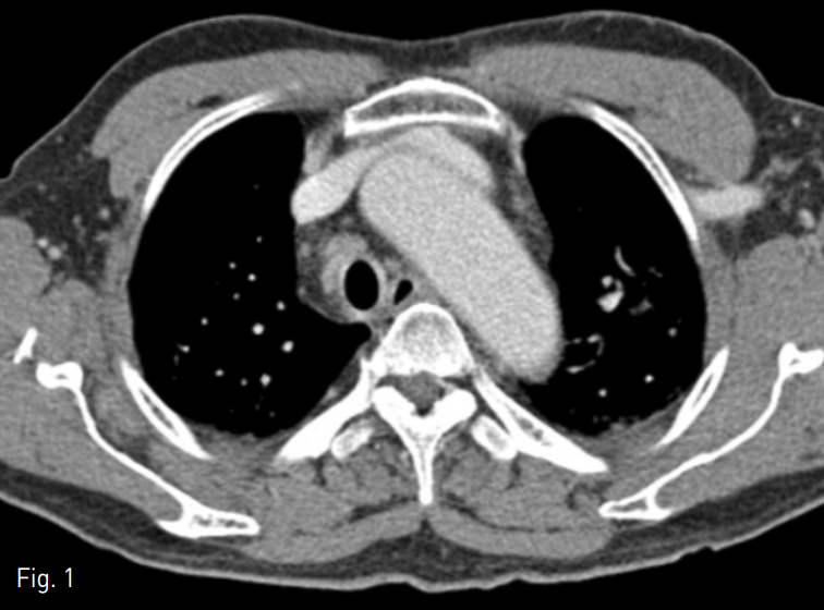
CT상 복장뼈와 대동맥 사이에서 무명정맥이 눌리고 있음 (Fig. 1).
Fig. 1
A 72-year-old man with left arm swelling after brachiocephalic AVF. An axial CT scan shows segmental narrowing in the left innominate vein, probably due to decreased space between the aortic arch and sternum
시술방법 및 재료
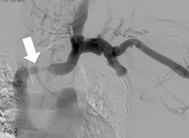
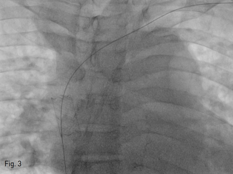
초음파 유도하에 좌측 상완부의 요측피정맥을 천자한 후 6 Fr sheath (Terumo, Tokyo, Japan)를 삽입함. 5 Fr KMP catheter (Cook, Bloomington, USA)를 좌측 무명정맥에 위치시킨 후 시행한 정맥조영술에서 무명정맥의 협착을 확인할 수 있음 (Fig. 2). 이에 12mm-4cm Mustang balloon catheter (Boston Scientific, Massachussets, USA)를 이용하여 무명정맥의 협착에 대해 풍선 확장술을 시행하였음. 그 후 14mm-3cm Niti-S vascular stent (TaeWoon medical, Gimpo, Korea)를 협착부위에 설치하였으나 스텐트가 상대정맥 (superior vena cava)으로 이동함 (Fig. 3).
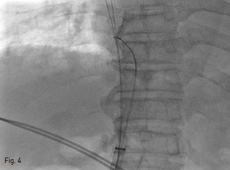
이에 우측 대퇴정맥을 천자한 후 18 Fr sheath (St.Jude medical, Minnetonka, USA)를 삽입함. 5 Fr KMP catheter (Cook, Bloomington, USA)와 0.035" hydrophilic guide wire (Terumo, Tokyo, Japan)를 이용하여 스텐트 내강을 통과시켜 wire를 거치시키고, 좌측 상완부에 삽입된 sheath를 통한 0.035" hydrophilic guide wire를 스텐트 내강을 거쳐 우측 대퇴정맥에 삽입된 sheath로 끄집어 냄.
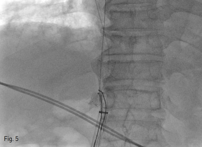
이후 Amplatz gooseneck snare (ev3, Minneapolis, USA)를 이용하여 스텐트의 중간부위를 포획하였으나 스텐트의 말단부가 sheath를 통과하기에는 충분하게 접히지 않음 (Fig. 4). 이에 또 다른 Goose neck snare를 이용하여 스텐트의 말단부를 포획하였고 스텐트를 femoral sheath 내부로 끄집어내어 몸 밖으로 성공적으로 제거 할 수 있었음 (Fig. 5).
Fig. 2
Fistulography revealed segmental narrowing in the left innominate vein (arrow).
Fig. 3
The stent migrated in to the superior vena cava because of insufficientstent fixation.
Fig. 4
The stent was captured with a nitinol loop.
Fig. 5
The stent was captured by a second nitinol snare delivered from the femoral vein and removed percutaneously through a femoral sheath
고찰
동정맥루를 통해 투석을 시행하는 환자에서 중심정맥 협착이 발생하는 빈도는 빈번하며 이 경우, 풍선 확장술과 더불어 스텐트 삽입이 도움이 될 수 있다. 이 외에도 혈관 스텐트는 다양한 혈관의 협착이 있는 경우, 경피적 혈관 성형술의 필요 불가결한 치료법의 하나로 사용된다. 그러나 간혹 스텐트가 maldeployment 혹은 migration 될 수가 있으며 때때로 수술적으로 이를 제거하거 나 우회술을 받아야 하는 경우도 있다.
Goose neck snare를 이용한 혈관 내 이물질의 제거는 비교적 안전하고, 효과적이며, 널리 사용되고 있는 방법이며 maldeployment 혹은 migration된 스텐트의 제거나 위치 조정에 있어서도 Goose neck snare는 유용하게 사용될 수 있다.
그러나 Nitinol 스텐트 같은 경우 자가 확장성이 높고 mesh의 크기가 크기 때문에 snare로 포획 시에 스텐트가 모래 시계의 형태로 변하여 스텐트를 구경이 작은 catheter나 sheath로 끄집어 내기 어려울 수가 있다. 이러한 경우 본 증례와 같이 구경이 큰 sheath를 이용하거나 또 다른 snare로 스텐트의 말단부를 포획하면 비교적 용이하게 스텐트를 몸 밖으로 제거 할 수 있겠다.
참고문헌
1. Bagul NB, Moth P, Menon NJ, Myint F, Hamilton G. Migration of superior vena cava stent. J Cardiothorac Surg. 2008 Mar 10;3:12.
2. Poludasu SS, Vladutiu P, Lazar J. Migration of anendovascular stent from superior vena cava to the right ventricular outflow tract in a patient with superior vena cava syndrome. Angiology. 2008 Feb-Mar;59(1):114-6.
3. Gabelmann A, Krämer SC, Tomczak R, Görich J. Percutaneous Techniques for Managing Maldeployed or Migrated Stents. J Endovasc Ther. 2001 Jun;8(3):291-302.
Citations
Citations to this article as recorded by