중심단어
gastric varix, embolizalion, portopulmonary venous anastomosis
임상소견
환자는 12년 전 bariatric surgery 이후 발생한 문맥 혈전에 의해 위식도 정맥류(gastroesophageal varix)로 수 차례 내시경적 식도정맥류지혈술(EVL)을 받았고, 간이식에 대하여 면담 후 intractable esophageal varix에 대한 색전술을 시행하기로 하였다. 이전 수 차례 시행한 EVL로 인하여 추가적인 내시경적 지혈이 어려웠고, 만성적인 문맥 혈전 및 cavernous transformation, gastrorenal shunt의 부재 등으로 인하여 TIPS 및 BRTO를 시행하기 어려운 상황이었다. 따라서 경피적으로 접근하여 비장정맥을 통하여 NBCA 와 lipiodol을 이용한 정맥류의 색전 및 비장동맥의 부분 색전술을 시행하기로 계획하였다.
진단명
Systemic embolism after transsplenic esophageal varix embolization
영상소견
복부 전산화 단층촬영에서 간문맥 혈전으로 인한 주문맥의 위축, cavernous transformation이 있으며 paraesophageal, esophageal, splenic varix가 보였다.
시술방법 및 재료
초음파와 투시 유도 하에 국소 마취 후 비장 정맥을 21G Chiba needle(TSK laboratory, Soja, japan)로 puncture한 후 6-Fr sheath(Radiofocus, Terumo,
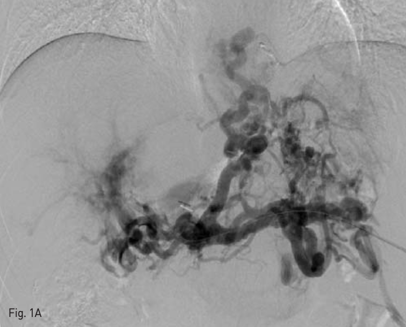
Tokyo. Japan)를 삽입하였다. 5-Fr Davis catheter(Cook medical, Bloomington, IN, USA)를 이용하여 정맥조영술을 시행하였으며, multiple feeder로부터 flow를 받는 esophageal varix가 조영 되었다(Fig. 1A).
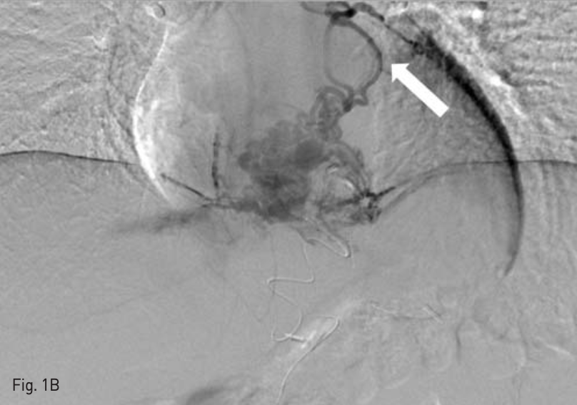
이후 feeder를 microcatheter(Progreat, Terumo, Tokyo, Japan)및 microwire(Transcend. Cook medical, Bloomington, IN, USA)를 이용하여 selection하여 1:5 and 1:8 NBCA - lipiodol mixture를 이용하여 embolizalion 시행하였는데, 소량의 NBCA - lipiodol mixture가 pulmonary artery로 migration한 것이 보였다(Fig. 1B).
Embolizalion을 시행한 후 추적 정맥조영술에서 esophageal varix를 통한 flow는 대부분 소실된 것을 확인하였다. 이후 우측 대퇴동맥을 초음파 유도 하에 puncture하여 4-Fr sheath Radiofocus, Terumo, Tokyo, Japan)를 삽입하고 RH catheter(Cook medical, Bloomington, IN, USA)를 이용하여 splenic artery branch를 selection하여 partial embolization을 시행하고자 하였으나 환자가 갑자기 심한 두통을 호소하여 시술을 종료하였다.
추적관찰
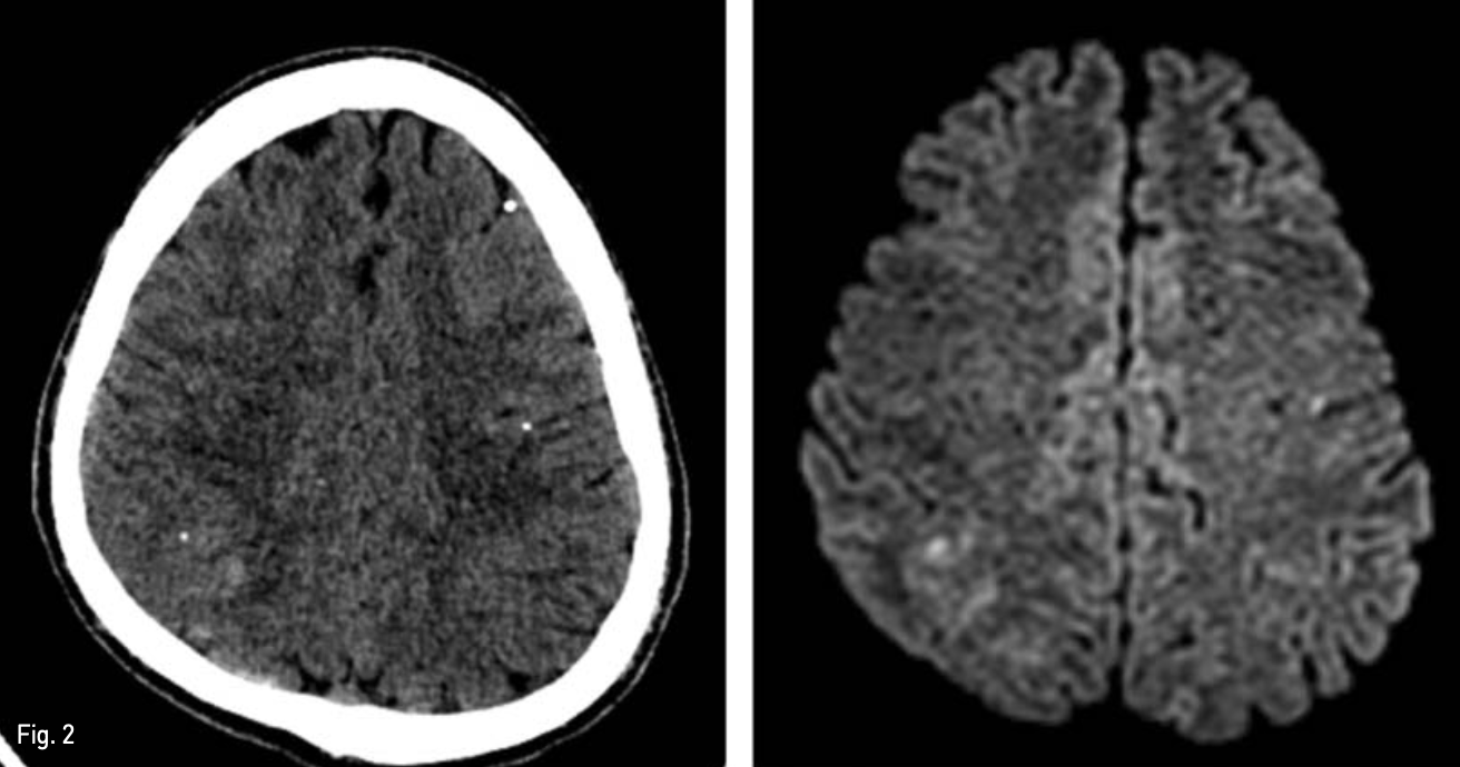
색전술 시행 후 환자는 심한 두통 및 양측성 시야 결손을 호소하였고, 색전술 시행 1일 후 추가적으로 촬영한 brain CT 및 MRI 상 다발성 급성 뇌경색 소견을 보였다(Fig. 2). 이후 보존적인 치료를 받으면서 환자의 모든 증상이 호전되었고 퇴원하였다.
Fig. 1
A 41-year-old man with intractable esophageal varix following multiple session of endoscopic variceal ligation.
A. Percutaneous transsplenic portography shows diffuse development of tortous esophageal varix with multiple collateral channels.
B. Venography performed from the microcatheter which was located at the proximal side of esophageal varix, shows PPVA (arrow), which was a dilated vessel running cranially and is connected to the left inferior pulmonary vein.
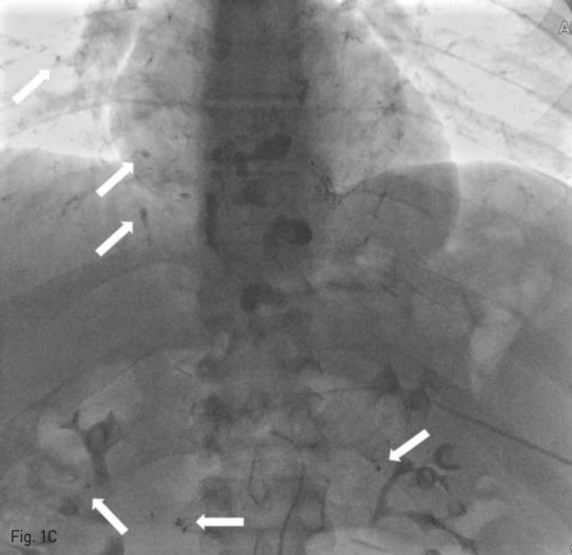
C. After injection of NBCA - lipiodol mixture, a radiographic image shows systemic embolism of NBCA - lipiodol to bilateral pulmonary artery and omental artery territory(arrows).
Fig. 2
An axial CT scan obtained 1 day following embolization, shows multifocal scattered lipiodol density in brain (left), and Diffusion weighted brain MR shows multiple diffusion restriction spot suggesting acute in farction (right).
고찰
Portopulmonary venous anastomosis(PPVA)는 문맥 고혈압 환자에서 발생할 수 있는 collaleral pathway 중 하나로 paraesophageal vein 과 left or right pulmonary vein을 연결하는 통로가 된다. 보고 문헌에 따라 다르나 PPVA의 발생 빈도는 약 20~ 30%로 드물지 않고, PPVA가 있으면 BRTO를 비롯한 esophageal-gastric varix embolizalion 시 치명적인 합병증을 일으킬 수 있는데, 그 이유는 다른 portasystemic collateral pathway와 달리 직접적인 right to left shunt를 일으켜 전신적인 색전을 유발할 수 있기 때문이다.
본 증례는 간문맥의 만성적인 폐색에 의하여 PPVA를 비롯한 다발성의 collateral pathway가 발달되어 있으면서 gastrorenal shunt가 없는 위식도 정맥류를 경피적 경비장 접근을 통하여 NBCA로 색전술을 시행 하던 중 NBCA가 PPVA를 통하여 전신순환으로 유출되어 전신 색전의 합병증이 발생하였던 경우이다.
이와 같은 합병증의 발생을 막기 위해서는 시술 전 CT를 시행하여 PPVA의 유무를 파악하고, 가능하다면 sclerosing agent를 주입하기 선 PPVA를 미리 색전하여야 한다. 특히 색전 물질로 주로 쓰이는 ethanolamine oleate나 50% glucose solution 또는 ethanol은 소량이 유출되더라도 좌심실 내의 다량의 혈액과 섞여 회석되면서 색전능을 잃는 데 반해, particle type agent나 N-butyl-cyanoacrylate(NBCA)는 소량이 유출되더라도 paradoxical embolism을 유발할 수 있어 주의가 요망된다.
참고문헌
1. Ko JM, Ahn MI, Han DH, Jung JI, Park SH. Dynamic CT and MRA findings of a case of portopulmonary venous anastomosis (PPVA) in a patient with portal hypertension: a case report and review of the literature. Acta radiologica (Stockholm, Sweden : 1987). 2011;52(5):566-9.
2. Miura H, Yamagami T, Tanaka O, Yoshimatsu R. Portopulmonary venous anastomosis detected at balloon-occluded retrograde transvenous obliteration for gastric varices. Journal of vascular and interventional radiology : JVIR. 2013;24(1):131-4.
3. Yamagami T, Yoshimatsu R, Miura H, Yamada K, Minami M. Successful balloon-occluded retrograde transvenous obliteration of gastric varix via pericardiacophrenic vein after embolization of portopulmonary venous anastomosis. Journal of vascular and interventional radiology : JVIR. 2013;24(1):137-9.
4. Kariya S, Komemushi A, Nakatani M, et al. Portopulmonary venous anastomosis in balloon- occluded retrograde transvenous obliteration for the treatment of gastric varices. Journal of gastroenterology and hepatology. 2014;29(7):1522-7.
Citations
Citations to this article as recorded by