중심단어
Gastric varices, BRTO, Gastrocaval shunt
임상소견
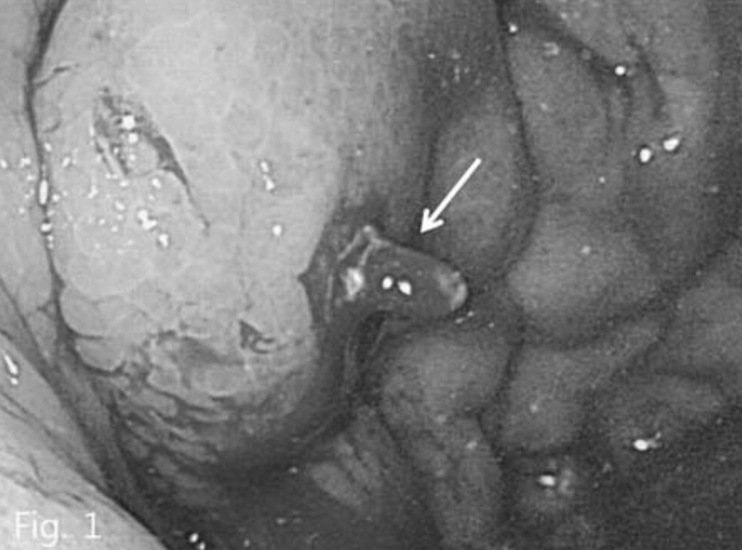
B형 간염에 의한 간경화 환자로 내원 하루 전 2차례의 토혈이 있어 내원하였다. 위내시경에서 식도 정맥류와 위정맥류가 동시에 관찰되었으며 내시경적 결찰술을 시행하였으나 그 후에도 위정맥류 부위에서는 지속적인 출혈이 있었다(Fig. 1).
Fig. 1
Endoscopy shows large gastric varices with active bleeding (arrow).
진단명
Gatric varices with gastrocaval shunt
영상소견
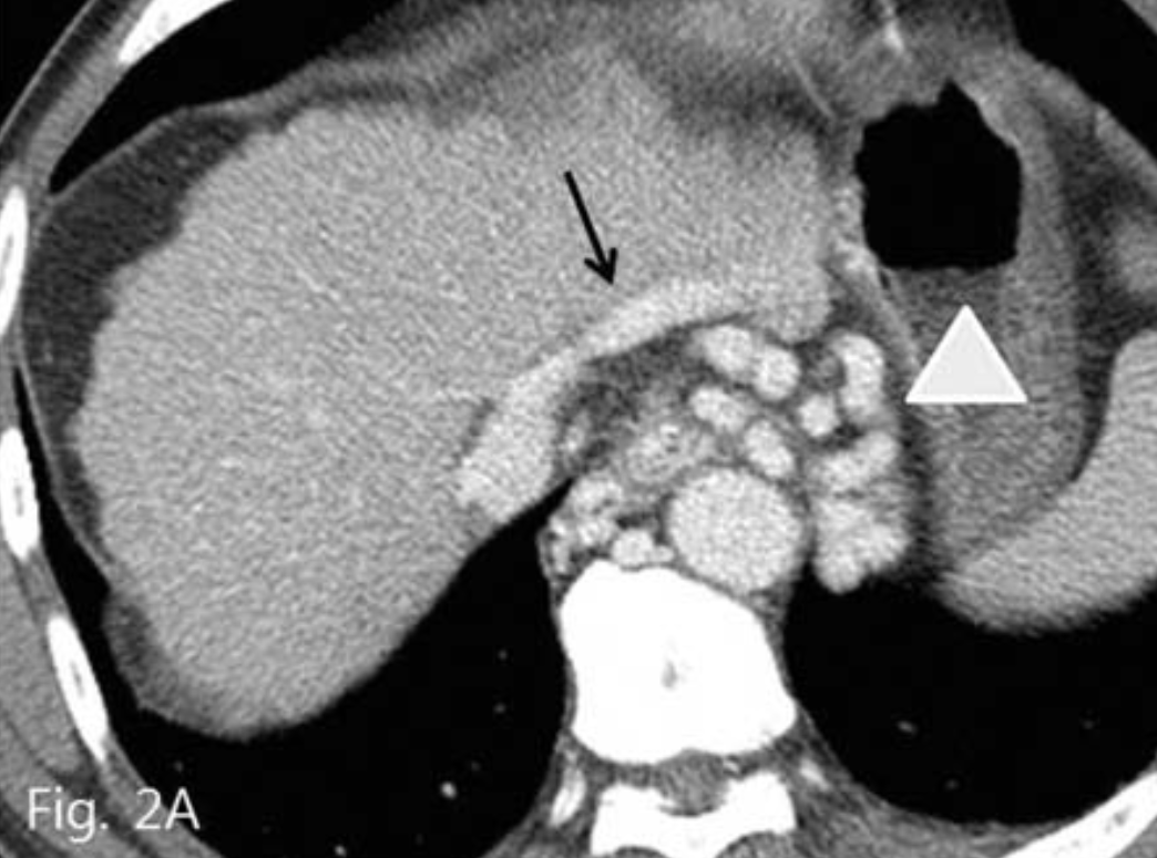
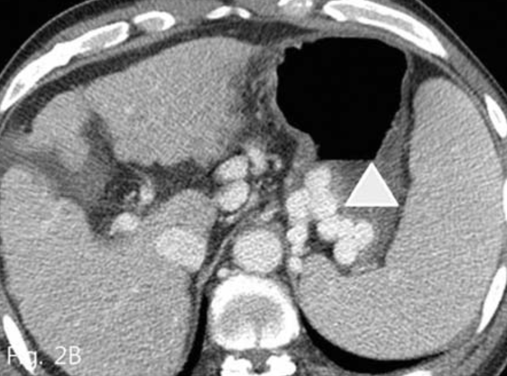
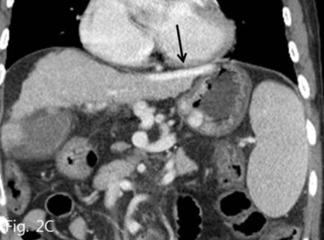
복부 전산화 단층촬영(CT)에서 간경화 및 비장비대의 소견과 함께 위식도 정맥류가 관찰되었다. 위정맥류의 경우 위신 단락 (gastrorenal shunt)은 보이지 않았으며, 대신 현저하게 늘어나 있는 위하대정맥 단락 (gastrocaval shunt)이 확인되었고 이것이 위정맥류의 주 유출정맥으로 생각되었다(Fig. 2A-C).
Fig. 2
Contrast enhanced CT (A~C) shows esophageal and gastric varices (arrowhead) and gastrocaval shunt (arrow).
시술방법 및 재료
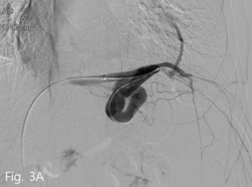
우측 총대퇴정맥을 통해 7-Fr sheath(Flexor Check-Flo; Cook, Bloominglon, IN, USA)를 삽입 후 5-Fr Cobra catheter(Cook)와 0.035-inch guidewire(Terumo, Tokyo, Japan)로 위대정맥단락를 동해 위정맥류 내부로 catheter를 진입시켰다. 시행한 정맥조영술에서 위정맥류 일부와 왼쪽 횡경막하 정맥이 보였으며 조영제는 위대정맥단락과 심장막정맥 (pericardial vein)을 통해 유출되고 있었다(Fig. 3A).
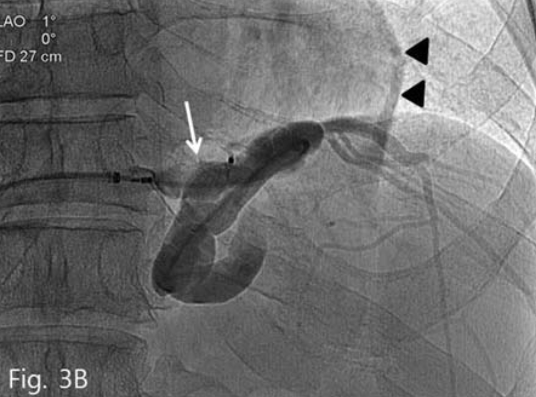
8-Fr sheath를 5-Fr catheter와 wire의 support 하에 gastrocaval shunt내로 진입시킨 후 wire를 남기고 catheter를 제거하였다. 그 후 sheath를 통해
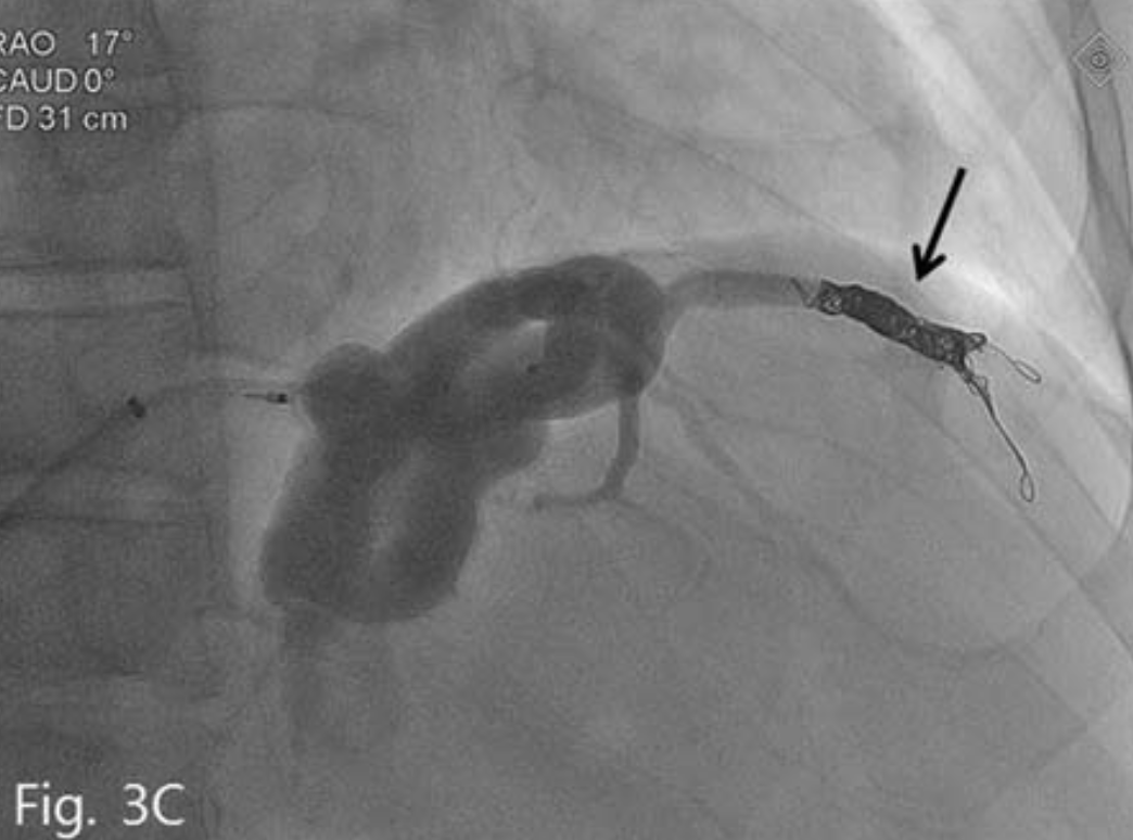
16mm vascular plug II(Amplatzer Vascular plug; AGA Medical, Golden Valley, Minn)를 삽입하였다. 그 후 wire를 따라 5-Fr catheter를 vascular plug를 지나 위치시키고 정맥조영술을 시행하였으며 주 유출정맥인 위대정맥단락은 막혔으나 대신 심장막정맥을 통한 곁가지 정맥배출이 좀 더 명확해졌다(Fig. 3B), 그래서 4개의 microcoil(Vortex; Boston Scientific, Cork, Ireland)로 이를 색전하였다(Fig. 3C). 그 후 5-Fr catheter를 통해 gelatin sponges(Gelfoam:
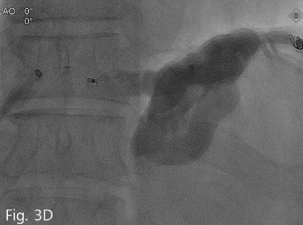
Upjohn, Kalamazoo, Mich)로 위정맥류내를 충분히 채우고 일부는 유입정맥까지 역류시킨 후 plug를 분리하고 시술을 마쳤다(Fig. 3D).
Fig. 3
A. Venogram shows gastrocaval shunt, gastric varices, left inferior phrenic vein and pericardial vein.
B. Vascular plug placement (arrow) is performed in gastrocaval shunt Left inferior phrenic vein and pericardial vein (arrowhead) is more prominently visualized.
C. Left inferior phrenic vein is embolized with microcoil (arrow). And then gastric varices is completely filled with gelfoam mixture.
D. Vascular plug is success fully detached.
추적관찰
시술 후 환자는 더 이상의 토혈이나 흑색변을 보이지 않았으며 일주일 후 퇴원하였다.
고찰
Balloon-occluded retrograde transvenous obliteration(BRTO)는 위정맥류 출혈 환자에게 있어서 위신단락이나 위하대정맥 단락이 있는 경우 사용할 수 있다. 하지만 기존에 사용되었던 5% ethanolamineolate(EO)의 경우 부작용으로 신독성, 폐부종, 심장쇼크, 파종성 혈관 내 응고 등이 보고되고 있으며 특히 풍선 카테터가 파열되었을 경우 색전증, 치료실패, 정맥류 재출혈 등이 발생할 수 있다.
또한 시술자마다 시간은 다양하나 30분에서 24시간까지 경화요법을 시행하여야 하며 EO의 용량도 40cc 이하로 사용하도록 권장되고 있다.
Plug-assisled retrograde transvenous obliteralion(PARTO)는 2013년 처음으로 소개된 방법으로 파열되기 쉬운 풍선카테터 대신 vascular plug를 사용하며 색전 물질로는 EO대신 gelfoam을 사용한다. 이를 통해 시술시간을 현저히 감소시킬 수 있으며 좀 더 쉽고 안전하게 시술을 시행할 수 있다.
위하대정맥단락은 위신단락이 없는 환자에게서 가장 흔한 유출 정맥으로서 이 환자에서도 위신단락이 없이 위하대정맥단락과 또다른 유출정맥인 심장막정맥을 통한 배출경로도 함께 확인할 수 있었다. 이에 저자들은 위신단락을 통한 PARTO 방법을 이용하여 위하대정맥단락을 통해 시술을 시행하였으며 또 다른 유출정맥인 심장막정맥은 coil로 색전하였고 이를 통해 성공적으로 위정맥류를 치료할 수 있었다.
따라서 본 증례는 위신단락이 없이 위하대정맥 단락 이 있는 환자에게서도 PARTO가 효과적이며 쉽게 시행할 수 있다는 것을 보여준다고 하겠다.
참고문헌
1. 대한인터벤션영상의학회. 인터벤션영상의학, 1st Ed., 일조각 2012;365-375.
2. Gwon DI, Ko GY, Yoon HK, et al. Gastric varices and hepatic encephalopathy; treatment with vascular plug and gelatin sponge-assisted retrograde transvenous obliteration-a primary report. Radiology 2013;268:281-287.
3. Araki T, Hori M, Motosugi U, et al. Can balloon- occluded retrograde transvenous obliteration be performed for gastric varices without gastrorenal shunts?. J Vasc Interv Radiol 2010;21:663-670.
Citations
Citations to this article as recorded by