중심단어
Liver, Abscess, Drainage, Stents
임상소견
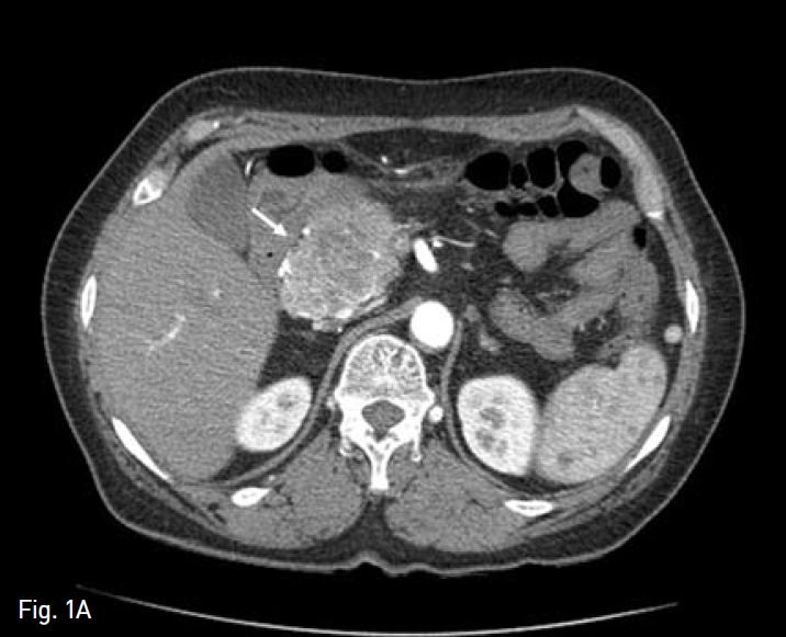
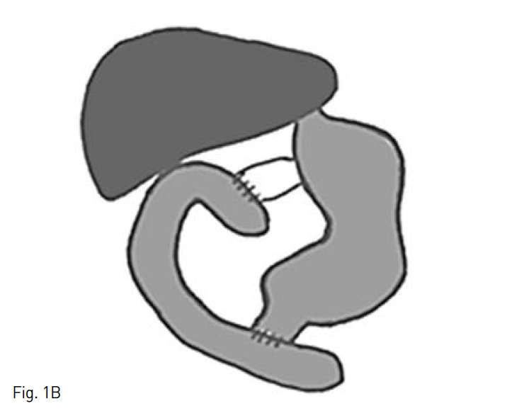
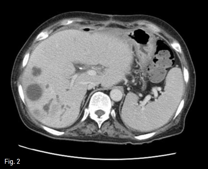
췌장 두부에 낭선종(Cystadenoma)으로 유문 보존 췌십이지장 절제술(PPPD)를 시행받은 환자로(Fig. 1A,B), 수술 2개월 후 지속적인 발열이 있어 내원하여 시행한 복부 전산화 단층 촬영상 간 우엽의 다발성 간농양과 함께 우엽 담관의 확장 소견이 보였고(Fig. 2), 경피경간 담도 배액술(PTBD)(Fig. 3A) 및 농양 배액술(Fig. 3B)을 시행받고, abscess content를 배양하여
시행한 병리학직 검사에서 E. coli가 연속해서 2차례 검출 되었다. 광범위항생제를 포함한 내과적 치료를 약 4개월 간 받았으며, 이후 증상의 호전을 보였고, 추적 관찰하여 시행한 복부 전산화 단층 촬영 상 대부분의 농양의 크기가 줄어늘고 특히 상대적으로 농양의 분포가 많았던 간우엽의 크기 감소와 꼬리엽의 보상적 비대가 보였다(Fig. 4). 더 이상 abscess content에 미생물 배양은 되지 않았으며, 퇴원하였다.
그러나, 20일 후 다시 우상복부 복통을 호소하며 응급실 내원하였다. 재 방문시 WBC는 퇴원시 9,500/mm3에서 12,100/mm3으로 증가하였으며, CRP 역시 퇴원 시 5.96mg/dL에서 9.38mg/dL로 상승하였다.
Fig. 1
Contrast-enhanced axial abdominal CT image A shows a bulky, low attenuated mass lesion with hypervascularity in pancreatic head portion (white arrow). So, our surgeon performed pylorus-preserving pancreaticoduod enectomy with Traveso method (schema in B).
Fig. 2
Follow-up axial abdominal CT image, obtained 2 months after operation, shows multiple small low attenuated lesions with in trahepatic bile duct dilatation.
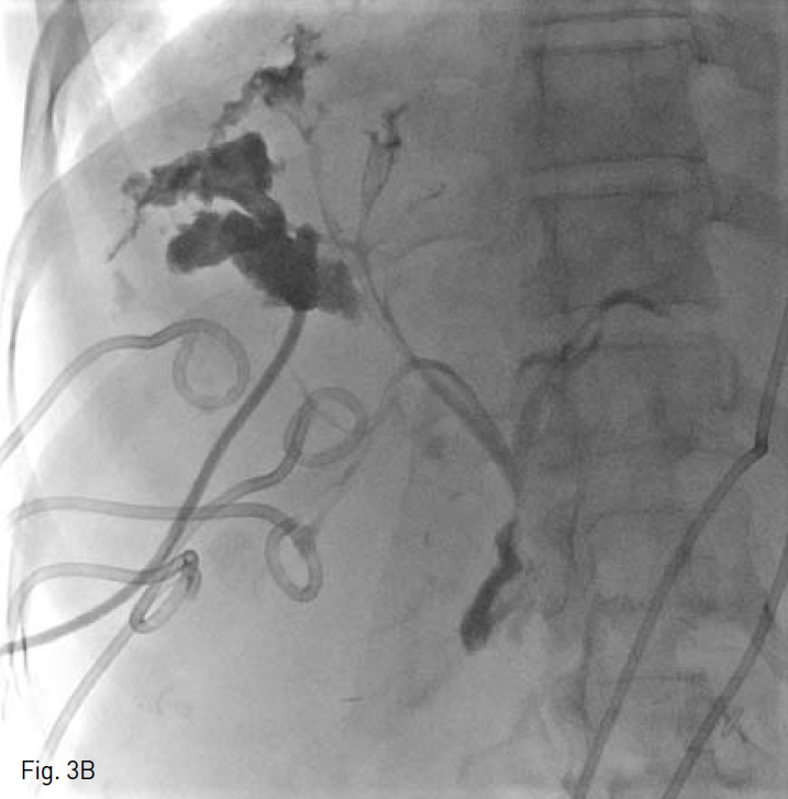
Fig. 3
Cholangiogram during right percutaneous biliary drainage A and cavitogram during percutaneous abscess drainage B show communications between abscess cavities and bile duct.
Fig. 4
Follow-up axial abdominal CT image, obtained 4 months after medical and interventional treatment, shows size reduction of multiple abscess and decompression of bile duct. Afterthen, the patient discharged.
진단명
유문 보존 췌십이지장 전제술을 받은 환자에서 발생한 반복적인 역류성간농양
영상소견
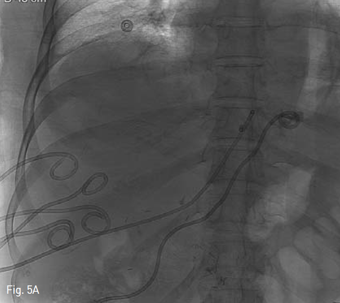
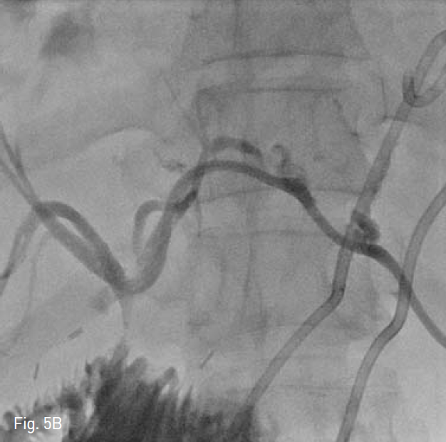
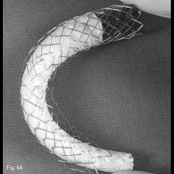
응급실 재방문 후 시행한 복부 전산화 단층촬영상 양측 간엽에 1~3cm크기의 다발성 간농양이 15일 전 퇴원 시 보다 다소 크기의 증가가 보이고, 농양 배액을 위해 다수의 농양 배액술(Fig. 5A) 및 담즙 우회를 위해 좌측 경피경간 담도 배액술(Fig. 5B)이 시행되었다. 이후 내과 및 외과 의료진과 상의 후 역행성 간농양에 대한 Anti-reflux designed, GD type stenl(Fig. 6A)를 계획하였다.
시술방법 및 재료
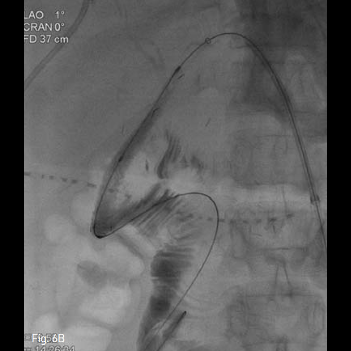
이전 시행된 좌측 경피경간 담도 배액관을 동해 담관 조영술을 시행하여 총담관 - 십이지장 분합부를 확인하고, 0.035-inch guide wire(Terumo, Tokyo, Japan)와 5-Fr KMP catheter(Cook, Bloomington, IN, USA)를 이용하여, wire를 원위부 십이지장에 거치 후, 분합부를 가로질러 직경 10mm, 길이 8cm의 Anti-reflux designed, GD type biliary stent(TaeWoong Medical, Gimpo, Korea)를 설치하였다(Fig. 6B). 시술 후 담관조영술을 통해 스텐트의 개방성과 적절한 위치를 확인 한 후, 추적 관찰을 위해 다시 8.5-Fr pigtail형drainage catheter(Cook, Bloomington, IN, USA)을 거치하였다.
Fig. 5
After 20 days, the patient revisited our hospital with abdominal pain. We performed multiple percutaneous abscess drainage to both hepatic lobes A and left percutaneous biliary drainage B.
Fig. 6
Anti-reflux stent consist of inner covered stent and outer uncovered stent A. Finally, we performed biliary stenting to efferent bowel loop across choledocho-duodenostomy site B.
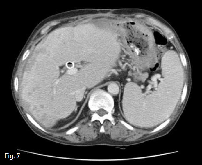
Fig. 7
Follow-up axial abdominal CT image, obtained 5 months after stenting, shows size reduction of right hepatic lobe with minimal abscess pockets and com pensatory hypertrophy of caudate lobe.
추적 관찰
이후 환자는 Lab 결과는 지속적으로 호전되어 일주일 후 빌리루빈 수치는 1.9mg/dL, WBC 수치는 8,500/mm3, CRP는 2.73mg/dL이었다. 환자는 장기 간의 질병 경과로 입원 치로를 약 2달 받았으나, 결국 호전 퇴원하여, 현재 외래를 통하여 추적 관찰 중이다.
고찰
췌장-십이지장 전제술 이후 담도염이나 간농양의 빈도는 약 5%로 보고되고 있다(1). 이는 간동맥의 협착이나 폐색, 총담관 - 장루술의 협착으로 합병증으로 나타나며, 드물게는 짧은 Roux-Y limb, 공장-공장 분합부의 협착, 또는 공장-공장 문합부 이하 폐색 등 수술의 합병에 의한 담즙 역류에서 기인한다. 미생물학적 검사에 기초한 항생제 선택이 예후를 좋게 할 수 있으나, 분합부 협착은 경피적 확장술, 스텐트 또는 재수술로 해견할 수 있다.
담즙 역류는 유문부 괄약근의 기능 상실에서 기인하고, 이를 방지하기 위해 다양한 역류 방지 방식의 스텐트 디자인이 소개되이 왔다. Dua등은 플라스틱 스텐트의 십이지장 부위에 4cm의 풍낭 모양의 밸브를 선치한 역류 방지용 플라스틱 스텐트를 개발하였다(2). 이를 응용하여 Lee 등은 32명의 악성 담도 폐색 환자에서 통상의 비피복형 스텐트의 원위부에 2cm 크기의 역류 방지 밸브와 고정 철사를 연결하여, 유문부를 경유하는 스텐트 설치술을 시행하여 14.4 개월의 스텐트 개통률을 보고하였다(3).
저자들은 담도 폐색 환자에게 많은 연구를 하고 있는 Gwon (4) 등이 최근 개발한, 내부에 깔대기형 피복 스텐트와 이동 방지형 외부 비피복 스텐트른 복합한 역류 방지형 GD type 스텐트를 이용하여, 반복적인 역류성 담도염 및 간농양으로 장시간 치료받는 환자에게 적용하여 좋은 결과를 보였다.
참고문
1. Yeo CJ, Cameron JL, Sohn TA, et al. Six hundred fifty consecutive pancreaticoduodenectomies in the 1990s: pathology, complications, and outcomes.Ann Surg 1997;226:248-257.
2. Dua KS, Reddy ND, Rao VG, Banerjee R, Medda B, Lang I. Impact of reducing duodenobiliary reflux on biliary stent patency: An in vitro evaluation and a prospective randomized clinical trial that used a biliary stent with an antireflux valve. GastrointestEndosc 2007;65:819-828.
3. Lee KJ, Chung MJ, Park JY, et al. Clinical advantages of a metal stent with an S-shaped anti-reflux valve in malignant biliary obstruction.Dig Endosc 2013;25:308-312.
4. Gwon DI, Ko GY, Ko HK, Yoon HK, Sung KB.Percutaneous transhepatic treatment using retrievable covered stents in patients with benign biliary strictures: midterm outcomes in 68 patients.Dig Dis Sci 2013; 58:3270-3279.
Citations
Citations to this article as recorded by