중심단어
Foreign body removal
임상소견
환자는 췌장암(Pancreatic ductal adenocarcinoma) 의심 하에 2016년 1월 15일 유문보존 췌십이지장 절제술(Pylorus preserving pancreaticoduodeneclomy)을 시행받았다. 수술 후 9일째 병실에서 배액관(Jackson Pratt drain tube)의 shortening을 시행하다가 잘려진 배액관이 복강 내로 들어가서 이를 중재적 시술로 제거할 수 있을지 혈관 조영실로 의뢰되었다.
진단명
Foreign body in peritoneal cavity
영상소견
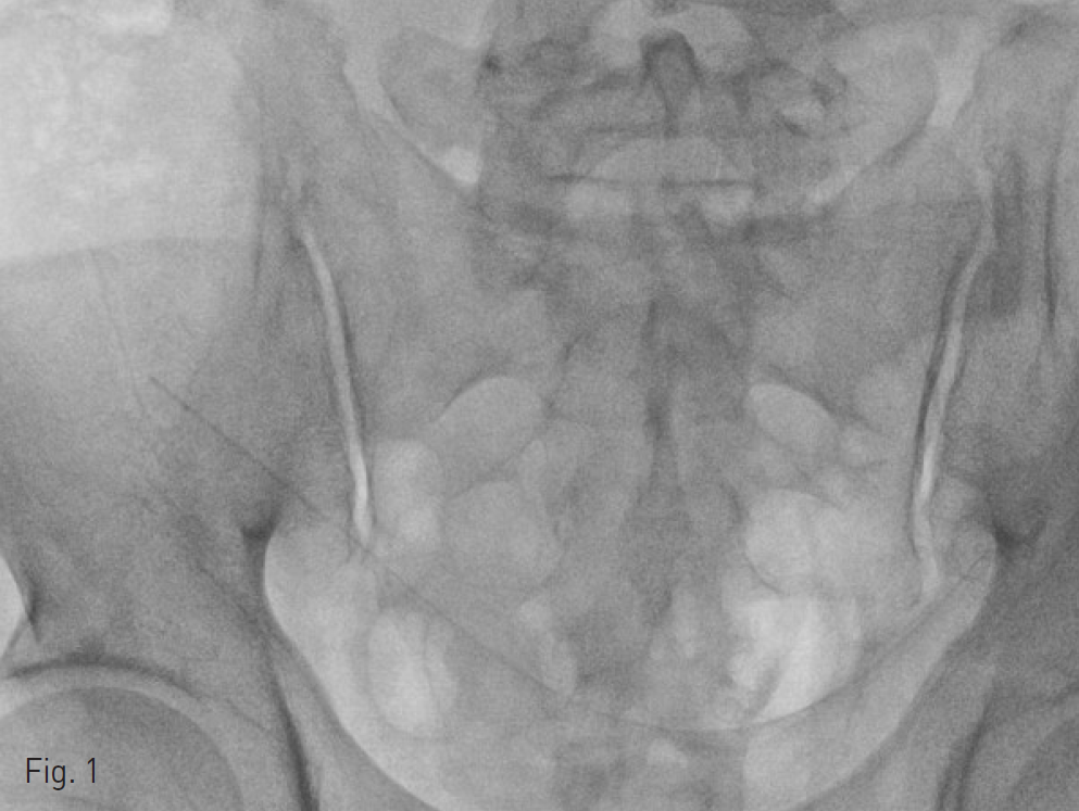
Fluoroscopy 및 cone-beam CT상에서 잘려진 배액관이 골반강 내에 위치하고 있음.
시술방법 및 재료
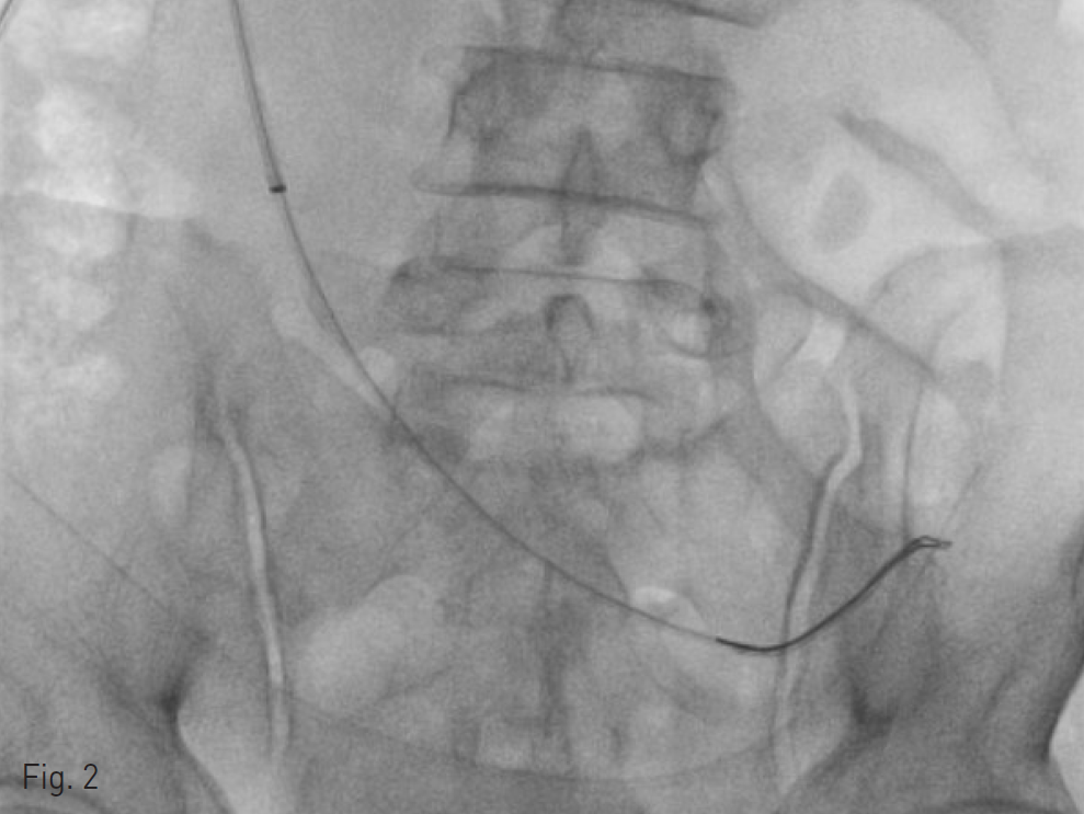
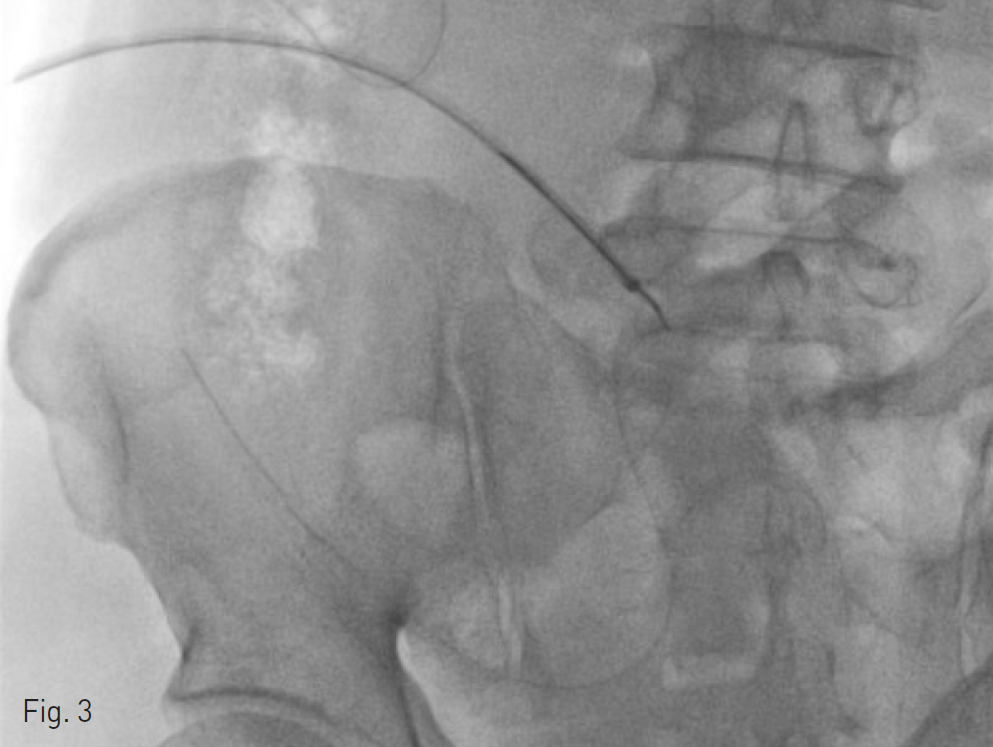
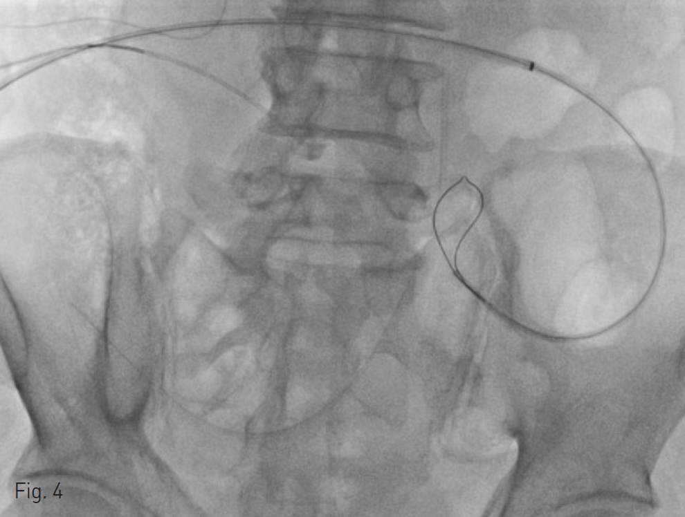
배액관이 삽입되었었던 우하복부위로 0.035-inch guide wire(Radiofocus, Terumo, Tokyo, Japan)와 5-Fr catheter(KMP, Cook, Blomington, USA)를 이용하여 골반강 내로 진입한 후 8-Fr sheath(Flexor check-flo introducer, Cook, Bloomington, USA)를 삽입하였다. 이를 통하여 snare(Amplatz goose neck snare, ev3, Plymouth, USA)를 삽입하였고, 수 차례 시도 후에 잘려진 배액관의 좌측 끝을 잡는 데에 성공하였다. 이후 snare로 잡은 배액관을 당겨서 제거하려고 하였으나 이 과정에서 bowel도 같이 당겨지는 소견을 보여서 bowel이나 mesentery 등이 snare에 같이 잡힌 것은 아닌지 확인하기 위해 cone-beam CT를 다시 한 번 시행하었다. Cone-beam CT 상 이러한 소견은 명확하지는 않았으나, fluoroscopy 소견을 고려하였을 때 bowel이나 mesentery 등이 snare에 같이 잡혀있을 가능성이 있었기 때문에 추가적으로 snare를 삽입하여 반대측으로 접근하여 안전하게 제거하는 것을 시도하였다. 우하복부의 sheath 삽입 부위를 통해 추가적으로 0.035-inch guide wire와 5-Fr catheter를 삽입하고 snare를 이용하여 배액관의 우측 끝을 잡으려고 시도했지만 실패하였다. 이에 다시 앞서 8-Fr sheath를 통하여 넣은, 배액관의 좌측 끝을 잡고 있던 snare를 몇 차례 풀면서 배액관을 당겨 더 이상 bowel이 같이 당겨지지 않는 것을 확인하고 배액관을 몸 밖으로 안전하게 제거하였다.
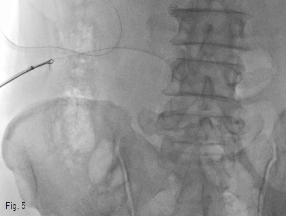
Fig. 1 ~ 5
Fig. 1. Jackson Pratt drain tube tip in the pelvic cavity.
Fig. 2. Capture of the left tip of Jackson Pratt drain tube with snare.
Fig. 3. When retrieving Jackson Pratt drain tube, small bowel was pulled along.
Fig. 4. Loosening of snare to release small bowel or mesentery, which was possibly captured by the snare
Fig. 5. Retrieval of Jackson Pratt drain tube with snare.
고찰
복강 내의 foreign body는 여리 가지 경로로 발생할 수 있으며, 이물질을 먹은 후 위장관에 천공이 발생하여 복강 내로 나오는 경우, 경피적으로 들이가는 경우, 질을 통헤서 들어가는 경우, 혹은 수술이나 검사 후에 남은 의인성(iatrogenic) 원인 등이 있을 수 있다. 의인성인 경우에는 거즈, 배액관, ventriculo peritoneal shunt catheter, intrauterine device 등이 비교적 흔한 원인으로 알려져 있다. 복강 내의 foreign body는 대부분 laparoscopy를 통한 수술적인 방법으로 제거를 하고 있으며 최근에는 single port laparoscopic procedure를 통한 최소 침습적인 방법으로 시행하는 경우가 보고되고 있다. 복강 내 foreign body는 bowel, mesentery 등의 구조물로 인해서 영상의학적인 인터벤션으로 접근하기 어려운 경우도 있겠으나 인터벤션은 laparoscopy 보다도 덜 침습적이고 전신마취 등의 위험성을 최소화 할 수 있는 방법인 만큼 본 증례에서와 같이 foreign body가 radiopaque하고 경피적으로 접근이 가능한 위치에 있을 경우 인터벤션을 통한 제거도 고려해보아야 할 것으로 생각한다.
참고문헌
1. Fujiwara T, Mitsunori Y, Kuramochi J, Hoshino N, Ono C, Nishioka Y, Nishimura H, Yaegashi K: Laparoscopic removal of a foreign body (a piece of wire) from the abdominal cavity: a case report and review of thirty two cases in Japan. J Jpn Endosc Surg 2007;12:415-419.
2. Kurita N, Shimada M, Nakao T, Chikakiyo M, Miyatani T, Higashijima J, Yoshikawa K, Nishioka M, Iwata T. Laparoscopic removal of a foreign body in the pelvic cavity through one port using a flexible cholangioscope. Dig Surg. 2009;26(3):205-8.
Citations
Citations to this article as recorded by