중심단어
Portal vein occlusion, Stent-graft, Ileal vein approach, Whipple’s operation, Pancreatic cancer
한글 초록
췌장암 환자로 4개월 전 Whipple 수술을 받은 환자에서 발생한 Portal vein occlusion에 대한 혈관적 치료 증례이다. 이 증례의 경우에는 경피적 경간 경로를 통해 폐색 부위를 통과하고자 다양한 방법을 시도하였으나 실패하였고, 이에 수술적 보조를 통해 회장 정맥 경로를 이용해 문맥 폐색부위에 접근하여 성공적인 시술을 시행하였다. 경피적경간문맥접근(percutaneous transhepatic portal approach)을 통해 간문맥 스텐트를 설치하는 방법이 일반적이나, 경우에 따라서는 상 장간막정맥(superior mesenteric vein)이나 하장간막정맥(inferior mesenteric vein)을 통한 시술이 시행되고 있다. 환자는 스텐트 삽입 후 3일 뒤 시행한 자기 공명영상에서 Portal vein의 flow가 유지되고 있었으며, 환자의 활력징후가 안정되고 복통 등의 증상이 호전되었으며 식이 능력이 회복되었다.
영문 초록
We report a case of stent graft placement through ileal vein for portal vein occlusion in a patient with pancreatic cancer. In general, percutaneous portal approach is preferred, but in some cases, the procedure is performed through the superior mesenteric vein or the inferior mesenteric vein. The flow of portal vein was maintained in the MRI performed 3 days after the stent insertion. The patient's vital signs were stabilized, abdominal pain and symptoms were improved, and the dietary capacity was restored.
Case report
증례
54세/여자
임상소견
Pancreatic cancer 환자로 4개월 전 Whipple 수술을 받았다. 당시 환자는 splenic vein과 superior mesenteric vein이 합류하는 부근의 portal vein에 tumor invasion이 있어 portal vein resection 및 Y graft interposition을 함께 시행하였다. 이후 환자는 RUQ pain을 호소하며 복수가 차는 등의 증상을 보였고 CT 추적 관찰에서 portal vein의 severe narrowing이 있는 상태로 점차 그 정도가 심해졌으며, 이에 대하여 혈관 내 치료가 의뢰되었다
진단명
Portal vein total occlusion, s/p Whipple’s operation with portal vein resection and Y graft interposition, s/p Pancreatic cancer with portal vein invasion.
영상소견
수술 후 추적 CT에서 주문맥(main portal vein)과 상장간막정맥(superior mesenteric vein)의 abrupt narrowing이 보이며, 이 두 구조물 사이의 혈관 구조물이 보이지 않아 total occlusion이 의심되는 상태였다.
시술방법 및 재료
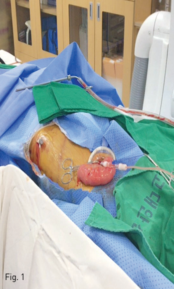
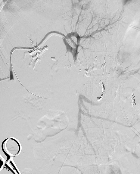
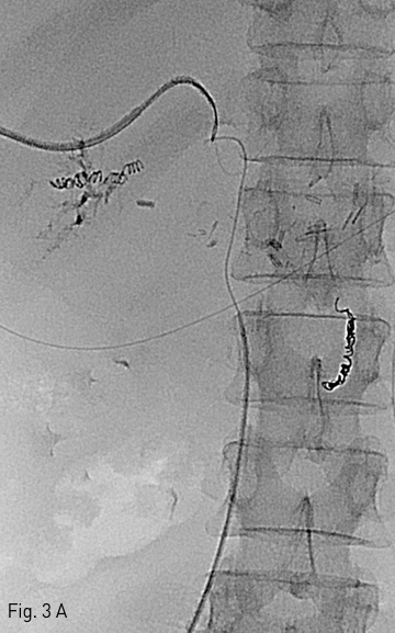
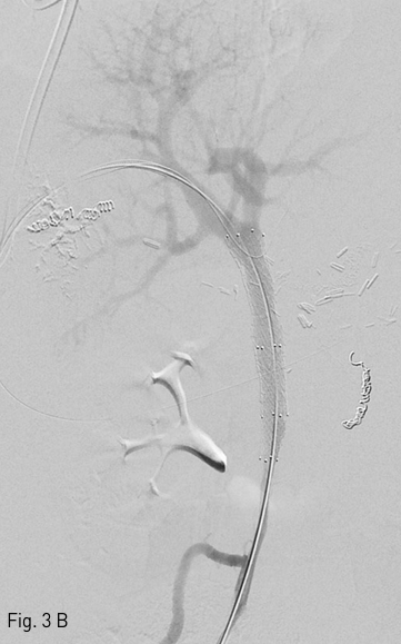
우측 간문맥 분지(Right portal vein branch)를 초음파 유도하에 천자하여 5-Fr sheath (Arrow sheath, Arrow international, PA, USA)를 삽입한 후 문맥 조영술을 시행하였다. 주문맥(main portal vein)의 total occlusion 소견이 보였으며, 이 부위를 통과하기 위해 0.035-inch 유도철선(Radiofocus guidewire, Terumo corp, Tokyo, Japan)과 CXI 유도철선(CXI, Cook medical, IN, USA), CTO wire (Astato 30, Asahi intecc, Aichi, Japan)으로 수 차례 통과를 시도하였으나 실패하였다. 이에 당일엔 tract에 코일 색전술을 하고 시술을 종료하였고, 이틀 후 외과와 마취과의 협진 하에 환자를 전신마취 시킨 후 시술하기로 하였다. 우측 간문맥 분지를 같은 방식으로 천자한 후 5-Fr sheath(Arrow sheath, Arrow international, PA, USA)를 삽입하였다. 외과 협진 하에 복벽을 절개하고 회장(ileum)을 노출시켜 회장 정맥 (ileal vein)을 천자하였다 (Fig.1, 2) 이후 회장정맥에 7-Fr sheath(Radiofocus introducer II, Terumo corp, Tokyo, Japan)를 삽입하고 5-Fr catheter (KMP catheter, Cook medical)와 위해 0.035-inch 유도철선(Radiofocus guidewire, Terumo corp)를 이용하여 성공적으로 폐색 부위를 통과한 후 간문맥 경로의 sheath로 snare(Amplatz goose neck snare kit, Covidien, Dublin 2, Ireland)를 삽입하여 통과시킨 wire를 잡아 경로를 확보하였다(Fig.3A). 확보된 경로의 유도철선을 따라 4x80 mm balloon (Mustang, Boston scientific, MA, USA)을 통과시켜 풍선확장술을 시행한 후, 정상 상장간막정맥(superior mesenteric vein)과 만나는 distal portion에는 8x40 mm self-expandable stent(Zilver flex, Cook medical)를 삽입하고, 주문맥(main portal vein) 폐색 부위에는 12x60 mm self-expandable stent(Zilver flex, Cook medical)를 삽입하였다(Fig.3B). 이후 풍선확장술을 시행하고 portogram을 통해 성공적인 개통을 확인한 후, 천자경로 코일색전술을 하고 시술을 종료하였다.
추적관찰
스텐트 삽입 후 3일 뒤 시행한 MR에서 Portal vein의 flow가 유지되고 있었으며, 환자의 활력징후가 안정되고 복통 등의 증상이 호전 되었으며 식이능력이 회 복되었다. 이후 안정적인 상태로 다른 합병증 없이 퇴원하였다.
Fig 1
After exposing the ileum, cannulation through ileal vein was made.
Fig 2
Angiogram taken from transportal & transileal route shows complete occlusion of main portal vein.
Fig 3A
(A) Guide wire via ileal vein which passes through obstructed lesion of portal vein was snared via percutaneous portal vein tract.
Fig 3B
(B) The stent is installed through the path of guide wire. DSA shows recanalization of the main portal vein
고찰
문맥 스텐트 삽입술은 간 췌장 수술 후 폐색이나 협착으로 인한 문맥 고혈압에 대해 안전하고 성공적인 치료 방법이 될 수 있다(1, 2). 경피적경간문맥접근 (percutaneous transhepatic portal approach)을 통해 간문맥 스텐트를 설치하는 방법이 일반적이나, 경우에 따라서는 상장간막정맥(superior mesenteric vein)이나 하장간막정맥(inferior mesenteric vein)을 통한 시술이 시행되고 있다(3, 4). 이 증례의 경우에는 경피적 경간 경로를 통해 폐색 부위를 통과하고자 다양한 방법을 시도하였으나 실패하였고, 이에 수술적 보조를 통해 회장 정맥 경로를 이용해 문맥 폐색부위에 접근하여 성공적인 시술을 시행하였다. 김 등은(Kim et al. 2007) 간 이식 수술 중, 또는 수술 후에 상장간막정맥이나 하 장간막정맥을 통한 접근으로 간문맥에 스텐트 설치를 한 후 평균 16개월간 95%의 개통률을 보였다고 보고한 바 있다(3).
참고문헌
1. Zhou ZQ, Lee JH, Song KB, et al. Clinical usefulness of portal venous stent in hepatobiliary pancreatic cancers. ANZ J Surg 2014;84:346-352
2. Kim KR, Ko GY, Sung KB, Yoon HK. Percutaneous transhepatic stent placement in the management of portal venous stenosis after curative surgery for pancreatic and biliary neoplasms. AJR Am J Roentgenol 2011;196:W446-450 74
3. Kim YJ, Ko GY, Yoon HK, Shin JH, Ko HK, Sung KB. Intraoperative stent placement in the portal vein during or after liver transplantation. Liver transpl 2007;13:1145-1152
4. Schellhammer F, am Esch JS, Hammerschlag S, Knoefel WT, Furst G. Surgical access to jejunal veins for local thrombolysis and stent placement in portal vein thrombosis. Cardiovasc Intervent Radiol 2008;31:S185-187
Citations
Citations to this article as recorded by