중심단어
lymphangiography, embolization, Noonan syndrome, chyle, chylous
한글 초록
14세 남자 누난 증후군 환자가 2개월 전부터 발생한 불수의적인 혈성 및 유미성 요도 분비물을 주소로 내원하였다. 증상은 선 자세에서 악화되고 누운 자세에서는 완화되었다. 음경꺼풀(foreskin)이 부풀어있었고 음경, 음낭 및 서혜부의 피부는 림프부종 소견을 보였다. 양측 서혜부 림프절 경유 리피오돌 림프조영술을 시행하였을 때 허벅지의 안쪽 및 음낭 쪽을 향하는 림프액의 역류가 보였고 골반 내 장골동맥 주변 림프관은 매우 비대되어 있었다. 비정상적인 요도-림프관 연결은 보이지 않았으며 주입된 리피오돌은 음낭 피부를 거쳐서 귀두 부근의 피부에 분포하고 있었다. 임상 및 검사 소견을 고려, 역류성 서혜부 림프부종으로 진단하고 유미성 림프액의 역류를 막기 위해서 양측 서혜부 림프관에 미세도관을 삽입하고 접착제(glue)를 이용한 색전술을 시행하였다. 이후 분비 증상은 없어졌고 서혜부 및 음낭 피부의 림프부종 소견도 호전되었다.
영문 초록
A 14-year-old boy who was previously diagnosed to have Noonan syndrome presented with involuntary urethral discharge of chylous fluid which started 2 months ago. The symptom aggravated in standing position or during activity and relieved in supine position. The scrotal and inguinal skin showed lymphedematous change. Inguinal intranodal lymphangiography using Lipiodol was performed for both diagnostic and therapeutic purposes. It revealed abnormal flow direction of the lymphatic fluid inside the pelvic lymphatic vessels which were abnormally hypertrophied. No direct fistulous communication between lymphatic vessels and urethra was identified on the CT taken after the lymphangiography. A hypothesis was made that the lymphedema and urethral discharge are caused by the reflux of chylous lymphatic fluid into the scrotal area due to the lymphatic hypertrophy and consequent valve malfunction. Based on the hypothesis, bilateral inguinal lymphatic vessels were cannulated using microcatheters and embolized using NBCA and Lipiodol mixtures to occlude the reflux. The chylurethra stopped immediately after the treatment and the lymphedema in the scrotal skin was also normalized. There was no complication related to the embolization procedure.
Introduction
누난 증후군은 1000명-2500명 당 1명꼴로 발생하는 비교적 흔한 선천성 유전질환으로 특징적인 얼굴모양, 저신장, 흉곽 모양 변형, 선천성 심장질환의 임상적 양상을 통해 진단하게 된다. 이 질환을 가진 환자의 약 20% 정도에서 림프계통의 다양한 이상을 동반하는데 말초 림프부종이 가장 흔한 형태이다. 이 밖에도 수종, 유미흉, 폐 림프관확장증, 위장관 림프관확장증, 하지 림프관 형성저하증, 흉곽의 이상 림프관, 흉관 저형성 또는 무형성, 고환 림프관확장증 등이 보고되어 있으며 이들에 대한 치료는 어려운 것으로 알려져 있다. 본 기고에서는 유미성 요도 분비물을 주소로 내원한 환자에 대해서 색전술을 성공적으로 치료한 증례에 대해서 보 고 하고자 한다.
Case report
증례
14세 / 남자
임상소견
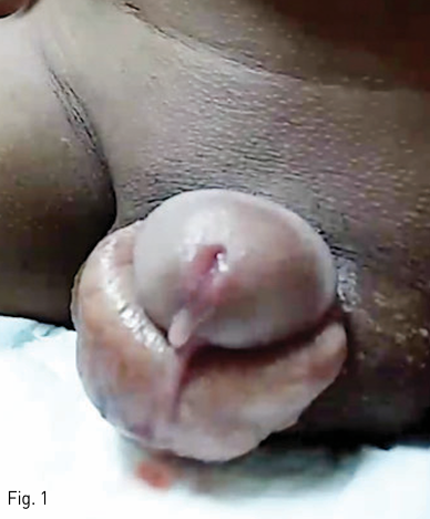
누난 증후군으로 진단된 환자가 2개월 전부터 발생한 혈성 및 유미성 요도분비물을 주소로 내원하였다. 소변과 관련성이 없고 수의적 조절이 불가능한 분비물이 요도에서 흘러나와서 뚝뚝 떨어지는 양상이었다 (Fig. 1). 일상적인 활동을 할 경우 하루에 2-3장의 기저귀를 필요로 하였다. 누워있는 자세에서 호전되기 때문에 환아는 활동을 피하고 누워있으려는 경향을 보였다. 신체검진상 음경 꺼풀이 크게 부풀어있었으며 음낭, 음경, 서혜부 피부가 부종성 변화를 보였다(Fig. 1).
진단명
Non-traumatic Chylous Urethral Discharge
영상소견
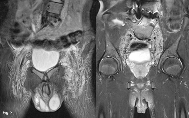
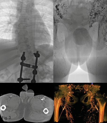
MRI T2 강조 영상에서 서혜부 림프절 및 림프관과 골반 내 장골동맥 주변 림프관의 비대 소견을 보였다 (Fig. 2). 양측 서혜부 림프절 경유 리피오돌 림프조영술에서도 상기 비대소견이 있었으나 이에 더하여 허벅지의 안쪽 및 음낭 쪽을 향하는 비정상적인 림프액의 역류가 보였다. 한편 흉관 및 좌측 쇄골하정맥으로의 림프액의 합류는 정상이었다. Spot radiography 및 C-arm CT를 이용하여 주입된 리피오돌의 분포를 정밀하게 관찰하였으나 전립선 요도에서부터 음경 요도에 이르기까지의 범위에서 비정상적인 요도-림프관 연결을 의심할 만한 소견은 보이지 않았다 (Fig. 3). 주입된 리피오돌은 음낭 피부를 거쳐서 귀두 부근의 피부 및 피하조직에 분포하고 있었다. 임상 및 검사 소견을 고려하여 누난증후군과 연관된 림프계 형성이상으로 인해 소장에서 생성된 유미 림프액이 하지, 서혜부, 음낭 피부 방향으로 역류하면서 발생한 림프부종이 있고, 압력이 높아진 림프관내 림프액이 요도로 배액되는 것 으로 진단하였다.
시술방법 및 재료
소장으로부터의 유미성 림프액 역류를 막기 위해서 서혜부 및 허벅지 안쪽으로 향하는 림프관에 대한 색전 시술을 계획하였다. 서혜부 림프절 경유 리피오돌 림프 조영술을 다시 시행하여 양측의 비대된 서혜부의 림프관을 조영하고 이를 투시 유도하에 21 gauge 5 cm 바늘(Cook, Bloomington, IN)로 직접 천자하여 유미 림프액의 유출을 확인하였다. Meister 미세 유도 철사 (0.016 inch, Asahi Medical, Japan)와 Progreat alpha 미세 도관 (2.0 Fr, Terumo, Japan)을 림프관에 삽입하고 골반과의 경계 부위까지 전진시킨 다음 접착체 [N-butyl cyanoacrylate (Histoacryl, B Braun)와 리피오돌 (Lipiodol Ultrafluid, Guerbet, France)의 1:5 혼합물]을 이용하여 색전술을 시행하였다(Fig. 4).
추적관찰
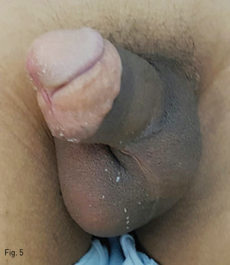
시술 후 양측 서혜부의 경등도의 통증 외에 특별한 합병증은 없었다. 시술 이후 분비 증상은 없어졌고 신체검진 상 서혜부 및 음낭 피부의 림프부종 소견도 호전 되었다 (Fig. 5).
Fig 1
Milky fluid was discharged from the penile orifice involuntarily. The discharge was not related to the urination. More than 2-3 diapers were needed a day. The symptom was improved in lying position. Skin texture of the penile and scrotal area was abnormally dark and edematous.
Fig 2
T2 weighted MR coronal images showed hypertrophic inguinal lymph nodes as well as lymphatic vascular structures in the pelvic retroperitoneum. Scrotal skin showed edematous change. However, there was no evidence of direct lympho-urethral fistula.
Fig 3
(Top) Bilateral inguinal intranodal lymphangiography using Lipiodol was successfully performed. Venous return via the thoracic duct was intact. However, the lymphatic vessels in the inguinal and pelvic area showed abnormally hyperactive peristaltic movement. In addition, the reversed flow of Lipiodol from the inguinal lymph node to the skin of both medial thigh and scrotum was also noted (dotted circle). (Bottom) However, no large lympho-urethral fistula which can be directly embolized or surgically resected was detected even in the cone-beam CT examination.
Fig 4
(Top left) A prominent lymphatic vessel (arrow) in left thigh area was directly punctured using 21 gauge needle under the fluoroscopic guidance. (Top right) Chylous fluid was gushed out from the needle immediately after the puncture of the vessel. (Bottom) A microwire (0.016 inch, Meister, Asahi Medical) and a microcatheter (2.0 Fr, Progreat alpha, Terumo) were advanced along the vessel up to the pelvis level. Then, the lymphatic vessels in the pelvic and inguinal area were embolized using 1:5 NBCA:Lipiodol mixture to occlude the reflux of the chylous fluid from small bowel to the scrotal skin. The same procedure was performed for the right side.
Fig 5
Milky urethral discharge stopped immeidately after the embolization procedures. (Left) A medical photo taken 10 days after the procedure showed the normalization of the skin texture in the scrotal area and decreased size of the penis compared to the initial status, which can be seen an image at the top left of this poster.
고찰
수술 후 발생하는 다양한 림프액 유출증에 대해서 색전술을 비롯한 인터벤션 치료가 안전하고 효과적이라는 사실이 최근 널리 받아들여지고 있지만, 수술이나 외상과 관련이 없이 자발적으로 발생하는 비외상성 림프액 유출증에 대해서는 아직까지 성공적으로 치료한 증례 보고가 매우 드물다. 본 증례에서는 누난 증후군 과 관련된 림프계의 선천성 이상이 있는 환자에서 발생한 비외상성 유미성 림프액의 요도 유출증에 대한 인터벤션 치료를 보고하였다. 림프계는 동맥 및 정맥계과 함께 전신 체액 순환의 일부분을 차지할 뿐만 아니라 면역관련 세포의 이동 및 지방의 소화흡수 과정에서도 중요한 역할을 한다. 소장 점막에서 흡수된 지방의 운반체인 카일로미크론 성분은 소장의 림프액을 우유와 같은 흰색의 탁한 액체로 보이게 하며 이를 유미성 림프액이라고 한다. 따라서 전신의 어느 부위에서든 유미성 림프액의 유출이 관찰될 경우 그 기원이 소장의 림프계라는 사실을 인지하고 유출부위와의 잠재적인 연결 및 유출 경로를 찾아야만 효과적인 치료가 가능하다. 본 증례의 경우 요도를 통해서 유출되는 물질이 유미성 림프액으로 확인 되었기 때문에 어떤 경로로 소장에서부터 요도까지 유미림프액이 이동하는지를 밝히는 것이 중요하였다. 일어선 자세에서 유출 증상이 심해지며 중간 경로에 해당하는 골반 및 복부의 동맥 주변 림프관이 비대되었고 리피오돌 림프조영술에서 서혜부 림프액 흐름의 역류가 증명되었다는 점을 종합적으로 고려하여 소장에서 생성된 유미림프액이 하지, 서혜부, 음낭 피부 방향으로 역류하면서 림프관내 압력이 높아졌고 이로인해 해당 부위의 림프 부종이 발생하였으며 최종적으로 일부 미세 연결을 통해서 림프액이 요도로 배액되는 것으로 병리기전을 추리할 수 있었다. 이는 마치 고환정맥 판막 이상으로 인한 정맥의 역류로 정계 정맥류가 발생하는 것과 유사한 기전이라고 할 수 있다. 정계정맥류의 인터벤션 영상의학 치료법이 고환정맥에 대한 색전술 또는 경화요법을 통한 역류의 차단이라는 점에 착안하여 본 증례에서도 서혜부 및 음낭 피부로 상부의 림프액이 역류하여 압력을 전달하는 것을 차단하기 위한 시도를 하였다. 이를 위해서 투시 유도 하에 리피오돌로 조영된 하지의 림프관을 셀딩거기법 으로 직접 천자하여 미세도관을 삽입하고 림프관의 상류로 도관을 삽입하였으며 이후 접착제를 이용한 색전술을 시행하였다. 이와 같이 동맥 또는 정맥에 대한 인터벤션에서 이미 광범위하게 사용되고 있는 각종 인터벤션 영상의학 기법을 림프계 인터벤션에 응용함으로써 기존의 지식과 방법으로는 진단 및 치료가 어려웠던 비외상성 림프액 유출증 중 일부 질환을 치료할 수 있었다.
참고문헌
1. Hur S, Shin JH, Lee IJ, et al. Early experience in the management of postoperative lymphatic leakage using lipiodol lymphangiography and adjunctive glue embolization. J Vasc Interv Radiol 2016;27:1177-1186
2. Romano AA, Allanson JE, Dahlgren J, et al. Noonan syndrome: clinical features, diagnosis, and 100 management guidelines. Pediatrics 2010;126:746-759
3. Matsumoto T, Yamagami T, Kato T, et al. The effectiveness of lymphangiography as a treatment method for various chyle leakages. Br J Radiol 2009;82:286-290
Citations
Citations to this article as recorded by