중심단어
Percutaneous gastrostomy, embolization, complication, skin necrosis
한글 초록
최근 경동맥 색전술은 다양한 출혈성 질환, 혈관질환 및 종양의 치료에 널리 이용되고 있다. 수술적인 치료에 비해 덜 침습적인 방법으로 효과적인 치료를 할 수 있어 그 장점이 있으나 드물게 심각한 주요 합병증을 야기할 수 있는 것으로 알려져 있다. 본 증례에서는 경피적 위루술을 시행한 뒤 시술 부위를 통해 도수 압박에 반응하지 않는 외부 출혈이 있어 내흉동맥에서 기시하는 복벽동맥 가지에서 선택적으로 n-butyl cyanoacrylate와 ethiodized oil 혼합액을 이용하여 색전술을 시행하였다. 환자는 시술 후 4주 째 시술 부위의 상처가 지속되어 다시 병원을 찾았으며, 피부 괴사 가있어 지속적인 국소 소독을 유지해 주었고 충분히 치유되었다.
영문 초록
Recently transarterial embolization is widely used to treat various hemorrhagic disease, vascular disease and tumors. When compared to surgical treatment, transarterial embolization has strong point in less invasiveness, but rarely it can cause serious major complications. In this case report, transarterial embolization was performed to control external bleeding after percutaneous gastrostomy which was not responding to manual compression. The procedure was performed selectively via epigastric artery branch arising from internal mammary artery with mixture of n-butyl cyanoacrylate and ethiodized oil. The patient revisited the hospital complaining unhealing wound around the gastrostomy site, and skin necrosis at the embolized territory was diagnosed. The wound fully regenerated with conservative management with topical dressing.
Introduction
최근 출혈에 대한 치료법으로 혈관내 접근을 통한 경동맥 색전술은 다양한 임상 상황에서 널리 이용되고 있다. 경동맥 색전술은 지혈을 위한 수술적인 치료법에 비하여 덜 침습적인 방법으로 비교적 안전하게 시행할 수 있으나 드물게 심각한 주요 합병증을 야기할 수 있다. 저자들은 경피적 위루술을 시행하고 나서 발생한 출혈에 대한 경동맥 색전술 후 발생한 피부 괴사의 증례를 경험하여 보고한다.
Case report
증례
77세 / 남자
임상소견
6년전 비인두암으로 항암화학요법 및 동시항암화학 방사선치료를 받은 적이 있었고, 1년전 관상동맥협착증으로 2군데의 혈관에 약물 방출 스텐트 설치술을 받았으며, 이어 복부 대동맥류로 인조혈관 스텐트 설치술을 받았다. 고형물 경구 식이는 불가능한 상태였으며, 이중 항혈전제를 복용하고 있었다. 경피적 위루술 시행이 의뢰되었다.
진단명
Skin Necrosis Following Embolization due to Abdominal Wall Bleeding after Percutaneous Gastrostomy
시술방법 및 재료
경피적 위루술을 시행하기 위해 통상적인 방법에 따라 진통제 (Fentanyl citrate 50 ㎍) 정맥주사를 시행하였다. 이어 구강을 통해 5-Fr 카테터를 이용하여 위 장으로 진입한 뒤 공기를 주입하여 팽창시켰으며, 투시 유도하에 16G 천자침을 이용하여 위장을 천자하고 2개의 T-fastener를 설치하였다. 위고정이 적절히 시 행되었음을 확인한 뒤 다시 위장을 천자하고 0.035 inch 유도철사를 삽입한 뒤 18-Fr까지 단계적으로 확장하고 18-Fr 도뇨관을 삽입하고 풍선을 확장시킨 뒤 복벽에 고정하였다. 환자는 시술 후 약 6시간이 지났을 때 위루술 시행한 자리를 통한 출혈이 점차 늘어나며 압박 지혈을 시도하였음에도 지속되어 조영증강 CT를 촬영하고 색전술이 의뢰되었다. 의뢰 당시 생체징후는 혈압 137/80mmHg, 맥박 86/분으로 시술 전과 비교하여 큰 변화 없이 안정적이었다. 환자는 시술 부위에 거즈를 이용한 압박 드레싱을 한 상태로 영상 검사를 시행하였으며, 동맥기 영상에서 조영 전 영상에서는 관찰되지 않는 피부 바깥으로의 조영제 유출이 관찰되어 진행성 출혈이 진단되었다 (Fig. 1). 초음파 유도 하에 미세천자 세트를 이용하여 우측 총대퇴동맥을 천자하고 5-Fr sheath를 삽입하였다. 5-Fr 카테터와 유도철사를 이용하여 좌측 내흉동맥을 선택하고 2.0-Fr 미세 도관(Progreat Alpha; Terumo, Tokyo, Japan)을 이용하여 내흉동맥의 원위부로 진입한 뒤 혈관 조영검사를 시행하였다. 혈관조영검사에서 내흉동맥 원위부에서 기시하는 복벽동맥에서 조영제 유출이 관찰되었으며 (Fig. 2A), 다시 미세도관을 이용하여 출혈을 보이는 혈관에 가능한 한 가까이 진입한 뒤 n-butyl cyanoacrylate (NBCA; Histoacryl; B. Braun, Melsungen, Germany)와 ethiodized oil (Lipiodol Ultra; Guerbet, Paris, France)을 1:2로 혼합한 뒤 주입하여 색전술을 시행하였다 (Fig. 2B). 색전술 후 시행한 혈관조영검사에서 출혈을 보이던 혈관이 성공 적으로 색전된 것을 확인하였으며, 다른 혈관을 통한 진행성 출혈은 관찰되지 않았다(Fig. 2C).
추적관찰
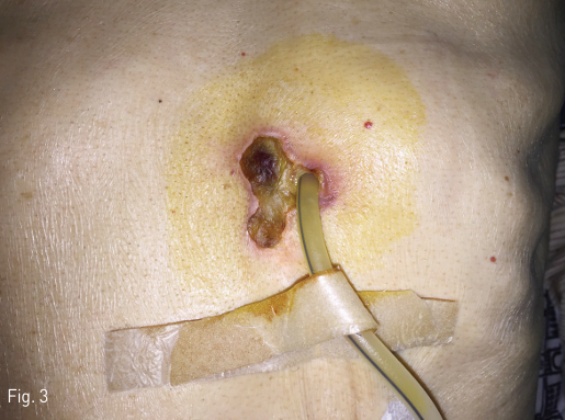
환자는 4주 후 삽입부 상처가 잘 낫지 않는다고 호소하였고, 육안적으로 삽입부 주변 피부 괴사 소견을 보였다(Fig. 3). 상처 부위 국소 소독 외에 추가 치료 없이 피부 병변은 치유되었다.
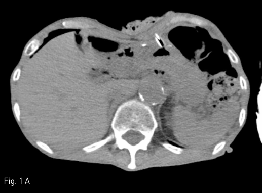
Fig 1A
Contrast-enhanced CT study was performed to find out the presence of active bleeding. Comparing the pre-contrast (A) and arterial phase (B) images, contrast extravasation (arrow) between the skin and the packing gauze (arrowheads) is noted around the gastrostomy site.
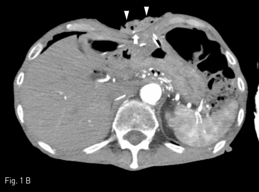
Fig 1B
Contrast-enhanced CT study was performed to find out the presence of active bleeding. Comparing the pre-contrast (A) and arterial phase (B) images, contrast extravasation (arrow) between the skin and the packing gauze (arrowheads) is noted around the gastrostomy site.
Fig 2A
Left internal mammary arteriography was performed after selection with 5-Fr angiographic catheter and microcatheter system, which revealed active contrast extravasation from a branch of superior epigastric artery (A, arrowhead).
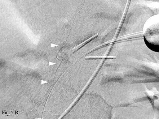
Fig 2B
After further selection of the bleeding branch with a microcatheter (B, arrowheads), embolization was performed with 1:2 mixture of n-butyl-cyanoacrylate (NBCA) and ethiodized oil.
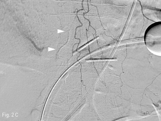
Fig 2C
On follow-up arteriography, artifact by the NBCA-ethiodized oil mixture is noted (C, arrowheads), and there was no evidence of further active arterial bleeding.
Fig 3
The patient revisited the clinic complaining unhealing wound around the gastrostomy site. About 3 x 2.5-cm sized skin necrosis was noted at the medial aspect of the gastrostomy tube.
고찰
경피적 위루술은 내시경적 접근, 혹은 영상유도하 경피적 접근을 통해 시행할 수 있으며, 기술적인 성공률은 95% 이상으로 보고되고 있다. 시술 후 주요 합병증 으로 흡인, 출혈, 복막염, 심부 감염, 위루관 이탈, 괴사성 근막염, 종양 이식, 사망 등이 발생 가능하며, 유병률은 0.4-22.5%로 보고되고 있다. 시술 관련 사망률은 0-2%로 보고되며, 30일 사망률은 심한 동반질환을 가진 환자군에서 주로 보고되며 6.7-26%로 알려져 있다. 주요 합병증으로서 출혈은 0-2.5% 정도로 보고되 고 있다. Society of Interventional Radiology의 권고안에 따르면 경피적 위루술은 중등도의 출혈 위험을 가지는 시술로 분류되어 있으며, 시술 후 합병증을 줄이기 위해 INR을 1.5 미만으로 조절하고, 혈소판 수치 50,000/㎕ 이상으로 교정, clopidogrel 5일 이상 중단, low molecular weight heparin은 한 차례 중단하며 aspirin은 중단할 필요가 없는 것으로 권고하고 있다. 환자는 관상동맥에 약물 방출 스텐트를 삽입한 과거력이 있으며 이중 항혈전제를 복용하고 있었으나 임상의의 판단에 따라 충분한 기간 동안 약물을 중단하지 못하고 시술을 진행하여서 출혈의 위험이 컸던 것으로 판단할 수 있다. 또한 기술적으로 출혈의 위험을 감소시키기 위해 복벽동맥 주행 경로를 피할 수 있도록 좌우측 복직근 사이의 위치나 좌측 복직근보다 더 편측의 위치를 선택하여 위장을 천자하는 것이 권장된다. 그러나 본 증례에서는 위장을 충분히 팽창시켰음에도 불구하고 위장이 충분히 흉강 아래쪽으로 하강하지 않아 명치에 매우 가깝게 천자할 수밖에 없었으며, 복직근을 통과하는 경로로 위루술을 시행하여 출혈의 위험이 큰 경우였다. 경동맥 색전술 후 합병증으로 발생하는 피부 병변이 발생하는 상황으로 인터벤션 영상의학 분야에서는 간암이 있는 환자에서 경동맥 화학색전술 후 발생하는 것이 흔히 알려져 있다. 이는 간암에서 기생적 측부 순환 발달로 인한 것으로 흔히 늑간동맥, 겸상동맥, 내흉동맥을 통해 치료할 때 피부 손상의 가능성이 있다. 그러나 이런 경우에도 일시적인 홍반, 발진 등 심하지 않은 형태의 합병증이 대부분이며, 대부분 보존적인 치료로 호전된다. 그 외에도 색전술 후 피부 합병증은 치료적 목적으로 시행하는 색전술을 필요로 하는 다양한 질병들에서 보고되고 있는데, 동정맥기형, 외상성 골반 골절, 산후 출혈, 자궁 근종 등이 있다. 색전술 중 피부 합병증은 주입한 색전 물질, 혹은 항암제가 동맥 순환의 최말단까지 도달하여 피부 순환을 차단하거나 자극하여 발생하는 것으로, 입자형 색전 물질은 합병증을 일으킬 정도로 말단으로 도달하지 못하고 미세 순환은 색전술 후에도 측부 순환을 통해 유지되어서 그 위험성이 비교적 낮지만, 본 증례에서 사용한 NBCA를 비롯하여 알코올, Onyx 등의 액상 색전 물질은 입자형에 비해서 더 작은 혈관으로 침투할 수 있어 상대적으로 그 위험이 높다. 또한 매우 작은 가지에서 출혈이 있는 경우 미세 도관 선택 과정에 발생할 수 있는 혈관 연축, 혹은 미세 도관 진입 자체에 의하여 혈관 내의 자유 혈류가 차단될 수 있으며, 이와 같은 상황에서는 자유 혈류가 유지된 상태에서 색전술을 시행하는 것 보다 색전 물질이 더 깊이 침투할 수 있고 측부 순환을 차단할 수 있어 색전 효과는 더 좋을 수 있으나 이와 함께 허혈성 합병증의 위험도도 높게 되며, 본 증례도 이와 같은 경우에 해당한다고 볼 수 있다. 본 증례에서는 허혈성 피부 병변은 일시적인 홍반, 발진 등의 흔히 볼 수 있는 것보다 심한 형태로 괴사가 발생하였으나 국소 소독만 유지하면서 충분히 치유되었고 피부 이식 등 수술적인 치료를 필요로 하지는 않았다.
참고문헌
1. Shin JH, Park AW. Updates on Percutaneous radiologic gastrostomy/gastrojejunostomy and jejunostomy. Gut Liver 2010;4:S25-31
2. Patel IJ, Davidson JC, Nikolic B, el al. Consensus guidelines for periprocedural management of coagulation status and hemostasis risk in percutaneous image-guided interventions. J Vasc Interv Radiol 2012;23:727-736
3. Itkin M, DeLegge MH, Fang JC, et al. Multidisciplinary practical guidelines for gastrointestinal access for enteral nutrition and decompression from the Society of Interventional Radiology and American Gastroenterological Association (AGA) Institute, with endorsement by Canadian Interventional Radiological Association (CIRA) and Cardiovascular and Interventional Radiological Society of Europe (CIRSE). J Vasc Interv Radiol 2011;22:1089-1106
4. Smith MT, Johnson DT, Gipson MG. Skin necrosis resulting from nontarget embolization of the falciform artery during transarterial chemoembolization with drug-eluting beads. Semin Intervent Radiol 2015;32:22-25
5. Li Z, Cai Z, Dai S, Deng H, Li Z, Ji F. Cutaneous necrosis associated with transcatheter arterial chemoembolization of the intercostal artery. J Vasc Interv Radiol 2014;25:1141-1148
6. Do YS, Yakes WF, Shin SW, et al. Ethanol embolization of arteriovenous malformations: interim results. Radiology 2005;235:674-682
7. Al-Thunyan A, Al-Meshal O, Al-Hussainan H, Al Qahtani MH, El-Sayed AA, Al-Qattan MM. Buttock necrosis and paraplegia after bilateral internal iliac artery embolization for postpartum hemorrhage. Obstet Gynecol 2012;120:468-470
Citations
Citations to this article as recorded by