중심단어
Pancreas, Insulinoma, Artery, Vein
한글 초록
68세 여자 환자가 컴퓨터 단층 촬영과 자기 공명 영상상 인슐린 종으로 의심되는 병변의 특성화와 정확한 위치 확인을 위해 의뢰되었다. 저자들은 동맥 자극 후 정맥혈 추칠 술을 시행하였고, 그 결과 비장 동맥 자극 후 추출한 정맥혈에서 혈중 인슐린과 씨-펩타이드 수치의 증가를 확인하여 병변이 췌장 체부와 미부에 있다 는 결과를 보고하였다. 이후 수술 중 병변의 위치가 일치함을 확인하였고, 원위부 췌장절제술 후 임상증상이 호전되었다.
영문 초록
A 68-year-old female was referred for characterization and accurate localization of lesions, which are suspected insulinoma on computed tomography and magnetic resonance imaging. The authors performed arterial stimulation with venous sampling. As a result, we observed increased serum insulin and C peptide levels in the venous sample extracted after splenic artery stimulation and concluded that the lesions were in the pancreatic body and tail. Thereafter, the lesions were found to be in agreement with the position of the lesion during the operation, and the clinical symptom improved after distal pancreatectomy.
Introduction
인슐린종은 혈중 인슐린 수치가 6mU/mL 이상이고 공복시 혈당 수치가 45mg/dL 이하이면서 저혈당 증세가 나타나면 의심한다. 수술로 90%에서 완치가 가능하 나 많은 경우 크기가 2cm 미만으로 위치를 찾는데 어려움이 있다. 이에 많은 imaging modality가 사용되나 다양한 민감도와 특이도를 가지고 있어 종종 CT로 찾을 수 없는 경우도 37% 정도로 보고되었다. 그러나 인슐린종의 동맥자극 정맥채혈술은 그 민감도와 특이도 모두 90%에 달한다.
Case report
증례
68세/ 여자
임상소견
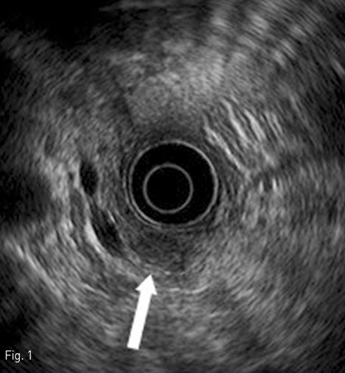
고혈압 병력이 있는 환자로 식후 발생하는 syncope 를 증상으로 내분비내과로 내원 후 시행한 lab 상 endogenous hyperinsulinemia와 hypoglycemia가 있었으며, 이후 시행한 EUS 상 pancreas tail에 19mm 크기의 lobulated mass 소견이 있고, 이는 hypoechoic halo가 동반되어 있는 hypoechoic mass로 관찰됨. 아울러 pancreatic duct의 확장은 보이지 않았음(Fig.1 ).
진단명
Pancreatic insulinoma
영상소견
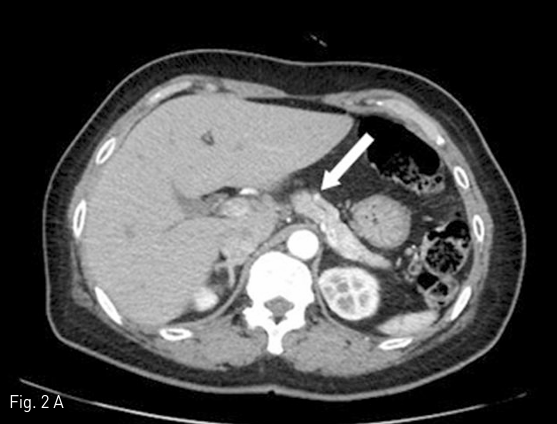
진단 위해 시행한 pancreas dynamic CT 상 pancreas tail 부분에 6mm size 이하의 multiple enhancing lesions이 보여 R/O neuroendocrine tumor 하에 (Fig. 2), insulinoma의 감별진단 및 localization을 위해 arterial stimulation with venous sampling (ASVS)를 시행함.
시술방법 및 재료
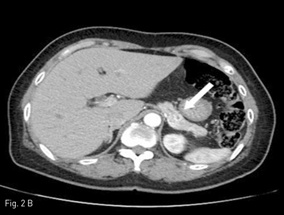
먼저 우 경정맥을 천자하여 sampling 용 5Fr Cobra catheter (Cook, Bloomington, IN)를 right hepatic vein에 거치하였다. 5Fr Röshe hepatic catheter (Cook)와 2.0 Fr microcatheter (Terumo, Tokyo, Japan)를 이용하여 gastroduodenal artery, splenic artery, SMA를 superselection 하여 baseline sampling을 실시하고, 10% calcium gluconate (DaeHan, Seoul, Korea)를 5ml씩 정량으로 투여한 후 30초, 60초, 120초에 hepatic vein blood sampling을 시행하였다. 각각의 sampling은 5분간의 간격을 두고 시행되었다(Fig. 3).
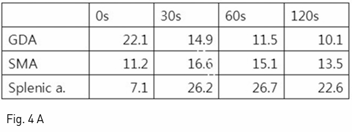
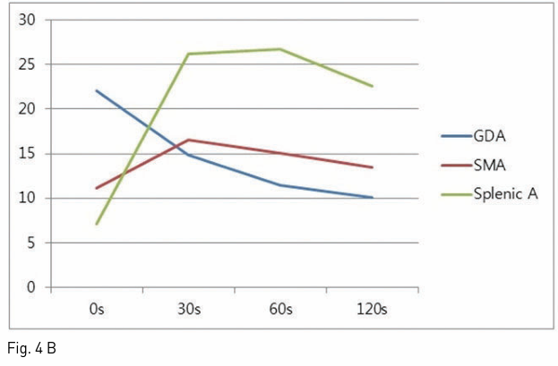
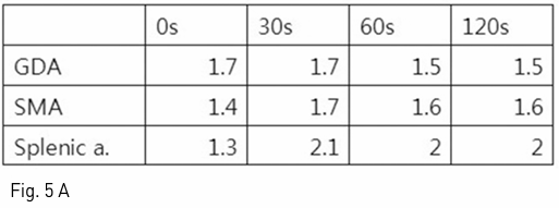
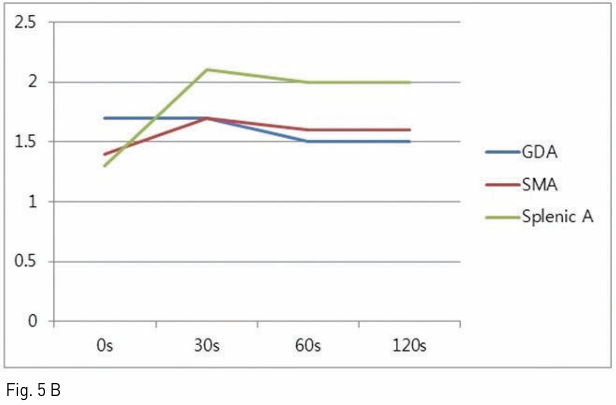
시술 결과 Gastroduodenal artery, splenic artery, SMA 각각에 10% calcium gluconate를 5ml 정량을 투여 후 0초, 30초, 60초, 120초 후 정맥혈채혈로 나온 insulin과 C-peptide 값을 측정하였으며, splenic artery 자극 후 얻은 정맥혈에서 투여 후 30초에 insulin이 26.2uIU/ml로 기저치의 3배 이상 증가하고, C-peptide 또한 2.1ng/ml로 증가함을 보였다. 이로써 insulinoma의 위치가 pancreas body 이하 tail 쪽에 위치함을 추정하였다(Fig. 4, 5).
추적관찰
Hypoglycemia 외에 다른 syncope 일으킬 만한 요인 없음을 확인하고, distal pancreatectomy 시행 중 병변의 위치가 일치함을 재차 확인하였으며, complication 없이 hypoglycemia 호전되어 정상 혈당이 지속되어 퇴원하였다.
Fig 1
Endoscopic ultrasonogram shows a hypoechoic nodule (arrow) with hypoechoic halo in pancreatic body.
Fig 2A
Pancreatic dynamic CT images show multiple small, hypervascular tumors in pancreatic body (arrow in A) and tail (arrow in B) portion.
Fig 2B
Pancreatic dynamic CT images show multiple small, hypervascular tumors in pancreatic body (arrow in A) and tail (arrow in B) portion.
Fig 3
A splenic angiogram shows subtle hypervascular tumor staining around mid-portion of splenic artery (arrow). Catheter for venous sampling in right hepatic vein was showed also (arrowheads).
Fig 4A
각 혈관 stimulation후 시간대별 얻은 venous sampling의 insulin 수치 (A) 및 변화도 (B)
Fig 4B
각 혈관 stimulation후 시간대별 얻은 venous sampling의 insulin 수치 (A) 및 변화도 (B)
Fig 5A
각 혈관 stimulation후 시간대별 얻은 venous sampling의 C-peptide 수치 (A) 및 변화도 (B)
Fig 5B
각 혈관 stimulation후 시간대별 얻은 venous sampling의 C-peptide 수치 (A) 및 변화도 (B)
고찰
과거에는 경피적으로 간내문맥을 천자하여 카테터를 splenic vein, SMV, 간문맥의 여러 부위에서의 인슐린 값으로 위치를 정하는 방법을 사용하였으나 정확도가 낮고, 합병증 유발률이 비교적 높아, 최근에는 동맥자극 정맥 채혈검사를 시행한다. 췌장을 공급하는 동맥에 칼슘을 주입하면 인슐린종에서 인슐린의 분비가 증가하고 분비된 인슐린은 문맥을 통하여 간정맥으로 돌아 나오는데 이를 채혈하여 측정한다. 보조적으로 인슐린 전구물질인 pro-insulin의 분해 산물인 C-peptide 값을 같이 측정하면 정확도를 높일 수 있다. 췌장의 동맥혈 공급은 크게 3 부위로 나누어 이루어지며, 췌장 두부의 상부와 경부는 위십이지장동맥에서 분지한 상췌십이지장동맥에서 혈액을 공급받고, 췌장 두부의 하부와 구상돌기는 상장간막동맥에서 기시하는 하체십이지장동맥에서 혈액을 공급받으며, 췌장의 체부와 미부는 비장동맥에서 분지하는 등췌장동맥, 대췌장동맥, 꼬리췌장동맥에서 혈액을 공급받는다. 시술 시 각 동맥마다 적어도 5분 이상의 시간을 두어야 하며, 선택하여 자극하는 혈관의 순서는 중요하지 않다. 30초와 60초에 얻은 인슐린 값이 다른 부위에 비하여 2배 이상 증가할 경우 의미 있는 결과로 판정한다.
참고문헌
1. Mattsson C, Young WF Jr. Primary aldosteronism: diagnostic and treatment strategies. Nat Clin Pract Nephrol 2006;2:198-208
2. Kempers MJ, Lenders JW, van Outheusden L, et al. Systematic review: diagnostic procedures to differentiate unilateral from bilateral adrenal abnormality in primary aldosteronism. Ann Intern Med 2009;151:329-337
3. Chung MJ, Choi BI, Han JK, Chung JW, Han MC, Bae SH. Functioning islet cell tumor of the pancreas. Localization with dynamic spiral CT. Acta Radiol 1997;38:135-138
4. Defreyne L, König K, Lerch MM, et al. Modified intra arterial calcium stimulation with venous sampling test for preoperative localization of insulinomas. Abdom Imaging 1998;23:322-331
Citations
Citations to this article as recorded by